CASE REPORT
Copyright ⓒ 2009 Korean Neurological Association 151
Print ISSN 1738-6586 / On-line ISSN 2005-5013 10.3988/jcn.2009.5.3.151 J Clin Neurol 2009;5:151-152
Anti-Ri-Antibody-Associated Paraneoplastic Syndrome in a Man with Breast Cancer Showing
a Reversible Pontine Lesion on MRI
Heeyoung Kim, MD; Youngmin Lim, MD; Kwang-Kuk Kim, MD
Department of Neurology, Asan Medical Center, University of Ulsan College of Medicine, Seoul, Korea
Received November 18, 2008 Revised February 5, 2009 Accepted February 5, 2009 Correspondence Kwang-Kuk Kim, MD Department of Neurology, Asan Medical Center, University of Ulsan College of Medicine, 388-1 Pungnap-dong,
Songpa-gu, Seoul 138-736, Korea Tel +82-2-3114-3440 Fax +82-2-474-4691 E-mail [email protected]
BackgroundaaParaneoplastic neurological disorders associated with anti-Ri-antibodies, which are typically present with opsoclonus-myoclonus-ataxia. Most cases with anti-Ri-antibodyasso- ciated paraneoplastic syndrome due to breast cancer occur in women - its occurrence in men is extremely rare.
Case ReportaaWe present herein the case of a male patient with breast cancer who had atypical anti-Ri-antibody-associated paraneoplastic syndrome presenting as complete horizontal ophth- almoplegia, left trigeminal sensory symptoms, and truncal ataxia. Following the diagnosis of paraneoplastic syndrome, chemotherapy and immunomodulating treatment including intrave- nous immunoglobulin and oral prednisolone were administered. Although the patient was ne- gative for serum anti-Ri-antibodies 14 weeks later, his symptoms persisted.
ConclusionsaaTo our knowledge, this is the first case report of ophthalmoplegia without op- soclonus-myoclonus in a male anti-Ri-antibody-positive patient with breast cancer.
J Clin Neurol 2009;5:151-152 Key Wordsaabreast cancer, anti-Ri-antibody, paraneoplastic syndrome.
Introduction
Paraneoplastic neurological disorders associated with anti-Ri- antibodies (also known as anti-neuronal nuclear antibodies type II) mainly present with opsoclonus-myoclonus-ataxia.1 Ophthalmoplegia without opsoclonus is very rare.2,3 Although anti-Ri-antibodies have been reported in patients with gyneco- logical tumors and small-cell lung cancer,1,4 they are mostly identified in patients with breast cancer. Most cases due to breast cancer occur in women. Its occurrence in men is ex- tremely rare; there is thus far only one reported case, in which paraneoplastic opsoclonus-myoclonus was found in a man with breast cancer and anti-Ri-antibodies.5 We present herein the case of a male patient with breast cancer who had atypical anti-Ri-antibody paraneoplastic syndrome presenting as com- plete horizontal ophthalmoplegia, left trigeminal sensory symp- toms, and truncal ataxia.
Case Report
A 52-year-old man was referred to a hospital with subacute
onset of binocular diplopia, left facial numbness, and gait disturbance. He was diagnosed with breast cancer 1 month after the onset of symptoms and then underwent a right mod- ified radical mastectomy to remove an invasive ductal car- cinoma. Neurologic examination revealed left trigeminal hy- pesthesia and complete horizontal gaze palsy without ops- oclonus-myoclonus. His motor strength and deep tendon re- flexes were normal. Sensation was intact to all modalities.
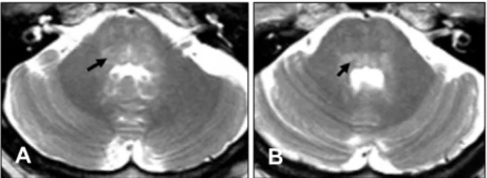
The patient exhibited a wide-based gait and could not per- form tandem gait. His cerebrospinal fluid showed normal cell counts and protein values, and negative cytology and viral markers. Immunofluorescence analysis revealed that the pa- tient’s serum contained anti-Ri-antibodies (serial dilutions revealed a specific antibody titer of 1 : 60), and was negative for anti-Hu, anti-Yo, and neuromyelitis optica antibodies. A brain magnetic resonance imaging (MRI) scan revealed a high-signal-intensity lesion in the pontine tegmentum that was not enhanced with gadolinium (Fig. 1). A brain magnetic re- sonance angiogram showed no significant steno-occlusive le- sion. Initial infusion of intravenous immunoglobulins (400 mg/kg/day for 3 days) was administered 4 months after
Anti-Ri Antibody Paraneoplastic Syndrome
152 J Clin Neurol 2009;5:151-152
symptom onset, followed by a maintenance dose (400 mg/
kg/day) every 2 months for 1 year. Concurrent chemotherapy of doxorubicin, cyclophosphamide, and dexamethasone was administered. After four cycles of chemotherapy, the patient’s serum tested negative for anti-Ri-antibodies, and a follow-up brain MRI showed neither abnormal signal change nor atro- phy in the brainstem. Further extensive searching, including whole-body positron-emission tomography, produced no evi- dence of tumor recurrence. However, this patient’s neurolo- gical deficit has persisted during the subsequent 3-year fol- low-up, in spite of intensive chemotherapy and immunomo- dulating treatment.
Discussion
Whilst opsoclonus-myoclonus ataxia syndrome is often used to describe anti-Ri-antibody-associated paraneoplastic dis- order, it has a wide spectrum of neurological manifesta- tions.6,7 The present case emphasizes the clinical heteroge- neity of this disorder. However, even in the absence of opso- clonus, oculomotor dysfunction is usually prominent,8,9 and the brainstem is a major site of autoimmunity. The main cli- nical features in our patient were brainstem syndrome involv- ing the structures of the dorsal pons: the parapontine reti- cular formation, abducens nucleus, medial longitudinal fasci- culus, and trigeminal nucleus of the spinal tract.
In anti-Ri-antibody-positive patients, brain MRI findings are usually normal, with only a few exceptions. There are reports of an abnormal signal in the dorsal midbrain,10 and atrophy of the cerebellar vermis1 on brain MRI, associated with anti-Ri-antibodies. In our patient, MRI disclosed sym- metric hyperintense lesions in the dorsal pons that were rever- sible with treatment. However, his symptoms persisted even
after the lesion had disappeared. Tumor removal and chemo- therapy did not improve the paraneoplastic abnormalities and the effect of immunoglobulin was also disappointing.
Thus, it appears that irreversible, rather than functional damage to the neurons causes the symptoms of anti-Ri-antibody-as- sociated paraneoplastic syndrome.
The underlying tumor in our case was an invasive ductal cell carcinoma of the breast. The incidence of breast caner in man is low in the literature, representing approximately 1%
of all breast cancer diagnoses.11 To our knowledge, this is the first case report of ophthalmoplegia without opsoclonus-myo- clonus in a male, anti-Ri-antibody-positive patient with breast cancer. The findings of this case support the differentiation of anti-Ri-antibody-associated paraneoplastic syndrome even in the absence of opsoclonus, and that it can also occur in male patients with breast cancer.
REFERENCES
1. Luque FA, Furneaux HM, Ferziger R, Rosenblum MK, Wray SH, Schold SC Jr, et al. Anti-Ri: an antibody associated with parane- oplastic opsoclonus and breast cancer. Ann Neurol 1991;29:241-251.
2. Escudero D, Barnadas A, Codina M, Fueyo J, Graus F. Anti-Ri-asso- ciated paraneoplastic neurologic disorder without opsoclonus in a pa- tient with breast cancer. Neurology 1993;43:1605-1606.
3. Ohmer R, Golnik KC, Richards AI, Kosmorsky GS. Ophthalmo- plegia associated with the anti-Ri antibody. J Neuroophthalmol 1999;
19:246-248.
4. Voltz R. Paraneoplastic neurological syndromes: an update on dia- gnosis, pathogenesis, and therapy. Lancet Neurol 2002;1:294-305.
5. Wirtz PW, Sillevis Smitt PA, Hoff JI, de Leeuw B, Lammers GJ, van Duinen SG, et al. Anti-Ri antibody positive opsoclonus-myoclonus in a male patient with breast carcinoma. J Neurol 2002;249:1710-1712.
6. Pittock SJ, Lucchinetti CF, Lennon VA. Anti-neuronal nuclear auto- antibody type 2: paraneoplastic accompaniments. Ann Neurol 2003;
53:580-587.
7. Kastrup O, Meyring S, Diener HC. Atypical paraneoplastic brainstem encephalitis associated with anti-ri-antibodies due to thymic carcinoma with possible clinical response to immunoglobulins. Eur Neurol 2001;
45:285-287.
8. Sutton IJ, Barnett MH, Watson JD, Ell JJ, Dalmau J. Paraneoplastic brainstem encephalitis and anti-Ri antibodies. J Neurol 2002;249:1597- 1598.
9. Ko MW, Dalmau J, Galetta SL. Neuro-ophthalmologic manifestations of paraneoplastic syndromes. J Neuroophthalmol 2008;28:58-68.
10. Case records of the Massachusetts General Hospital. Weekly clinic- opathological exercises. Case 9-1988. A 57-year-old woman with wor- sening opsoclonus. N Engl J Med 1988;318:563-570.
11. Giordano SH, Cohen DS, Buzdar AU, Perkins G, Hortobagyi GN.
Breast carcinoma in men: a population-based study. Cancer 2004;101:
51-57.
Fig. 1. T2-weighted magnetic resonance images showing bilat- eral symmetric hyperintense lesions in the pontine tegmentum. A:
Parapontine reticular formation (arrow) in the lower pons. B: Me- dial longitudinal fasciculus (arrow) in the mid pons.
A B