Airway management during general anesthesia in an intellectually disabled patient with undiagnosed tracheomalacia
Sooil Shin
1, Seungoh Kim
21Department of Advanced General Dentistry, School of Dentistry, Dankook University, Cheonan, Korea
2Department of Anesthesiololgy, School of Dentistry, Dankook University, Cheonan, Korea
In cases of intellectually disabled patients, there is sometimes difficult to obtain sufficient information due to the intellectual disorder, even though the patient has significant medical problems. Herein, we report a case of decreased oxygen saturation and inadequate air exchange during general anesthesia in an intellectually disabled patient. We also describe the subsequent management, including the diagnosis of tracheomalacia (TM) using bronchoscopy, and the management of airway compromise with manual and/or controlled respiration, which led to the prevention of complications.
Keywords: Airway management; Bronchoscope; Dental treatment; Tracheomalacia.
This is an Open Access article distributed under the terms of the Creative Commons Attribution Non-Commercial License (http://creativecommons.org/licenses/by-nc/4.0/) which permits unrestricted non-commercial use, distribution, and reproduction in any medium, provided the original work is properly cited.
Received: April 1, 2018•Revised: April 16, 2018•Accepted: April 16, 2018
Corresponding Author:Seungoh Kim, Department of Anesthesiology, School of Dentistry, Dankook University, 119 Dandaero, Dognam-gu, Cheonan, 31116, Korea Tel: +82-41-550-1863 E-mail: ksomd@dankook.ac.kr
Copyrightⓒ 2018 Journal of Dental Anesthesia and Pain Medicine
Tracheomalacia (TM) refers to a weakness in the support of the trachea, including reduction and/or atrophy of the longitudinal elastic fibers of pars membranacea, or im- paired cartilage integrity, which leads to symptoms of airway obstruction [1-2]. Diagnosis of the condition is often overlooked, as chest radiography appears normal [3]. In case of intellectually disabled patients, medical staffs are unable to obtain patient’s information because intellectually disabled patients have difficulty in communicating and cannot express in detail, about their discomfort, condition, and symptom. Moreover, these patients are not cooperative or refuse to undergo clinicopathologic and radiographic examination. As a result, inadequate medical information obtained before anesthesia and operation in most cases. Although the patients have normal vital signs before anesthesia, there
are cases in which the physiological changes are sharply exacerbated in anesthesia and operation.
In this case report, we outline the method used to solve the respiratory problem of TM using the bronchoscope, thereby preventing complications, which was not diag- nosed before anesthesia and operation due to lack of communication with the intellectually disabled patient and insufficient information.
CASE REPORT
A 27-year-old male patient (155 cm, 75 kg) with grade 3 intellectual disability presented with the chief com- plaints of severe dental caries and generalized calculus deposition. The patient was capable of minimal com-
Fig. 1. Panoramic Radiograph in a 27-year-old male patient was conducted before treatment.
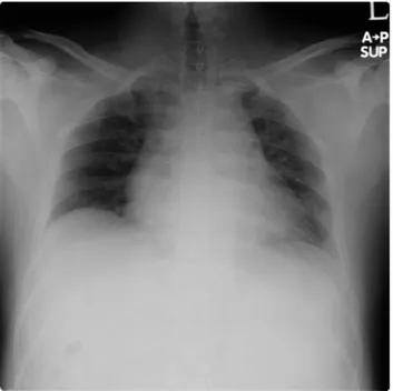
Fig. 2. Chest Radiograph shows normal lungs. Fig. 3. Bronchoscopic image shows crescent type of collapsed trachea.
munication, was uncooperative towards oral examination due to negative experience during previous dental treat- ments. He reported no significant medical history. At the first appointment, simple intraoral examination and panoramic radiography were performed, and chest radio- graphs were obtained in order to establish accurate diagnostic and treatment plans under general anesthesia (Fig. 1, 2). Radiological findings showed clear lungs, heart of normal size, and absence of definite abnormal lung infiltration.
The patient presented after 8 h of fasting. He was sedated with inhalation of O2 (50%) and N2O (50%) 4 L/min via mask as well as 4% sevoflurane gas (USP Baxter Inc.). After the loss of consciousness was confirmed, EKG, heart rate, blood pressure, respiratory
rate, end-tidal carbon dioxide (CO2), body temperature, and entropy level were all monitored. Subsequently, the patient’s SpO2 level declined to below 88-90%, and O2
(100%) 8 L/min mask ventilation was started. Three puffs of Ventolin Respiratory Solution (GlaxoSmithKline, Korea) were administered through the mask under the suspicion of an asthma attack. The patient had an episode of vomiting, and oral suction was performed. After that, SpO2 level recovered to 90-93%. A line was inserted, and atropine (Daewon Pharm. Co.) 0.5 mg i.v. and atracurium (Hana Pharm. Co., Ltd) 20 mg i.v. were administered for preventing bradycardia and muscle relaxation. Epine- phrine agent (Jeil Pharm. Co., Ltd.) was administered using a cotton bud for nasal intubation, and the trachea was intubated with a 24 cm 6.5 endotracheal tube (Sewoon Medical Co.,Ltd.). However, since the patient’s SpO2 level was relatively low at 91-93%, and the airway pressure was high, bronchoscopy of the trachea was performed. There was an area of narrowing, approxi- mately 3 cm long, at the end of endotracheal tube, and pulsatory motion was observed (Fig. 3). The position of the endotracheal tube was adjusted upon deeper reinsertion, after which SpO2 level was maintained above 94%.
Clinical and radiological evaluations of the patient showed missing #13, 21, and 23; severe dental caries of
#16, 15, 14, 12, 11, 22, 24, 25, 26, 35, 36, 37, 45, 46, and 47; and mild caries of #27, 34, and 44. Upon
Fig. 4. Oblique coronal reconstructed image shows prominent aortic arch (arrow) and focal indentation and luminal narrowing of trachea (arrowhead) due to extrinsic compression. Sagittal and curved multiplanar reconstructed image shows prominent aortic arch (arrow) and compression of trachea (black arrowhead) between aortic arch and thoracic spine.
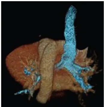
Fig. 5. 3D-Volume reconstructed image of heart, major arteries and trachea (blue colored structure) shows prominent aortic arch (arrow) and luminal narrowing of trachea (arrowhead) due to extrinsic compression.
consultation with the guardian, prosthodontic treatment in the patient was planned as follows: for the removable partial denture on maxilla with #17, and 27 abutments, and mandible with #34, and 44 abutments; and removal of all teeth of maxilla without abutments that were displaced due to severe dental caries, and posterior teeth of mandible without first premolars.
On the day of the first operation, extraction of #16, 15, 14, 12, 11, 45, 46, and 47 and suture with Vicryl 4-0 (Ethicon Inc.,) were performed.
After the dental treatment, N2O and sevoflurane gas were turned off, and the patient went into recovery with inhaling of O2 8 L/min. Upon spontaneous respiration, the patient’s SpO2 level decreased again to 85% and recovered to 95-99% with positive pressure ventilation.
After the recovery of consciousness, extubation was attempted, but SpO2 level declined again to 88-90% and was maintained at 95-99% in the recovery room with O2
100% 4 L/min inhalation via nasal cannula. Total anesthesia time was 180 min, and total operation time was 100 min. The patient was discharged after 4 h of monitoring, during which, he maintained SpO2 94-95%
at room air. Magnetic resonance imaging (MRI) was ordered for accurate diagnosis of the airway narrowing.
MRI findings showed prominent aortic arch and focal indentation and luminal narrowing of trachea due to
extrinsic compression between aortic arch and thoracic spine (Fig. 4, 5).
The procedure for general anesthesia was the same as the first visit. Bronchoscopy findings showed a pulsatory motion within the trachea, consistent with the findings of the bronchoscopy performed during the first treatment.
Sufficient positive pressure ventilation was implemented during induction and recovery, in order to prevent the
narrowing of the area affected by TM during spontaneous respiration, which resulted in stable vital signs including SpO2.
Under the 2nd , 3rd general anesthesia, restorative treatment of #34, root canal therapy and crown preparation of #27, extraction of #22, 24, 25, 26, 35, 36, and 37.
Other dental treatments, including caries removal and restorative treatment of #34, 44, were relatively simple and administered on an outpatient basis. The observation of mandibular movement and the design of removable partial denture for the readjustment of articulation were also performed on an outpatient basis, due to greater ease of treatment with the patient’s consciousness intact. The patient’s fear of dental treatment reduced with each clinic visit, and the patient became very cooperative with greater understanding of the treatment process. He also adjusted well to the use of removable partial denture and showed good masticatory function and esthetic recon- struction on follow-up.
Consent: Informed consent was obtained from the guardian of patient for this case report.
DISCUSSION
Malacia is abnormal softening of biological tissue and, generally refers to cartilage or bone in medical terminology. TM refers to a softness of the tracheal cartilage, resulting in expiratory collapse, leading to symptoms of airway obstruction [2]. Czyhlarx first described the postmortem finding of an abnormally large trachea and bronchi in 1897 [4]. In 1932, Mounier-Kuhn was the first to report a case of isolated tracheal enlargement in an adult [5]. The incidence of this disorder is unclear. Jokinen et al. reported a 23% incidence in 214 patients examined for a history of chronic bronchitis [6].
Other studies reported 4.5% incidence of pulmonary disorders in 2150 patients examined through bron- choscopy [7].
TM is classified as congenital and acquired forms.
Congenital TM is the most common anomaly in premature infants as a consequence of inadequate maturity of trachea cartilage. Moses et al. and Cohen recognized that although the innominate artery was in normal position, it was often implicated in acquired TM [8,9]. This manifests as pulsatile movement of an abnormally soft and weak trachea parallel to the normally placed artery and is often considered a primary form of the disease [10]. Acquired TM that results from degeneration of previously normal cartilage is more common than congenital TM. Feist et al. identified tracheostomy or intubation with endotracheal tubes as the most common cause of acquired TM [11]. Also, chronic external compression of the trachea results in acquired TM including malignancy, benign tumors, cysts, abscesses and aortic aneurysm etc. [2].
Symptoms may include dyspnea, hemoptysis, and chronic cough, but it can also be asymptomatic. Routine anterio-posterior and lateral chest radiographs often show no abnormality [11]; hence bronchoscopic visualization of the trachea remains the diagnostic gold standard.
Our patient had no sign and symptom suggesting TM in preoperative examination; he had no relevant medical history except overweight, he had undergone uneventful surgery with tracheal intubation, and his preoperative chest radiograph was normal.
Moreover, it was difficult to obtain accurate information about the patient, since we had to rely on the guardian. Hence, we could not have suspected the existence of TM.
During loss of consciousness after the induction of sedation, his SpO2 level decreased rapidly during spontaneous respiration and recovered by performing positive pressure ventilation. This is because spontaneous respiration causes intrathoracic negative pressure which led to further narrowing of the airway affected by TM;
whereas, controlled respiration through mechanical ventilation can supply mixed gas to the lungs through positive pressure, leading to expansion of the airways.
After the intubation with ETT, airway problem due to tracheomalacia was not as evident due positive pressure
ventilation, but a similar phenomenon recurred during recovery when the patient was transitioning from controlled respiration to spontaneous respiration under a semi-conscious state. Such a phenomenon can be explained as “paradoxical respiration,” wherein the intrathoracic space decreases during inspiration and increases during expiration [12]. This phenomenon may appear during spontaneous respiration in an unconscious patient under sedation. In case of TM, a negative intrathoracic pressure is formed during the inspiratory phase of spontaneous respiration and the increased airway resistance due to airway narrowing compromises efficient gas movement, leading to rapid decline in SpO2 level and increased pressure of the airway. In our patient, the patency of the airway was maintained with manual or controlled respiration, and stable SpO2 was achieved.
Under subsequent general anesthesia, airway was maintained with sufficient mask ventilation during induction and recovery, and all treatment was uneventful.
We report a case of TM diagnosed during general anesthesia in an intellectually disabled patient due to difficult communication leading to inadequate preo- perative examination, which was promptly managed by dilating the narrowed airway with sufficient positive pressure ventilation during induction and recovery, which allowed the resolution of decreased SpO2 and prevention of complications.
AUTHOR ORCIDs
Sooil Shin: https://orcid.org/0000-0002-7203-749X Seungoh Kim: https://orcid.org/0000-0001-7288-8457
NOTES: There are no financial or other issues that might lead to conflict of interest.
REFERENCES
1. Johnson TH, Mikita JJ, Wilson RJ, Feist JH. Acquired tracheomalacia. Radiology 1973; 109: 576-80.
2. Carden KA, Boiselle PM, Waltz D, Ernst A. Tracheo- malacia and tracheobronchomalacia in children and adults:
an in-depth review. Chest 2005; 127: 984-1005.
3. Wright CD. Tracheomalacia. Chest Surg Clin N Am 2003;
13: 349-57.
4. Czyhlarz ER. Ueber ein pulsiondivertikel der trachea mit bemerkungen uber das verhalten der elastichen fasern an normalen tracheen und brounchein. Zentralbl Allg pathol 1897; 18: 721-8.
5. Mounier-Kuhn P. Dilation de al trachee: Constantations rudui-graphiques et bronchoscopiques. Lyons Medical 1932; 150: 106-9.
6. Jokinen K, Palva T, Sutinen S, Nuutinen J. Acquired tracheobronchomalacia. Ann Clin Res 1977; 9: 52-7.
7. Jacobs IN, Wetmore RF, Tom LW. Tracheobroncho- malacia in children. Arch Otolaryngol Head Neck Surg 1994; 120: 154-8.
8. Mose CA, Izukawa T, Trusler GA. Innominate artery compression of the trachea. Arch Otolaryngol 1975; 101:
733-8.
9. Cohen D. Tracheopexy-tracheal suspension for severe tracheomalacia. Aust Paediatr J 1981; 17: 117-21.
10. Creenholz DK, Karrer FM, Lilly JR. Contemporary surgery of tracheomalacia. J Pediatr Surg 1986; 21: 511-4.
11. Feist JH, Johnson TH, Wilson RJ. Acquired tracheomalacia:
etiology and differential diagnosis. Chest 1975; 68: 340-5.
12. Proctor H. The stove-in chest with paradoxical respiration.
Br J Surg 1955; 42: 622-33.
![Bid Invitation [Security Guard]](data:image/gif;base64,R0lGODlhAQABAIAAAP///wAAACH5BAEAAAAALAAAAAABAAEAAAICRAEAOw==)