INTRODUCTION
For many years, Porcelain fused to metal (PFM) restorations have represented the most widely used restorative technique in fixed prosthodontics and this popularity may have attributed to the clinical longevity and accepted esthetics. However, these restorations are constantly exposed to challenges such as marginal leakage, which leads to the demise of the tooth pulp.1,2 When the crowns or bridges are going to be retained, there is very little information regarding the best way to restore the endodontic access opening. Clinicians routinely use either amal- gam, composite resin or resin modified glass ionomer cement.
Material of choice is critical, since coronal leakage of bacte-
ria or their by products are an important factor in the failure of non-surgical root canal treatment (NSRCT).1
The newer generations of dentin bonding agents have better adhesion and marginal integrity in the laboratory but clinical data and long term results still need to be evaluated.3-5The advan- tage of silane coupling agents appears to enhance the bond strength by promoting a chemical bond between the adhesive and the adherent.6The dye leakage model has been used to deter- mine if any of the dental materials in current clinical use has the ability to prevent coronal leakage in restored endodon- tic access openings in permanently fixed crowns following NSRCT.7Exposing the samples to thermocycling speeds up the diffusion of water between the filling materials and the
The effect of thermocycling on the bonding of different restorative materials to access opening through
porcelain fused to metal restorations
Mohammed M. AL-Moaleem1, BDS, MSc, PhD, Farhan Khalid Shah1, BDS, MDS, Nausheen Saied Khan2, BDS, MSc, Amit Porwal3*, BDS, MDS
1Department of Prosthodontics, College of Dentistry, King Khalid University, Abha, Saudi Arabia
2Dental Public Health, Year II , Kings College London, UK
3Department of Prosthodontics, Pacific dental college and Hospital, Udaipur, India
PURPOSE. Porcelain fused to metal (PFM) crowns provide the best treatment option for teeth that have a large or defective restoration. More than 20% of teeth with PFM crowns or bridges require non-surgical root canal treatment (NSRCT). This may be due to the effect of restora- tive procedures and the possible leakage of bacteria and or their by-products, which leads to the demise of the tooth pulp. Thus, this study was planned to compare the ability of the restorative materials to seal perforated PFM specimens. MATERIALS AND METHODS. The study eval- uates the ability of amalgam, composite or compomer restorative materials to close perforated PFM specimen’s in-vitro. Ninety PFM speci- mens were constructed using Ni-Cr alloys and feldspathic porcelain, and then they were divided into 3 groups: amalgam (A), composite + Exite adhesive bond (B) and compomer + Syntac adhesive bond (C). All the PFM samples were embedded in an acrylic block to provide complete sealing of the hole from the bottom side. After the aging period, each group was further divided into 3 equal subgroups according to the ther- mocycling period (one week for 70 cycles, one month for 300 cycles and three months for 900 cycles). Each subgroup was put into containers containing dye (Pelikan INK), one maintained at 5℃and the other at 55℃, each cycle for 30 sec time. The data obtained was analyzed by SPSS, 2006 using one way ANOVA test and student t-test and significant difference level at (P<.01). RESULTS. The depth of dye penetration was measured at the interfaces of PFM and filling materials using Co-ordinate Vernier Microscope. The lowest levels of the dye penetration for the three groups, as well as subgroups were during the first week. The values of dye leakage had significantly increased by time intervals in sub- groups A and C. CONCLUSION. It was seen that amalgam showed higher leakage than composite while compomer showed the lowest lev- el of leakage. [J Adv Prosthodont 2011;3:186-9]
186
KEY WORDS: Porcelain fused to metal restorations; Restorative dental materials; Thermocycling effect
Corresponding author: Amit Porwal
Pacific dental college and hospital, Debari, Udaipur, 313024, India Tel. 919950547337: e-mail, [email protected]
Received May 16, 2011 / Last Revison June 26, 2011 / Accepted September 8, 2011
ⓒ 2011 The Korean Academy of Prosthodontics
This is an Open Access article distributed under the terms of the Creative Commons Attribution Non-Commercial License (http://creativecommons.org/licenses/by- nc/3.0) which permits unrestricted non-commercial use, distribution, and reproduction in any medium, provided the original work is properly cited.
http://dx.doi.org/10.4047/jap.2011.3.4.186 ORIGINAL ARTICLE J Adv Prosthodont 2011;3:186-9
ceramic.8This study was planned in order to evaluate the abil- ity of filling materials such as amalgam, composite and com- pomer, to close access openings in PFM specimens.
MATERIALS AND METHODS
Ninety PFM specimens were constructed from green wax of 0.4 mm in thickness which were punched with 25 mm diam- eter ring and in the center with 6 mm diameter ring. Each of the 90 samples was invested, burned out and casted according to manufacturer’s instructions. The metal samples were sep- arated from their sprues, leaving 2 mm to facilitate handling during acrylic block constructions. All the samples were sandblasted with aluminum oxide particle to remove the excess oxide layer. Opaque and body porcelain were then applied according to manufacturer’s instructions (The opaque layer around the perforation was 0.3 ± 0.1 mm while the body porcelain was 2.0 ± 0.3 mm in thickness). The PFM samples were filled with wax to prevent inward movement of the acrylic into the perforation during acrylic mold construction. The depths of the PFM samples were 2.0 ± 0.5 mm (Fig. 1).
The samples were divided into 3 equals groups according to the filling material.
Group A: The PFM perforations were filled with amalgam Septalloy NG-70 (Septodont, France) according to the man- ufacturer’s instructions, regarding mixing and processing time. After 48 hours, the amalgam fillings were polished with green silicon polishing burs (Vivadent, Liechenstein) (with one bur for 10 samples).
Group B: The PFM perforations were filled with Tetric-Ceram composite (Vivadent, Liechenstein). The walls of the PFM per- forations were treated with 37% phosphoric acid gel for 60 sec, washed with a copious amount of water for 15 sec, dried with oil-free air spray for 10 sec. The bonding agent, Excite (Vivadent, Leichtenstein), was applied to the treated walls of PFM samples using Vivadent applicator brush for 10 sec
and finally, Tetric-Ceram was applied into the PFM perfora- tion in one increment. After adapting resin with plastic instru- ments, the filling and perforation were covered with celluloid strip through which the light curing was done for 40 sec by hold- ing the light emission window close to the strip and the fillings.
Group C: The PFM perforations were filled with Compoglass- F compomer (Vivadent, Liechenstein). The walls of the PFM perforations were treated with 37% phosphoric acid gel for 60 sec, rinsed and dried thoroughly and then the silane liquid Monobond-S (Vivadent, Liechenstein) was applied to the ceramic surface and left for 60 sec. The bonding agent, Syntac adhesive was applied with a brush, left for 2 sec, and dispersed with oil-free air spray until no movement of the liquid was visible. Then, with plastic instruments, sufficient amount of compomer was filled into the PFM perforations and light curing was done for 40 sec by holding the light emission window as close as possible to the celluloid strip and the fill- ing. The fillings of group B and C were finished with gray sil- icon composite finishing burs and polished with green silicon polishing burs (one bur for 10 samples). The manual pressure during condensation of the amalgam and the adaptation of com- posite as well as the compomer was 5 ± 1 kg.
All the samples were incubated in normal physiological saline for one week at 37℃. Each group was further subdivided into three equal subgroups according to the thermocycling peri- od (one week for 70 cycles, one month for 300 cycles and three months for 900 cycles). The dye used was Pelikan INK. The samples were put long enough in the dye solution to increase hydraulic pressure to enhance the dye penetration. Thermocycling was carried out to investigate the effect of temperature change on the adaptation of restorative materials to PFM interfaces and walls. The thermocycling device cycles the samples between two water bathes, one maintained at 5 ± 2℃ and the other at 55 ± 2℃. Each cycle was done for 30 sec in each bath.
After completing the thermocycling, the samples were removed from the water bath and allowed to dry for 2 hrs at room tem- perature to facilitate the dye fixation.
All the samples were sectioned by a double-ended dia- mond wheel disk mounted on hand piece under water spray.
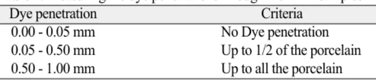
The disk sectioned at the midline of the samples passing through the center of the filling materials (Fig. 1). The depth of dye penetration was measured from both sides for each PFM and filling materials interfaces using Co-ordinate Vernier Microscope (Griffin & George, UK) at × 20 magnification and scores calculated (Table 1). The data obtained was analyzed
187
The effect of thermocycling on the bonding of different restorative materials to access opening through porcelain fused to metal restorations
J Adv Prosthodont 2011;3:186-9
AL-Moaleem MM et al.
Fig. 1. PFM sample mounted in acrylic block.
Table 1. Measuring the dye penetrations through the PFM samples
Dye penetration Criteria
0.00 - 0.05 mm No Dye penetration
0.05 - 0.50 mm Up to 1/2 of the porcelain 0.50 - 1.00 mm Up to all the porcelain
by SPSS, 2006 using one way ANOVA test and student t-test and significant difference level at (P<.01).
RESULTS
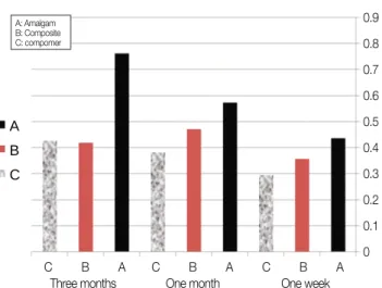
Two surfaces were obtained after sectioning of each sample (Fig. 1). The four readings gained from each sample were obtained together and then divided by four to get the mean pen- etration level. From Fig. 2, it is clear that the lowest level of the dye penetration occurred during the first week in all three groups including the subgroups. The value of dye leak- age had significantly increased by time intervals in sub- groups A and C. There was no significant difference in the time intervals between one month and three months in subgroups B and C.
DISCUSSION
The aim of this study was to determine if amalgam, composite or compomer has the ability to prevent coronal leakage in restored endodontic access opening in PFM specimen’s in-vitro.
Septalloy NG-70 amalgam capsules (high copper alloy admixed type) were used in this study because amalgam has been widely used as a restorative material for more than a cen- tury in the field of dentistry.9Tetric-Ceram has an advanced com- posite technology used for anterior and posterior fillings. It is a highly dispersed hybrid material that has continuous fluoride ion releasing effect and contains ceramic particles in its com- position.4,10Compomer is a hybrid material composed of a sin- gle hydrophobic resin that is filled with acid-leachable glass particles bonded with functional primer and light cured, which results in its higher mechanical strength, improved
marginal seal by hygroscopic expansion and improved bond- ing ability after storage in water.4,11Therefore, when two dis- similar materials are to be attached, whether micromechani- cal or chemical is chosen, the use of silane coupling agents could increase attachment.12
An interpretation of the results indicated that all restorative materials showed leakage but the dye did not reach the met- al part of the PFM, and this is due to low difference in linear coefficient of thermal expansion (LCTE) between Ni-Cr alloy and amalgam or composite, this is in agreement with the previous studies1,7,13,14
Group A (with highly significant difference) had the high- est dye penetration compared to other groups and this may be due to the high difference between LCTE of porcelain and amal- gam15 or due to the contact angel that formed between the PFM walls and the amalgam, which is considerably sufficient for the dye particles to penetrate.15
Our finding agreed with those1who concluded that amalgam is not the material of choice for restoration of access opening through the PFM crowns, but did not agree with those13who founded that the use of lathe-cut and admixed amalgam would reduce the marginal microleakage.
Dye penetration of group B (with significant difference) indi- cated that microleakage of Tetric-Ceram composite increased as the number of thermocycling increased (Fig. 2). This increase with aging may be related to disintegration of the bond- ing agent and gap formation at the composite filling and PFM interfaces.15This could be explained by the initial poly- merization shrinkage of Tetric-Ceram which resulted in a pulling back of the restoration and adhesive bonding toward the light source resulted in gap formation and increased microlekage.15The incorporation of water into the composite may compensate the initial shrinkage of the Tetric-Ceram and resulted in secondary expansion, which could maintain the same level of leakage with time lapses.10,16The use of Exite, 5th generation adhesive system which has both hydrophilic and hydrophobic groups, results in good bond with hydrophobic porcelain. Although our results did not agree with those7,13who concluded that microleakage of Tetric-Ceram is very high dur- ing the first 48 hrs, it shows great reduction in leakage with aging due to water sorption.
Group C indicated a significant difference among samples thermocycled for week and samples thermocycled for one or three month. This is because compomer does not under-go dimen- sional changes by expansion and did not improve the marginal seal after storage in water for a week,11but during one or three months absorption of water and hygroscopic expansion of com- pomer has resulted in improvement of bonding ability after stor- age in water. On the other hand, there is no significant difference between samples thermocycled for one and three months and this may be due to the effect of silane bonding agent, which minimizes the gap formation at compomer and PFM interfaces.
188
The effect of thermocycling on the bonding of different restorative materials to access opening through porcelain fused to metal restorations
J Adv Prosthodont 2011;3:186-9 AL-Moaleem MM et al.
Fig. 2. The mean values of dye penetration in mm for all groups and sub- groups (time intervals).
A B C
A: Amalgam B: Composite C: compomer
0.9 0.8 0.7 0.6 0.5 0.4 0.3 0.2 0.1 0 C B A C B A C B A
Three months One month One week
Also, compomer is a single component hydrophobic resin, com- posed of combination of both composite and GI, so the LCTE of it is in the range of GI and anterior or posteriors com- posite, which is similar to that of hydrophobic porcelain.3,15Some authors8,11have reported that Compoglass-F produced smoother surfaces than conventional GI cements, which lead to the reduction of susceptibility to bacterial adhesion that comes as a consequence of gap formation.
It is obvious from Fig. 2 that increasing the number of thermocycling resulted in more dye penetration in all subgroups, which may be due to excessive contraction of amalgam dur- ing the first days and the polymerization shrinkage of Tetric- Ceram and Compoglass-F and this corresponds with Grobler et al.17 Septalloy NG-70 amalgam subgroups showed the highest level of dye penetration because the time provided is not long enough and the high-copper alloys did not readily pro- duced corrosion product. Our results did not agree with those7,14 who said that the dual-cured composite and glass ionomer had the greatest level of dye penetration in PFM crowns comparing with that of amalgam.
There is no significant difference between one and three months in subgroup B and C; this may be due to polymerization shrinkage of Tetric-Ceram and Compoglass-F during the first days, and water absorption and secondary expansion afterwards. In subgroups C, etching and silane application pri- or to placement of dentin bonding was significantly effective as it pulled resin-based composite toward walls of the PFM result- ing in reduction of microleakage and better marginal integri- ty in vitro.4,5,18Several authors3,17have concluded that compomer is alleged to produce less shrinkage and compensate for internal stress by its elasticity and through water resorption there- fore, it has the potential to minimize the initial shrinkage due to polymerization.
This in vitro and short time study only simulates the actual used condition and it should not be used alone to predict the clinical performance of these materials, and therefore, more long time studies are necessary in the future.
CONCLUSION
From this study, it may be concluded that all the restorative materials showed leakage at different levels. Amalgam showed higher leakage than both Tetric-Ceram and Compoglass- F. Compoglass-F showed the lowest level of leakage compared to the other types of filling materials.
REFERENCES
1. Trautmann G, Gutmann JL, Nunn ME, Witherspoon DE, Shulman JD. Restoring teeth that are endodontically treated through existing crowns. Part II: Survey of restorative materials commonly used. Quintessence Int 2000;31:719-28.
2. Kappert HF. Modern metal ceramic system. Dent News 1998;11:29-40.
3. Hickel R, Dasch W, Janda R, Tyas M, Anusavice K. New direct restorative materials. FDI Commission Project. Int Dent J 1998;48:3-16.
4. Haller B. Recent developments in dentin bonding. Am J Dent 2000;13:44-50.
5. Puckett AD, Fitchie JG, Inman CC, Dellinger TM, Karns L.
Microlekage of a compomer compared to conventional and hybrid ionomer. Quintessence Int 2001;32:49-54.
6. Gu¨ler AU, Yilmaz F, Ural C, Gu¨ler E. Evaluation of 24-hour shear bond strength of resin composite to porcelain according to surface treatment. Int J Prosthodont 2005;18:156-60.
7. Trautmann G, Gutmann JL, Nunn ME, Witherspoon DE, Berry CW, Romero GG. Restoring teeth endodontically treated through existing crowns. Part IV. Material usage and prevention of dye leakage. Quintessence Int 2001;32:33-41.
8. Hakimeh S, Vaidyanathan J, Houpt ML, Vaidyanathan TK, Von Hagen S. Microleakage of compomer class V restorations: ef- fect of load cycling, thermal cycling, and cavity shape differences.
J Prosthet Dent 2000;83:194-203.
9. Ozer F, Unlu¨N, Oztu¨rk B, Sengun A. Amalgam repair: evalu- ation of bond strength and microleakage. Oper Dent 2002;27:199- 203.
10. Alavi AA, Kianimanesh N. Microleakage of direct and indirect composite restorations with three dentin bonding agents. Oper Dent 2002;27:19-24.
11. Irie M, Suzuki K. Marginal seal of resin-modified glass ionomers and compomers: effect of delaying polishing procedure after one- day storage. Oper Dent 2000;25:488-96.
12. Ozcan M, Vallittu PK. Effect of surface conditioning methods on the bond strength of luting cement to ceramics. Dent Mater 2003;19:725-31.
13. Briseño Marroquin B, Kremers L, Willershausen-Zo¨nchen B, Mu¨cke A.Microleakage of gold casting repairs with different ma- terials as quantified by a helium gas system. Oper Dent 1995;
20:197-203.
14. Trautmann G, Gutmann JL, Nunn ME, Witherspoon DE, Berry CW, Romero GG. Restoring teeth endodontically treated through existing crowns. Part III. Material usage and prevention of bacterial leakage. Quintessence Int 2001;32:27-32.
15. Craig RG. Restorative dental materials (10thed). St. Louis;
Mosby, 1997.
16. Irie M, Suzuki K. Effects of delayed polishing on gap formation of cervical restorations. Oper Dent 2002;27:59-65.
17. Grobler SR, Rossouw RJ, van Wyk Kotze TJ. In vitro, relative microleakage of five restorative systems. Int Dent J 1999;49:47- 52.
18. Leinfelder KF. Is it possible to control the directional shrinkage of resin-based composites? J Am Dent Assoc 2001;132:782-3.
189
The effect of thermocycling on the bonding of different restorative materials to access opening through porcelain fused to metal restorations
J Adv Prosthodont 2011;3:186-9
AL-Moaleem MM et al.