ABSTRACT
BACKGROUND: This study evaluated whether blunted autonomic activity as measured by heart rate recovery (HRR) was associated with increased arterial stiffness, especially increased exercise-induced arterial stiffness, in normotensive patients without overt atherosclerosis.
METHODS: One hundred fifty-four normotensive patients without overt atherosclerosis who had undergone a treadmill exercise test were consecutively enrolled. HRR was measured at one minute after exercise. Brachial-ankle pulse wave velocity (baPWV) at rest was measured, and carotid arterial stiffness indices at rest (CSI at rest) and after exercise (CSI after exercise) were assessed.
RESULTS: Patients with slow HRR were older and tended to be male, and they had diabetes, higher resting and peak systolic blood pressures, higher resting heart rate, lower peak heart rate, lower metabolic equivalents, increased baPWV, and increased CSIs at rest and after exercise. HRR was inversely associated with baPWV and CSI after exercise when established cardiovascular risk factors were adjusted as confounding factors, and HRR was associated with CSI after exercise when resting systolic blood pressure and metabolic equivalent of tasks on cardiovascular risk factors were added as confounding factors.
CONCLUSIONS: Sympathovagal imbalance demonstrated by slow HRR was associated with increased arterial stiffness and, above all, was closely associated with exercise-induced arterial stiffness in normotensive patients without overt atherosclerosis. This phenomenon might have been observed because blunt carotid arterial vasomotion following exercise results from autonomic dysfunction as well as vascular endothelial dysfunction.
Keywords: Arterial stiffness; Exercise physiology; Heart rate recovery; Pulse wave velocity
Original Article
Received: Dec 19, 2018 Revised: Feb 11, 2019 Accepted: Mar 31, 2019 Address for Correspondence:
Hui-Jeong Hwang, MD, PhD Department of Cardiology, Kyung Hee University Hospital at Gangdong, 892 Dongnam-ro, Gangdong-gu, Seoul 05278, Korea.
E-mail: [email protected] Copyright © 2019 Korean Society of Echocardiography
This is an Open Access article distributed under the terms of the Creative Commons Attribution Non-Commercial License (https://
creativecommons.org/licenses/by-nc/4.0/) which permits unrestricted non-commercial use, distribution, and reproduction in any medium, provided the original work is properly cited.
ORCID iDs In-Ho Yang
https://orcid.org/0000-0002-2179-1767 Hui-Jeong Hwang
https://orcid.org/0000-0002-3577-524X Hong Ki Jeon
https://orcid.org/0000-0002-7142-958X Il Suk Sohn
https://orcid.org/0000-0001-8004-5185 Chang-Bum Park
https://orcid.org/0000-0002-4008-3897
In-Ho Yang , MD1, Hui-Jeong Hwang , MD, PhD1, Hong Ki Jeon , MD1,
Il Suk Sohn , MD, PhD1, Chang-Bum Park , MD, PhD1, Eun-Sun Jin , MD, PhD1, Jin-Man Cho , MD, PhD1, Chong-Jin Kim , MD, PhD1, and Taek-Gu Lee , MD2
1Department of Cardiology, Kyung Hee University Hospital at Gangdong, Kyung Hee University, Seoul, Korea
2Department of Surgery, Chungbuk National University Hospital, Cheongju, Korea
Slow Heart Rate Recovery Is Associated with Increased
Exercise-induced Arterial Stiffness in
Normotensive Patients without Overt
Atherosclerosis
Eun-Sun Jin
https://orcid.org/0000-0003-1182-8244 Jin-Man Cho
https://orcid.org/0000-0003-3696-3557 Chong-Jin Kim
https://orcid.org/0000-0002-7460-2835 Taek-Gu Lee
https://orcid.org/0000-0002-3765-5655 Conflict of Interest
The authors have no financial conflicts of interest.
INTRODUCTION
In healthy subjects, the heart rate (HR) falls rapidly after cessation of exercise as the parasympathetic tone is enhanced and catecholamine level drops, whereas the heart rate recovery (HRR) after exercise in patients with impaired parasympathetic nervous systems, such as heart failure1) or ischemic heart disease,2) shows a marked attenuation of this drop.1) Arterial stiffness is increased in hypertension,3) but it can be also observed in pre-
hypertensive patients.4) Pulse wave velocity (PWV) is considered the gold standard method for estimation of arterial stiffness.5) The carotid arterial stiffness index (CSI) is another well- known parameter of arterial stiffness that can be used directly and non-invasively to measure the elastic property of the arterial wall.6)-8)
Several experimental studies have demonstrated that atherosclerosis reduces baroreceptor sensitivity,9)-11) which contributes to impairments in the parasympathetic nervous system.12) However, clinical evidence has been scarce as to whether the degree of autonomic modulation is related to arterial stiffness and a blunted vasoactive response following exercise, even in prehypertensive patients with an early atherosclerotic stage. Therefore, we investigated whether impaired vagal activity as demonstrated by slow HRR after symptom- limited treadmill exercise was associated with increased arterial stiffness, as assessed by PWV and CSI at rest, and a blunted vasoactive response following exercise, as assessed by CSI after exercise in normotensive patients without overt atherosclerosis.
METHODS
Study population
A total of 162 consecutively normotensive patients without overt atherosclerosis who underwent treadmill stress echocardiography (TSE) between June 2013 and April 2014 at one cardiology center (Kyung Hee University Hospital at Gangdong, Seoul, Korea) with typical or atypical chest pain or dyspnea and electrocardiographic abnormalities was screened. Patients also underwent brachial-ankle PWV (baPWV) measurements at rest and ultrasonographic examination of their carotid arteries at rest and after exercise. Among these 162 patients, eight demonstrated significant coronary artery disease and were excluded from this study.
The remaining 154 patients were ultimately enrolled. Patients who met the following criteria were also excluded: younger than 20 years of age; past medical history of hypertension;
previously documented coronary artery disease; non-sinus rhythm, such as atrial fibrillation;
valvular heart disease greater than moderate grade; left ventricular ejection fraction less than 50%; systemic diseases, including chronic obstructive pulmonary disease, renal failure, and hepatic failure; suspicious peripheral artery disease with clinical symptoms; ankle brachial index less than 0.9; and overt atherosclerosis with carotid plaque ≥ 1.5 mm. All patients provided written informed consent before undergoing TSE. This study was approved by the hospital ethics committee (KHNMC IRB 2013-01-114).
BaPWV
BaPWV (VaSera VS-1000, Fukuda Denshi Co., Tokyo, Japan) was performed before TSE according to the manufacturer's recommendation. Patients were examined in the supine position. Four pressure waveforms from blood pressure (BP) cuffs that were wrapped around both arms and ankles and connected to a plethysmographic sensor and an amorphous
sensor were stored for 10 seconds. Phonogram, BP, and HR were also simultaneously recorded. BaPWV was calculated by measuring the time for the pulse wave to travel between the brachial and posterior tibial arteries. The mean value of the right-sided and left-sided baPWVs was used for this analysis. All measurements were performed by experienced operators who were blinded to patient information.
TSE and ultrasonographic examination of the carotid arteries
Symptom-limited treadmill exercise was carried out according to the standard Bruce protocol.13) After cessation of exercise, patients were moved to the left decubitus position for an assessment of wall motion abnormalities. BP and HR were measured before exercise, at the end of each stage, and at 1 min after exercise. The target HR was calculated as 220 – age.
HRR was defined as the difference between HR just before termination of exercise (peak HR) and HR at 1 minute after exercise.
CSIs were measured before exercise (CSI at rest) and within 5 minutes after exercise (CSI after exercise) on both common carotid arteries using high resolution B-mode ultrasound (iE33 Ultrasound with Linear Array Transducer, Philips, San Jose, CA, USA) in the supine position. Systolic and diastolic carotid luminal diameters were measured on the longitudinal carotid arterial image as the maximal and minimal carotid diameters during the cardiac cycle (Figure 1). Systolic and diastolic BPs were simultaneously measured using cuff sphygmomanometry of the brachial artery (Tango, SunTech Medical, Morrisville, NC, USA) during the ultrasound measurements. CSI was calculated as the ratio of the natural logarithm of (systolic BP/diastolic BP)/[(systolic carotid arterial diameter - diastolic carotid arterial diameter)/diastolic carotid arterial diameter] (Figure 1). The mean value of the right- sided and left-sided CSIs was used for this analysis. TSE and ultrasonographic examination of the carotid arteries were performed by one experienced operator who was blinded to all patient data to exclude inter-observer variability.
Statistical analysis
Statistical analyses were performed using SPSS for Windows, version 13.0 (SPSS Inc., Chicago, IL, USA). Continuous variables were expressed as the mean ± standard deviation.
Carotid stiffness index =
(Ds − Dd)/Dd In(Ps/Pd)
Ps = Systolic blood pressure Pd = Diastolic blood pressure Ds = Systolic carotid diameter Dd = Diastolic carotid diameter
Ds Dd
Figure 1. B-mode ultrasonographic systolic and diastolic images of the common carotid artery and a calculation formula of carotid stiffness index.
Categorical variables were expressed as group percentage. Pearson's correlation was utilized to demonstrate the correlations between indices of arterial stiffness and HRR. Simple and multiple regressions analyses to assess the relationships between indices of arterial stiffness and HRR were performed. The intra-observer variability for values of CSI at rest on Bland- Altman analysis was tested in 10 patients, and the coefficient of variability was 3.9%.14) Statistical significance was considered at a p value less than 0.05.
RESULTS
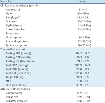
Baseline clinical characteristics, variables of the treadmill stress test, and arterial stiffness indices of the study population are described in Table 1. Table 2 shows univariate regression analyses between HRR and clinical variables. Slow HRR was correlated with patients who were aging, tended to be male (t-test, male: female = 41 ± 11 beats/min: 37 ± 11 beats/min, p = 0.024), and had diabetes (t-test, non-diabetes: diabetes = 40 ± 11 beats/min: 33 ± 12 beats/min, p = 0.008), higher resting and peak systolic BP, higher resting HR, lower peak HR, lower metabolic equivalents, increased baPWV, and increased CSIs at rest and after exercise. HRR had moderate correlation to baPWV (r = −0.331, p < 0.001) and CSI after exercise (r = −0.334, p < 0.001) and a weak correlation to CSI at rest (r = −0.269, p = 0.001) (Figure 2). No relationships were found between HRR and baseline laboratory findings, such as cholesterol, triglycerides, and high- and low-density lipoprotein cholesterol (data not shown). HRR was inversely associated with baPWV and CSI after exercise after adjusting for established cardiovascular risk factors of age, sex, body mass index, diabetes,
Table 1. Clinical characteristics and variables associated with the treadmill stress test and arterial stiffness indices of the study population
Variables Value
Clinical characteristics (n = 154)
Age (years) 53 ± 10
Male 80 (52%)
BMI (kg/m2) 24.1 ± 2.8
Diabetes 20 (13.0%)
Dyslipidemia 43 (27.9%)
Current smoker 33 (21.6%)
Symptoms
No symptom 6 (3.9%)
Atypical symptom 92 (59.7%)
Typical symptom 56 (36.4%)
Treadmill stress test
Resting SBP (mmHg) 114.2 ± 14.3
Resting DBP (mmHg) 66.3 ± 9.0
Resting HR (beats/min) 74.1 ± 11.7
Peak SBP (mmHg) 166.9 ± 24.3
Peak DBP (mmHg) 76.6 ± 17.2
Peak HR (beats/min) 163.6 ± 17.3
Target HR (%) 98.3 ± 8.9
MET 11.8 ± 1.9
HRR (beats/min) 38.9 ± 11.1
Arterial stiffness indices
baPWV (m/s) 13.2 ± 1.6
CSI at rest 3.21 ± 0.34
CSI after exercise 3.16 ± 0.32
baPWV: brachial ankle pulse wave velocity, BMI: body mass index, CSI: carotid arterial stiffness index, DBP:
diastolic blood pressure, HR: heart rate, HRR: heart rate recovery at 1 min after exercise, MET: metabolic equivalent of tasks, SBP: systolic blood pressure.
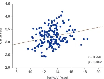
and current smoking but was not associated with CSI at rest (Table 3, Model 1). HRR was also associated with CSI after exercise after adjusting for model 1 + resting systolic BP and metabolic equivalent of tasks but was not associated with baPWV or CSI at rest (Table 3, Model 2). BaPWV had a weak correlation with CSI at rest (r = 0.250, p = 0.002, Figure 3).
CSI after exercise had positive correlation with baPWV (r = 0.363, p < 0.001) and CSI at rest (r = 0.514, p < 0.001) (Figure 4).
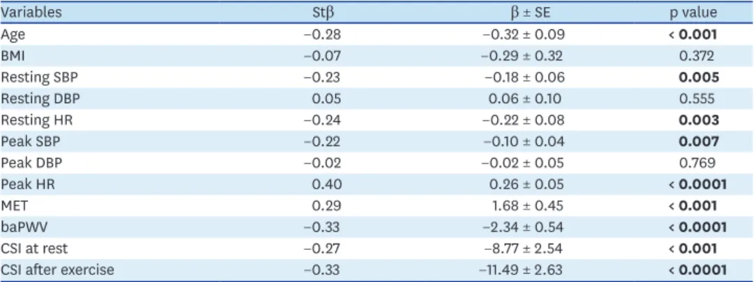
Table 2. Univariate analysis of associated variables for HRR
Variables Stβ β ± SE p value
Age −0.28 −0.32 ± 0.09 < 0.001
BMI −0.07 −0.29 ± 0.32 0.372
Resting SBP −0.23 −0.18 ± 0.06 0.005
Resting DBP 0.05 0.06 ± 0.10 0.555
Resting HR −0.24 −0.22 ± 0.08 0.003
Peak SBP −0.22 −0.10 ± 0.04 0.007
Peak DBP −0.02 −0.02 ± 0.05 0.769
Peak HR 0.40 0.26 ± 0.05 < 0.0001
MET 0.29 1.68 ± 0.45 < 0.001
baPWV −0.33 −2.34 ± 0.54 < 0.0001
CSI at rest −0.27 −8.77 ± 2.54 < 0.001
CSI after exercise −0.33 −11.49 ± 2.63 < 0.0001
baPWV: brachial-ankle pulse wave velocity, β: unstandardized coefficients, BMI: body mass index, CSI: carotid arterial stiffness index, DBP: diastolic blood pressure, HR: heart rate, HRR: heart rate recovery, MET: metabolic equivalent of tasks, SBP: systolic blood pressure, SE: standardized error, Stβ: standardized coefficients.
baPWV (m/s) 0
8 12 18 20
HRR (beats/min)
80
60
40
20
16
10 14
CSI at rest
2.0 2.5 3.0 3.5 4.0 4.5
CSI after exercise
2.0 2.5 3.0 3.5 4.0 4.5
p < 0.001 r = −0.331
p = 0.001 r = −0.269
p < 0.001 r = −0.334
Figure 2. Scatter plots describing the correlations of HRR with baPWV (left panel) and CSI at rest (middle panel) and after exercise (right panel). baPWV:
brachial ankle pulse wave velocity, CSI: carotid arterial stiffness index, HRR: heart rate recovery.
Table 3. Associations of HRR with atherosclerotic markers after adjusting for confounding variables: multivariate linear analyses
Variables Stβ β ± SE p value
Model 1*
baPWV −0.19 −1.35 ± 0.61 0.029
CSI at rest −0.13 −4.30 ± 2.68 0.111
CSI after exercise −0.249 −8.56 ± 2.86 0.003
Model 2†
baPWV −0.13 −0.91 ± 0.70 0.198
CSI at rest −0.12 −4.05 ± 2.61 0.124
CSI after exercise −0.26 −8.77 ± 2.78 0.002
baPWV: brachial-ankle pulse wave velocity, β: unstandardized coefficients, CSI: carotid arterial stiffness index, HRR: heart rate recovery, SE: standardized error, Stβ: standardized coefficients.
*Adjusting for established cardiovascular risks including age, sex, body mass index, diabetes, current smoking;
†adjusting for model 1 + resting systolic blood pressure, and metabolic equivalent of tasks.
DISCUSSION
This study demonstrated that HRR was closely and inversely associated with CSI after exercise but not CSI at rest. HRR was also weakly associated with baPWV. Several authors have previously reported that carotid atherosclerosis may alter the elastic properties of the arterial wall to collagen fibers and impair the vasoactive paracrine functions of the endothelium such as secretion of nitric oxide, and prostaglandin, thus may decrease the distensibility of the carotid sinus and baroreflex sensitivity.6)9)15)-17) Decreased baroreflex sensitivity contributes to the decrease in parasympathetic activity and increase in sympathetic activity that accompany the development and progression of cardiovascular disease.18) Indeed, Nasr et al.19) demonstrated that decreased baroreflex sensitivity was associated with carotid atherosclerosis. Chao et al.15) reported that baroreflex sensitivity was closely associated with HR variability, a typical parameter for estimating autonomic function. Similarly, Jae et al.20) demonstrated that HRR, which is another marker to estimate autonomic function,1)2) was impaired in patients with carotid atherosclerosis with stenosis > 25% and/or intima-media thickness > 1.2 mm. However,
baPWV (m/s)
2.0 8 12 18 20
CSI at rest
4.5
4.0
3.5
3.0
2.5
16
10 14
p = 0.002 r = 0.250
Figure 3. The correlation plot between baPWV and CSI at rest. baPWV: brachial ankle pulse wave velocity, CSI:
carotid arterial stiffness index.
A B
baPWV (m/s)
8 12 18 20
CSI after exercise
4.0
3.5
3.0
2.5
16
10 14
p < 0.001 r = 0.363
CSI at rest
2.0 3.0 4.5
CSI after exercise
4.0
3.5
3.0
2.5
4.0
2.5 3.5
p < 0.001 r = 0.514
Figure 4. The correlation plots of CSI after exercise with baPWV (A) and CSI at rest (B). baPWV: brachial ankle pulse wave velocity, CSI: carotid arterial stiffness index.
this association between HRR and carotid atherosclerosis might be clearly observed after overt atherosclerotic change. Indeed, HRR was not associated with CSI at rest in our study, which only enrolled patients with a low atherosclerotic burden of carotid plaque < 1.5 mm (average carotid intima-media thickness = 0.6 ± 0.1 mm). Meanwhile, HRR was weakly associated with baPWV when established cardiovascular risks were adjusted. However, the association between the two factors was not significant when systolic BP or metabolic equivalent of tasks were included as confounding factors. We surmise that this is because measurement of baPWV is pressure-dependent.5) Therefore, autonomic activity following exercise might not be closely associated with atherosclerotic change apart from established cardiovascular risks, at least in the early atherosclerotic stage.
Several studies have consistently shown that repetitive aerobic exercise training improved arterial stiffness.21)-23) On the contrary, the immediate impact of aerobic exercise on arterial stiffness might be complex and controversial according to the anatomic site at which the arterial stiffness was assessed, the timing of the measurement post-exercise, the intensity of the exercise, and characteristics of the patients.21)24) Seo et al.25) demonstrated that arterial stiffness measured by baPWV was decreased after maximal treadmill exercise. However, Liu et al.21) demonstrated that immediate carotid stiffness was increased after acute exercise training of moderate intensity, but the difference before and after exercise tended to decrease according to repetitive exercise training. Mutter et al.24) insisted that stiffness on the central and upper body peripheral arterial segments deteriorated for a short duration after exercise and then improved, while stiffness on the lower arterial segments decreased regardless of the post-exercise timing of the measurement. Some researchers suggested that the decrease in arterial stiffness after acute exercise is due to increased release of shear stress-induced nitric oxide or other vasoactive agents.26)27) However, Sugawara et al.28) demonstrated that immediate vasodilation after low intensity exercise was not associated with increased production of nitric oxide. Boutouyrie et al.29) also insisted that autonomic activity to modulate sympathetic tone during exercise might be a mechanism of decreased arterial stiffness after acute exercise. The results of this study do not allow us to confirm which hypothesis is right. However, the close association between HRR and CSI after exercise may exist because CSIs after exercise are modulated by autonomic activity as well as vascular endothelial function.
In this study, the correlation between baPWV and CSI at rest was weak. BaPWV is an
atherosclerotic marker to indirectly assess arterial stiffness in regional muscular components as well as the aortic, brachial, femoral and tibial arteries.30) On the other hand, CSI is a direct stiffness marker limited to the carotid artery. The arteries of the entire body are not homogeneously affected in the progression of atherosclerosis. In general, the carotid artery becomes stiffer than the femoral or radial arteries in association with aging and high BP.31)32) This study suggested that baPWV might be a more useful marker to estimate association with autonomic function derived by HRR than CSI at rest.
CSI at rest was not associated with HRR but was highly associated with CSI after exercise, even after adjusting for established cardiovascular risks and HRR (data not shown; Stβ = 0.412, p < 0.001). It might be suggested because the two markers measured arterial stiffness on the same anatomic site.
This study had some limitations. First, we measured brachial BP instead of central BP to calculate CSI. The BP measurement may have been overestimated because peripheral arterial
pulse pressure is amplified compared with central arterial pulse pressure.5) However, some previous studies have demonstrated that a stiffness index using peripheral BP had a very good correlation with indices using central BP33)34) and also was clinically useful.35) A second limitation is that we measured baPWV, but we did not measure carotid-femoral PWV (cfPWV) which has been traditionally used to evaluate aortic stiffness. Although the value of baPWV is higher than that of cfPWV because of the inclusion of the stiffness of the peripheral muscular artery,30) baPWV is known to have good correlation with cfPWV36) and aortic PWV measured by an invasive method.37) In addition, baPWV is easier to measure, so it has better intra- and inter-observer reproducibilities than cfPWV.38) Finally, because this was a cross-sectional study, we could not determine causation between arterial stiffness and HRR.
CONCLUSIONS
Sympathovagal imbalance indicated by slow HRR after exercise was associated with increased arterial stiffness in normotensive patients without overt atherosclerosis. Furthermore, the close association between HRR and exercise-induced carotid arterial stiffness might have been observed because autonomic function following exercise as well as vascular endothelial function determine carotid arterial vasomotion after exercise.
REFERENCES
1. Imai K, Sato H, Hori M, et al. Vagally mediated heart rate recovery after exercise is accelerated in athletes but blunted in patients with chronic heart failure. J Am Coll Cardiol 1994;24:1529-35.
PUBMED | CROSSREF
2. Kleiger RE, Miller JP, Bigger JT Jr, Moss AJ. Decreased heart rate variability and its association with increased mortality after acute myocardial infarction. Am J Cardiol 1987;59:256-62.
PUBMED | CROSSREF
3. Beltran A, McVeigh G, Morgan D, et al. Arterial compliance abnormalities in isolated systolic hypertension. Am J Hypertens 2001;14:1007-11.
PUBMED | CROSSREF
4. Gedikli O, Kiris A, Ozturk S, et al. Effects of prehypertension on arterial stiffness and wave reflections.
Clin Exp Hypertens 2010;32:84-9.
PUBMED | CROSSREF
5. Laurent S, Cockcroft J, Van Bortel L, et al. Expert consensus document on arterial stiffness:
methodological issues and clinical applications. Eur Heart J 2006;27:2588-605.
PUBMED | CROSSREF
6. Koskinen T, Juonala M, Kähönen M, et al. Relations between carotid artery distensibility and heart rate variability The Cardiovascular Risk in Young Finns Study. Auton Neurosci 2011;161:75-80.
PUBMED | CROSSREF
7. Reneman RS, Meinders JM, Hoeks AP. Non-invasive ultrasound in arterial wall dynamics in humans: what have we learned and what remains to be solved. Eur Heart J 2005;26:960-6.
PUBMED | CROSSREF
8. Van Bortel LM, Duprez D, Starmans-Kool MJ, et al. Clinical applications of arterial stiffness, Task Force III: recommendations for user procedures. Am J Hypertens 2002;15:445-52.
PUBMED | CROSSREF
9. Chapleau MW, Cunningham JT, Sullivan MJ, Wachtel RE, Abboud FM. Structural versus functional modulation of the arterial baroreflex. Hypertension 1995;26:341-7.
PUBMED | CROSSREF
10. Carretta R, Bardelli M, Cominotto F, et al. Relationship between mechanical properties of the carotid artery wall and baroreflex function in acutely treated hypertensive patients. J Hypertens 1996;14:1105-10.
PUBMED | CROSSREF
11. Randall OS, Esler MD, Bulloch EG, et al. Relationship of age and blood pressure to baroreflex sensitivity and arterial compliance in man. Clin Sci Mol Med Suppl 1976;3:357s-60s.
PUBMED
12. Katsube Y, Saro H, Naka M, et al. Decreased baroreflex sensitivity in patients with stable coronary artery disease is correlated with the severity of coronary narrowing. Am J Cardiol 1996;78:1007-10.
PUBMED | CROSSREF
13. Fletcher GF, Balady G, Froelicher VF, Hartley LH, Haskell WL, Pollock ML. Exercise standards. A statement for healthcare professionals from the American Heart Association. Writing Group. Circulation 1995;91:580-615.
PUBMED | CROSSREF
14. Bland JM, Altman DG. Statistical methods for assessing agreement between two methods of clinical measurement. Lancet 1986;1:307-10.
PUBMED | CROSSREF
15. Chao AC, Chern CM, Kuo TB, et al. Noninvasive assessment of spontaneous baroreflex sensitivity and heart rate variability in patients with carotid stenosis. Cerebrovasc Dis 2003;16:151-7.
PUBMED | CROSSREF
16. Sung J, Yang JH, Cho SJ, Hong SH, Huh EH, Park SW. The effects of short-duration exercise on arterial stiffness in patients with stable coronary artery disease. J Korean Med Sci 2009;24:795-9.
PUBMED | CROSSREF
17. Ekblom B, Kilbom A, Soltysiak J. Physical training, bradycardia, and autonomic nervous system. Scand J Clin Lab Invest 1973;32:251-6.
PUBMED | CROSSREF
18. La Rovere MT, Pinna GD, Raczak G. Baroreflex sensitivity: measurement and clinical implications. Ann Noninvasive Electrocardiol 2008;13:191-207.
PUBMED | CROSSREF
19. Nasr N, Pavy-Le Traon A, Larrue V. Baroreflex sensitivity is impaired in bilateral carotid atherosclerosis.
Stroke 2005;36:1891-5.
PUBMED | CROSSREF
20. Jae SY, Carnethon MR, Heffernan KS, et al. Slow heart rate recovery after exercise is associated with carotid atherosclerosis. Atherosclerosis 2008;196:256-61.
PUBMED | CROSSREF
21. Liu HB, Yuan WX, Wang QY, et al. Carotid arterial stiffness and hemodynamic responses to acute cycling intervention at different times during 12-week supervised exercise training period. Biomed Res Int 2018;2018:2907548.
PUBMED
22. Fujie S, Sato K, Miyamoto-Mikami E, et al. Reduction of arterial stiffness by exercise training is associated with increasing plasma apelin level in middle-aged and older adults. PLoS One 2014;9:e93545.
PUBMED | CROSSREF
23. Beck DT, Martin JS, Casey DP, Braith RW. Exercise training reduces peripheral arterial stiffness and myocardial oxygen demand in young prehypertensive subjects. Am J Hypertens 2013;26:1093-102.
PUBMED | CROSSREF
24. Mutter AF, Cooke AB, Saleh O, Gomez YH, Daskalopoulou SS. A systematic review on the effect of acute aerobic exercise on arterial stiffness reveals a differential response in the upper and lower arterial segments. Hypertens Res 2017;40:146-72.
PUBMED | CROSSREF
25. Seo J, Chung W, Kim S, Kim M, Zo J. Immediate impact of exercise on arterial stiffness in humans. World J Cardiovasc Dis 2013;3:40-5.
CROSSREF
26. Kingwell BA, Berry KL, Cameron JD, Jennings GL, Dart AM. Arterial compliance increases after moderate-intensity cycling. Am J Physiol 1997;273:H2186-91.
PUBMED
27. Endo T, Imaizumi T, Tagawa T, Shiramoto M, Ando S, Takeshita A. Role of nitric oxide in exercise-induced vasodilation of the forearm. Circulation 1994;90:2886-90.
PUBMED | CROSSREF
28. Sugawara J, Maeda S, Otsuki T, Tanabe T, Ajisaka R, Matsuda M. Effects of nitric oxide synthase inhibitor on decrease in peripheral arterial stiffness with acute low-intensity aerobic exercise. Am J Physiol Heart Circ Physiol 2004;287:H2666-9.
PUBMED | CROSSREF
29. Boutouyrie P, Lacolley P, Girerd X, Beck L, Safar M, Laurent S. Sympathetic activation decreases medium- sized arterial compliance in humans. Am J Physiol 1994;267:H1368-76.
PUBMED
30. Munakata M, Ito N, Nunokawa T, Yoshinaga K. Utility of automated brachial ankle pulse wave velocity measurements in hypertensive patients. Am J Hypertens 2003;16:653-7.
PUBMED | CROSSREF
31. Benetos A, Laurent S, Hoeks AP, Boutouyrie PH, Safar ME. Arterial alterations with aging and high blood pressure. A noninvasive study of carotid and femoral arteries. Arterioscler Thromb 1993;13:90-7.
PUBMED | CROSSREF
32. Boutouyrie P, Laurent S, Benetos A, Girerd XJ, Hoeks AP, Safar ME. Opposing effects of ageing on distal and proximal large arteries in hypertensives. J Hypertens Suppl 1992;10:S87-91.
PUBMED | CROSSREF
33. Tomiyama H, Kihara Y, Nishikawa E, et al. An impaired carotid sinus distensibility and baroreceptor sensitivity alter autonomic activity in patients with effort angina associated with significant coronary artery disease. Am J Cardiol 1996;78:225-7.
PUBMED | CROSSREF
34. Tomiyama H, Nishikawa E, Abe M, et al. Carotid arterial distensibility is an important determinant of improvement in autonomic balance after successful coronary angioplasty. J Hypertens 2000;18:1621-8.
PUBMED | CROSSREF
35. Giannattasio C, Failla M, Piperno A, et al. Early impairment of large artery structure and function in type I diabetes mellitus. Diabetologia 1999;42:987-94.
PUBMED | CROSSREF
36. Tanaka H, Munakata M, Kawano Y, et al. Comparison between carotid-femoral and brachial-ankle pulse wave velocity as measures of arterial stiffness. J Hypertens 2009;27:2022-7.
PUBMED | CROSSREF
37. Yamashina A, Tomiyama H, Takeda K, et al. Validity, reproducibility, and clinical significance of noninvasive brachial-ankle pulse wave velocity measurement. Hypertens Res 2002;25:359-64.
PUBMED | CROSSREF
38. Wilkinson IB, Fuchs SA, Jansen IM, et al. Reproducibility of pulse wave velocity and augmentation index measured by pulse wave analysis. J Hypertens 1998;16:2079-84.
PUBMED | CROSSREF