J. of Korean Shoulder and Elbow Society Volume 9, Number 2, December, 2006
※통신저자: 김 영 배*
서울특별시 강동구 둔촌2동 6-2 서울보훈병원 정형외과
Tel: 02) 2225-1352, Fax: 02) 487-0754, E-Mail: [email protected]
견봉 쇄골 관절 탈구에 사용된 K-강선의 경추부로의 이동 - 증례보고(2예) -
서울보훈병원 정형외과
이우승∙김택선∙윤정로∙김영배*∙서동훈∙권제호
—
— Abstract ——
Migration of K-wires from the Acromioclavicular Joint to the Neck - Case Report (2 cases) -
Woo-Seung Lee, M.D., Taik-Seon Kim, M.D., Jeong-Ro Yoon, M.D., Young-Bae Kim, M.D.*, Dong-Hoon Seo, M.D., Jae-Ho Kwon, M.D.
Department of Orthopedic Surgery, Seoul Veterans Hospital, Seoul, Korea
We report two cases of migration of K-wires from the acromioclavicular joint to the neck. A 73-year-old man com- plained of right shoulder pain for one month and had undergone orthopedic surgery because of acromioclavicular joint dislocation about 27 years earlier. Another 56-year-old man complained of left shoulder pain and neck pain for 5 years and had undergone orthopedic surgery because of acromioclavicular joint dislocation about 25 years earlier. In both cases, we took X-rays to look for the cause of shoulder pain and discovered broken and migrated K-wires in the neck. We removed the K-wires from the trapezius muscle and the paraspinal muscle respectively. K-wire fixation technique is simple and effective but should be followed up with X-rays periodically. In addition, we should warn patients of the possibility of migration of K-wire. And it is desirable for us to avoid using K-wire near major neu- rovascular structures like the sternoclavicular joint and the clavicle.
Key Words: Acromioclavicular joint, Migration, K-wire
K-강선은 1909년 Martin Kirschner가 골견 인(skeletal traction)에 사용한 후 오늘날까지 수족부 및 견주관절 부위 등의 골절 및 탈구의 수 술에 널리 사용되고 있다1,3,21). K-강선의 사용 후 합병증으로 주위 조직으로의 이동이 있는데 특히 견관절 부위의 수술 후 흔한 것으로 알려져 있다.
이 경우 임상 증상 없이 우연히 발견되거나 단순 통증만 일으키는 경우도 있으나, 주요 혈관 및 신 경의 손상을 유발하는 경우도 있다2,4-20,22-30)
. K-강 선의 이동 방향에 대하여는 저자들에 따라 발생 빈도의 보고가 다른데, 국내에서의 문헌보고들 (Table 1)2,4)에 따르면 경추부로, Lyons와 Rockwood15)의 보고(Table 2)에 따르면 주요 혈 관 방향으로 이동이 제일 많다고 하였다.
저자들은 견봉 쇄골 관절 탈구 수술 후에 사용 되었던 K-강선의 경추부로의 이동을 2례 경험하
였기에 문헌 고찰과 함께 보고하고자 한다.
증례 보고 1. 증례 1
73세 남자 환자가 내원 한 달 전부터 시작된 우 측 견관절부 통증을 주소로 내원하였다. 과거력상 55년 전 발생한 우측 견봉 쇄골 관절 손상으로 27년 전 수술적 치료를 시행받았으며 8년 전 경 추부 수술을 받았다. 이학적 검사상 우측 견관절 부 관절운동 범위는 능동적 전방거상 35도, 수동 적 전방거상 100도, 외회전 10도 및 내회전 우측 대퇴부의 소견을 보였으며, 우측 대결절부 압통, 충돌징후 양성 및 동통을 동반한 외회전 근력 약 화소견이 관찰되었다. 단순방사선 사진상 우측 견
Table 1. Reports of migration of Kirschner wire in journal of the Korean Orthopedic Association.
Articles No. of cases Location to which the K-wires used Location to which the K-wires migrated
Rho et al (86’) 2 ACjoint* Neck (Subcutaneous layer)
Neck (Under surface of Trapezius muscle)
Ha et al (94’) 2 AC joint Neck (Trapezius muscle)
Neck (Supraspinatus & Levator scapularis muscle)
Total 4
* AC joint : Acromioclavicular joint
Table 2. Reports of migration of pins written by Lyons FA and Rockwood CA Jr. (JBJS, 1990)
Reasons for which the pins inserted No. of cases
Sternoclavicular dislocation 21 (45%)
Fracture of the clavicle 10 (21%)
Acromioclavicular dislocation 18 (17%)
Others 18 (17%)
Total 47
Location to which the pins migrated No. of cases
Major vascular structure (Heart, subclavian, aorta etc) 17 (36%)
Lung or mediastinum 18 (38%)
Others (abdomen, orbit etc) 12 (26%)
Total 47
Morbidity & mortality No. of cases
Cardiovascular events and pericardial tamponade 18 (17%)
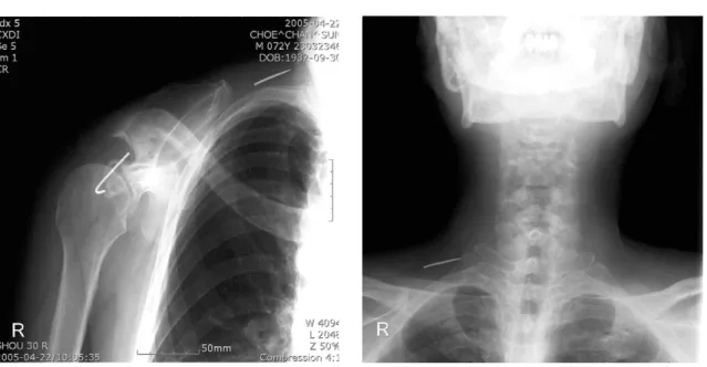
봉하 골극, 이소성 골화 소견을 보였으며, 파단된 K-강선이 우측 견봉 쇄골 관절부에 그리고 파단 된 K-강선의 일부가 경추부로 이동되어 있었다 (Fig. 1, 2). 우측 회전근개 증후군과 파단된 K- 강선의 우측 견봉 쇄골 관절부에서 경추부로의 이 동으로 진단을 하였고, 파단된 K-강선의 제거를 위한 수술을 시행하였다. 수술은 전신 마취하에 측와위로 시행하였고, 수술 소견상 경추부로 이동 한 K-강선은 우측 승모근의 전면에 위치하여 이 를 확인 후 제거하였다.
2. 증례 2
56세 남자 환자가 내원 5년 전부터 시작된 좌 측 견관절부와 경추부 통증을 주소로 내원하였다.
과거력상 25년 전 좌측 견봉 쇄골 관절 손상으로 수술적 치료를 시행받았으며 수년 전 경추부 수술 을 받았다. 이학적 검사상 좌측 견관절부 관절운 동 범위는 전방거상 140도, 외회전 40도, 내회전 제 1요추의 소견을 보였으며, 좌측 대결절부 압 통, 충돌징후 양성 및 통증을 동반한 외회전 근력
Fig. 1. Simple radiograph of the right shoulder shows a broken Kirschner wire at acromioclavicular joint and a broken K-wire migrated to the neck.
Fig. 2. Plain X-ray of the cervial spine shows a migrated and broken Kirschner wire at the neck.
Table 3. Analysis of cases on migration of Kirschner wires from the shoulder in articles other than the Korean Ortho- pedic Association. (except the article written by Lyons FA, Rockwood CA Jr. in 1990.)
Reasons for which the K-wires inserted No. of cases
Fracture of the clavicle 12 (41%)
Sternoclavicular dislocation 16 (21%)
Acromioclavicular dislocation 16 (21%)
Others 15 (17%)
Total 29
Location to which the K-wires migrated No. of cases
Major vascular structure (Heart, subclavian, aorta etc) 19 (31%)
Lung or mediastinum 17 (24%)
Others (Spinal cord, spleen etc) 13 (45%)
Total 29
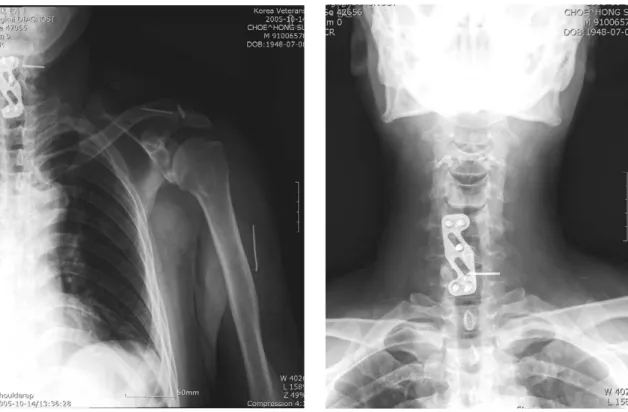
의 약화소견이 관찰되었으며 경추부 신전시 통증 을 호소하였다. 단순방사선 사진상 좌측 견봉 쇄 골 관절에 3개의 파단된 K-강선 및 경추부와 좌 측 상완에 좌측 견봉 쇄골 관절에서 이동한 파단 된 K-강선이 각각 하나씩 관찰되었다(Fig. 3, 4).
좌측 회전근 개 증후군과 파단된 K-강선의 좌측 견봉 쇄골 관절부에서 경추부와 좌측 상완으로의 이동으로 진단하였고 파단된 K-강선의 제거를 위 하여 이에 대한 수술을 시행하였다. 수술은 전신 마취하에 측와위로 시행하였고, 수술 소견상 경추 부로 이동한 파단된 K-강선은 경추부의 좌측 paraspinal muscle 속에 위치하였고 상완으로 이동한 파단된 K-강선은 상완근 속에 위치하여 이를 확인한 후 제거하였다.
고 찰
K-강선은 여러 가지 내고정물의 발전에도 불구
하고, 견관절 및 수족부 등의 골절과 탈구의 치료 에 많이 사용되고 있다. 그러나 전위력과 회전력 에 약하여 술 후 추가적인 고정 및 운동제한을 필 요로 하고, K-강선의 파단 또는 주위 조직으로 이동 등의 합병증이 발생할 수 있는데, 특히 견봉 쇄골 관절에서 흔한 것으로 알려져 있다. K-강선 이 외측부로 이동하는 경우 피부자극 및 감염의 원인이 되며, 내측부로 이동하는 경우에는 증상이 없는 경우가 많지만 드물게 혈관, 신경, 심장 및 폐 등으로 이동하여 심한 경우 사지마비나 사망을 초래할 수 있다15,24). 저자들은 내측부로 이동하는 파단된 K-강선 2예를 경험하여 보고하는 바이다.
K-강선의 내측부 이동은 견봉 쇄골 관절 탈구 에서 1943년 Mazet16)이 폐로의 이동을 보고한 이래로 많은 보고가 있었다2,4-15,17-20,22-30)
. 국내의 발 표된 문헌의 경우 견관절부에 사용한 K-강선의 이동은 견봉 쇄골 관절 탈구에 사용한 총 4예이었 고 모두 경추부로 이동하였다(Table 1). 외국문
Fig. 3. Plain X-ray of the left shoulder shows three Kirschner wires at the left acromioclavicular joint and two broken Kirschner wires to lateral aspect of the left arm and to the neck.
Fig. 4. Anteroposterior radiograph of the cervical spine shows a broken Kirschner wire migrated to the neck.
헌의 경우 1990년 Lyons와 Rockwood15)는 여러 종류의 강선(Hagie pin, threaded K-강선, K- 강선 포함)을 이용한 흉쇄 관절 탈구, 쇄골 골절 및 견봉 쇄골 관절 탈구의 치료에서 47예의 강선 의 이동을 분석 발표하였는데 쇄골하동맥 및 대동 맥 등 주요 혈관부로의 이동이 17예(36%), 폐와 종격부위로의 이동이 18예(38%) 등이었다 (Table 2). 그 외의 문헌들을 통합 분석하여 보 면 K-강선이 이동한 부위로는 주요 혈관부가 9예 (31%)로 가장 많았으며2,4-14,16-20,22-30)
경추부로 이 동한 K-강선으로 인하여 사지 마비를 일으키거나 안와, 복부대동맥 및 비장 등 다양한 부위로 이동 하는 등 치명적인 결과를 보이기도 하였다15,17,30). 저자들의 경우 우측 승모근의 전면 1예 및 경추부 의 paraspinal muscle 속 1예 등이었다. 이들 예에서는 K-강선이 아직 척추관을 침범하지 않아 신경학적 증상을 일으키지 않았지만 경추부의 운 동과 관련되는 통증을 호소하기도 하였다.
K-강선의 이동의 원인으로는 근육운동, 호흡운 동, 모세혈관작용, 국소 골 흡수, 중력 및 상지의 큰 관절운동범위 등 다양한 원인이 제시되고 있다17). 본 례에서는 지속적인 견관절 관절운동에 의해 K-강선 의 파단과 이동이 발생하였을 것으로 생각하였다.
본 증례의 환자들은 오래 전(각각 27년과 25 년)에 수술을 받은 시기는 기억을 하였으나, 수술 의 종류, 내고정물인 K-강선의 삽입 여부 및 이 의 이동 가능성에 대해서는 알고 있지 않았다. 그 러므로 K-강선의 이동을 방지하기 위하여 충분한 환자의 교육과 철저한 추시 관찰이 제일 중요한 방법이며, 그 외에는 굵은 K-강선 혹은 Thread- ed K-강선의 사용, 삽입 방향의 신중한 결정, 삽 입 후 외측단의 구부림, 수술 후 제거 전까지의 세심한 견관절 운동 교육 및 적절한 시기에 K-강 선의 제거 등이 있다15). 하지만 Threaded K-강 선을 사용하거나 외측단의 구부림 후에도 K-강선 이 파단되어 이동할 수 있으므로 수술 후 K-강선 의 제거 전까지 적극적인 추시관찰과 함께 환자의 교육에 중점을 두어야 할 것으로 사료된다.
Lyons와 Rockwood15)의 4예의 pin의 이동에 대한 보고에 의하면 pin이 사용된 부위 및 원인으 로는 흉쇄 관절 탈구가 21예(45%)로 가장 많았 으며, 8예(17%)에서 수술 전 후에 K-강선 이동
과 관련된 심혈관계 문제로 사망하였음을 보고하 였다(Table 2). 그리고 흉쇄 관절에서의 고정을 위한 pin의 사용은 절대적인 금기사항임을 강조 하였다. 하지만 다른 논문의 통합분석에서는 K- 강선이 사용된 부위로는 쇄골부가 총 29례 중 12 례(41%)로 가장 많았다5-14,16-20,22-30)
. 그러므로 주 요 혈관 및 신경 인접부인 흉쇄 관절부 및 쇄골 골절부 모두 고정물의 선택 시 K-강선의 사용은 피하는 것이 좋을 것으로 생각한다.
REFERENCES
01) 김김 정정 환환, 김김 종종 관관, 이이 상상 국국, 김김 영영 오오, 박박 재재 규규, 윤윤 종종 호
호: 급성 견봉 쇄골 관절 탈구의 수술적 치료. 대한 견주관절학회지,4:17-23, 2001.
02) 노노 성성 만만, 이이 우우 석석: 견봉 쇄골 관절에 삽입한 금속고 정물의 경부내이동(2 례 보고). 대한정형외과학회 지,21:499-501, 1986.
03) 문문 은은 선선, 김김 명명 선선, 배배 봉봉 현현, 최최 진진: 45세 이상의 제 3 형 견봉 쇄골 관절 탈구 환자의 수술적 치료 - 일차 적 쇄골 외측단 절제 술식과 고식적인 견봉쇄골 관 절 정복 술식의 비교 -. 대한견주관절학회지, 8:88- 96, 2005.
04) 하하 상상 호호, 유유 재재 원원, 이이 상상 홍홍, 표표 영영 배배, 신신 동동 민민: 견봉 쇄골관절에 삽입한 Kirschner 강선의 경부내 이동.
대한정형외과학회지, 29:1264-1267, 1994.
05) Chou NS, Wu MH, Chan CS, Lai WW and Lin MY: Intrathoracic migration of Kirschner wires. J Formos Med Assoc, 93:974-976, 1994.
06) Daus GP, Drez D Jr, Newton BB Jr and Kober R: Migration of a Kirschner wire from the sternum to the right ventricle. A case report.
Am J Sports Med, 21:321-322, 1993.
07) Fuster S, Palliso F, Combalia A, Sanjuan A and Garcia S: Intrathoracic migration of a Kirschner wire. Injury, 21:124-126, 1990.
08) Grauthoff H and Klammer HL: Complications due to migration of a Kirschner wire from the clavicle. Rofo, 128:591-594, 1978.
09) Janssens de Varebeke B and Van Osselaer G:
Migration of Kirschner’s pin from the right ster- noclavicular joint resulting in perforation of the pulmonary artery main trunk. Acta Chir Belg, 93:287-291, 1993.
10) Leonard JW and Gifford RW Jr: Migration of a Kirschner’s wire from the clavicle into the pul- monary artery. Am J Cardiol, 16:598-600, 1965.
11) Leppilahti J and Jalovaara P: Migration of Kirschner wires following fixation of the clav- icle--a report of 2 cases. Acta Orthop Scand, 70:517-519, 1999.
12) Lindsey RW and Gutowski WT: The migration of a broken pin following fixation of the acromioclavicular joint. A case report and review of the literature. Orthopedics, 9:413-416, 1986.
13) Liu HP, Chang CH, Lin PJ, Chu JJ, Hsieh HC, Chang JP and Hsieh MC.: Pulmonary artery perforation after Kirschner wire migration:
case report and review of the literature. J Trauma, 34:154-156, 1993.
14) Loncan LI, Sempere DF and Ajuria JE:
Brown-Sequard syndrome caused by a Kirschner wire as a complication of clavicular osteosynthe- sis. Spinal Cord, 36:797-799, 1998.
15) Lyons FA and Rockwood CA Jr: Migration of pins used in operations on the shoulder. J Bone Joint Surg, 72-A:1262-1267, 1990.
16) Mazet RJ: Migration of a Kirschner wire from the shoulder region into the lung, Report of two cases. J Bone Joint Surg, 25-A:477-483, 1943.
17) Naidoo P: Migration of a Kirschner Wire from the clavicle into the abdominal aorta. Arch Emerg Med, 8:292-295, 1991.
18) Nettles JL and Linscheid RL: Sternoclavicular dislocations. J Trauma, 8:158-164, 1968.
19) Ngarmukos C, Parkpian V and Patradul A:
Fixation of fractures of the midshaft of the clav- icle with Kirschner wires. Results in 108 patients. J Bone Joint Surg, 80-B:106-108, 1998.
20) Norrell H Jr and Llewellyn R: Migration of a threaded steinmann pin from an acromioclavicu-
lar joint into the spinal canal. a case report. J Bone Joint Surg, 47-A:1024-1026, 1965.
21) Peltier LF: A brief history of traction. J Bone Joint Surg, 50-A:1603-1617, 1968.
22) Potter FA, Fiorini AJ, Knox J and Rajesh PB:
The migration of a Kirschner wire from shoulder to spleen: brief report. J Bone Joint Surg, 70- B:326-327. 1988 .
23) Rajesh PB and Nair KK: Unusual migration of a Kirschner wire. Eur J Cardiothorac Surg, 5:164, 1991.
24) Regel JP, Pospiech J, Aalders TA and Ruch- holtz S: Intraspinal migration of a Kirschner wire 3 months after clavicular fractur fixation.
Neurosurg Rev, 25:110-112, 2002.
25) Sethi GK and Scott SM: Subclavian artery lac- eration due to migration of a Hagie pin. Surgery, 80:644-646, 1976.
26) Salvatore JE: Sternoclavicular joint dislocation.
Clin Orthop, 58:51-55, 1968.
27) Tristan TA and Daughtridge TG: Migration of metallinc pin from the humerus into the lung. N Engl J Med, 270:987-989, 1964.
28) Urban J and Jaskiewicz A: Spontaneous dis- placement of a Kirschner wire to the chest cavity after fixation of the acromioclavicular joint. Chir Narzadow Ruchu Ortop Pol, 49:399-402, 1984.
29) Worman LW and Leagus C: Intrathoracic injury following retrosternal dislocation of the clavicle. J Trauma, 7:416-423, 1967.
30) Yadav V and Marya KM: Unusual migration of a wire from shoulder to neck. Indian J Med Sci, 57:111-112, 2003.