Vol. 18, No. 4, September, 2006
서 론
골다공증은 대표적인 대사질환의 하나로, 골밀도 감소 및 골조직의 미세구조 파괴로 인한 골강도 저하와 골절의 위험성이 증가하는 근골격계 질환이다80). 인구의 고령화로 골다공증은 증가추세에 있으며, 골다공증성 골절로 인한 노인에서의 삶의 질 저하와 합병증들로 인한 사망률 및 의 료비 증가 등 여러 면에서 문제로 대두되고 있다3). 골다공 증에 대한 적절한 대처를 위해서는 골다공증의 병인에 대 한 명확한 이해가 필수적이라 하겠다. 이에 저자들은 최신 지견들에 근거하여 골다공증의 근본적인 병인론을 기술하 고자 한다.
본 론
성인의 골밀도는 태아에서 성장기에 걸쳐 형성된 최대골 량(peak bone mass)과 이후의 골소실에 의해 결정된다.
따라서 성장기까지의 최대골량 형성과정과 골의 재형성 (bone remodeling)과정에서의 이상은 골다공증의 유발요 인이 된다. 비록 유전적 요인이 최대골량 형성과 밀접한 관계를 가지는 것으로 알려져 있으나70), 칼슘 및 비타민D 섭취, 육체적 활동 등의 환경요인도 중요하다80). 사춘기까 지의 골량증가는 주로 성호르몬과 성장판에서의 성장에 기 인하며, 여성은 초경이후 5~10년간 골막의 부가성장 (appositional growth)에 의해 소량의 골량이 증가하는 반면 남성은 17~20세까지 골량이 증가되어, 성별간 최대 골량의 차이가 유발한다9). 30세 전후로 최대골량에 도달한 후에는 비교적 평탄한 골량의 감소가 관찰되나, 여성은 폐경기 에스트로겐 결핍으로 인한 급격한 골감소가 발생하 고, 남성은 뚜렷한 호르몬의 변화없이 50세 이후에도 비교 적 완만한 골감소를 보이게 된다29).
한편 골재형성은 조혈세포 전구체와 조골세포계열의 작 용에 의한 파골세포 및 염증세포 활성화기(activation phase), 파골세포 형성에 의한 3~4주간의 골흡수기 (resorption phase), 단핵구가 골표면에 배열되는 3~4일 간의 역전기(reversal phase) 그리고 조골세포에 의해 골 기질 및 칼슘이 침착되는 3~4개월간의 골형성기 (formation phase)로 분류되며, 골형성기는 이전 3단계 보다 많은 시간이 소요되므로 재형성율 증가는 골감소를 동반한다. 그러나 골흡수 증가에도 불구하고 전체 골량은 증가하는 사춘기의 예처럼 골재형성율 증가가 반드시 골량 의 감소를 의미하는 것은 아니며, 골흡수에 반응하는 골형 성의 장애가 동반될 때 골량이 감소하게 된다76).
이러한 골다공증의 병인은 국소 및 전신적 조절인자 뿐 만 아니라, 조절인자의 생성과 활성화에 관여하는 수용체, 신호전달경로(signal transduction pathway), 전사인자 (transcription factor)와 효소 등에 의해 이질성 (heterogeneity)을 띄게 된다.
1. 유전적 요인
쌍생아 연구를 통해 최대골량의 50%이상이 유전적 요인 에 의해 결정되며 부위에 따라 다소 양상의 차이를 보인다 고 알려져 있다78,79). 그러나 단일 유전자의 돌연변이에 의 해 골다공증이 야기되는 경우는 드물고, 작은 효과를 가진 각각의 유전자가 복합적으로 작용하는 다인성(polygenic) 질환으로 생각되며78), 남성이 여성보다 높은 최대골량을 형성한다. 이러한 유전적 영향은 최대골량이 형성되는 청 장년기 뿐 아니라 노령에서도 지속된다14).
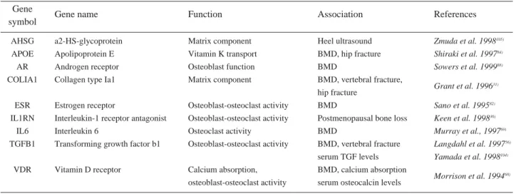
많은 연구들이 후보유전자(candidate gene)와 골밀도 및 골절의 상관관계에 초점을 맞춰 진행되었으며(Table 1), 현재까지 가장 광범위하게 연구되고 있는 것은 비타민 D수용체(Vitamin D receptor, VDR)와 type I collagen α유전자들이다89).
VDR의 다형성(polymorphism)은 광범위하게 연구되 어 왔으나, 다양한 이견을 보이고 있으며16,68), 이는 환경요 인 특히 칼슘과의 상호작용에서 일부 기인한다23). 또한 VDR 다형성은 칼시트리올(calcitriol) 치료반응의 차이와
※ 통신저자 : 김 신 윤
대구광역시 중구 삼덕동 50
경북대학교 의과대학 정형외과학교실 Tel: 82-53-420-5635
Fax: 82-53-422-6605 E-mail: [email protected]
골다공증의 병인론
백승훈∙김신윤
경북대학교 의과대학 정형외과학교실
도 관계있으며, 골밀도나 골교체율(bone turnover)과는 무관하게 골절위험도를 감소시키는데, 이는 낙상빈도의 변 화에 기인한 것으로 추측된다27).
또한 type I collagen α1 사슬을 코딩하는 유전자의 다 형성과 homocysteine 증가는 골밀도와 관계없이 골절위 험도를 증가시킬 수 있는 것으로 알려져 있으며, 이는 교 원질의 helix형성이나 교차결합(cross-linking)의 이상에 서 기인하는 것으로 생각된다64,66).
한편, 국내에서는 최근 저자들이 참여한 보건복지부지정 근골격계 유전체 연구센터에서도 골다공증과 관련된 여러 유전자들과 골밀도 및 골절과의 상관관계, 유관유전자들의 상호작용에 대한 여러 결과들이 보고하였다(Table 2).
2. 조골세포의 분화와 기능을 조절하는 전사인자
신호전달경로와 전사인자의 조골세포 분화 및 기능 조절 에 관한 최근의 연구는 골다공증 병인을 이해하는 데 있어 새로운 장을 열었다. 유전자 제거 연구(gene deletion study)를 통해 runt-related transcription factor 2 (Runx2)와 그 하향조절인자(downstream factor)인 osterix(Osx)가 조골세포 분화에 결정적인 역할을 한다는 사실이 밝혀졌다18,71).
Runx는 사람에서는 Runx1, 2, 3으로 분류되며, Runx2가 조골세포의 분화와 관계있는 것으로 알려져 있 다51). 이들 유전자는 virus연구자들에 의해 발견되어 Polyoma enhancer binding protein 2-α (Pebp2-α)
Table 1. Extensively investigated candidate-genes in osteoporosis.
Gene
symbol Gene name Function Association References
AHSG a2-HS-glycoprotein Matrix component Heel ultrasound Zmuda et al. 1998105)
APOE Apolipoprotein E Vitamin K transport BMD, hip fracture Shiraki et al. 199784)
AR Androgen receptor Osteoblast function BMD Sowers et al. 199988)
COLIA1 Collagen type Ia1 Matrix component BMD, vertebral fracture,
Grant et al. 199631) hip fracture
ESR Estrogen receptor Osteoblast-osteoclast activity BMD Sano et al. 199582)
IL1RN Interleukin-1 receptor antagonist Osteoblast-osteoclast activity Postmenopausal bone loss Keen et al. 199836)
IL6 Interleukin 6 Osteoclast activity BMD Murray et al., 199769)
TGFB1 Transforming growth factor b1 Osteoblast-osteoclast activity BMD, vertebral fracture Langdahl et al. 199756) serum TGF levels Yamada et al. 1998104)
VDR Vitamin D receptor Calcium absorption, BMD, calcium absorption
osteoblast-osteoclast activity serum osteocalcin levels Morrison et al. 199468)
Table 2. Investigated correlation between candidate-genes and BMD/fractures and interaction between the genes in osteoporosis at musculoskeletal disease genome research center.
A. Candidate genes in osteoporosis
Gene name Association References
Leptin receptor BMD Koh et al. 200247)
Estrogen receptor BMD Koh et al. 200246)
Interleukin 6 BMD Kim et al. 200340)
LDL receptor-related protein 5 BMD Koh al. 200445)
Interleukin-10 haplotype BMD Park et al. 200474)
OSCAR BMD Kim et al. 200541)
Semaphorin 7a BMD, vertebral fracture Koh et al. 200650)
Plexin A2 BMD, vertebral fracture Hwang et al.32)
B. Interaction between the candidate-genes
Gene name References
Leptin receptor and Estrogen receptor Koh et al. 200248) Estrogen receptor, Vitamin D receptor and TGF Koh et al. 200449)
또는 core binding factor-α(cbf-α)로 명명되었고, 혈액 종양 연구자들은 이들 유전자변이와 급성 골수성 백혈병 발병과의 연관성으로 acute myeloid leukemia(AML) 로 명명하였다. 이러한 다양한 명칭은 혼란을 줄이기 위해 Runx로 통일되었으며, Runx유전자에 대한 번호는 적중 (knock-out)에 의해 기능이 규명된 순서대로 부여되었다.
Runx2 유전자 발현을 자극하는 인자들로 TGF-β1, FGF, IGF, BMP-2, BMP-7, prostaglandin, PTH 등이 보고되었으며, 상이하고 복잡한 작용경로가 알려져있 다18). 또한 Runx2는 ligand for receptor activator of NF-κB (RANKL)유전자 및 Osteoprotegerin (OPG)유 전자의 전사조절부위에도 결합되는 것으로 보고되어, 조골 세포 및 파골세포의 분화는 매우 복잡한 상호조절 관계가 있는 것으로 생각된다. 흥미롭게도 Runx2의 과다발현 (overexpression)은 골량의 감소를 유발하는 반면 Runx2의 돌연변이는 골형성이상질환인 쇄골두개이골증 (cleidocranial dysostosis)을 야기한다42). 이러한 전사인 자들의 다형성이 골다공공증에 미치는 역할은 아직 완전히 규명되지 않은 상태이다28).
Osx는 zinc-finger 도메인을 포함하는 SP/KLF (krupple-like factor)계열의 전사인자로, 20개 이상의 전사인자군 중 사람에서는 Sp7에 해당한다. Zinc-finger 도메인은 promotor와 결합하여 유전자 발현을 조절함으로 써 조골세포 증식과 분화에 관여하며43), 결함시 연골내골 화와 막내골화의 이상을 야기하는 것으로 알려졌다. 또한 Osx 적중(knock-out)시 Runx2 적중과 유사한 표현형을 발현하나, Osx를 적중한 생쥐에서는 Runx2가 발현되지 만 그 역은 발현되지 않는 사실에서 Osx는 Runx2의 down-stream 유전자로 추정된다. 두 전사인자의 상호작 용 여부나 Osx 돌연변이와 연관된 유전질환은 아직 발견 되지 않고 있다71).
최근 조골세포 기능 조절에 있어 Wnt 전달경로의 중요 성이 주목을 받고 있으며, 동물시험을 통해 조골세포분화 와 기능발현에 필수적인 것으로 알려졌다10,30). Wnt 전달 경로가 조골세포기능을 변화시키는 정확한 기전은 아직 완 전하게 밝혀지지는 않았으나 caronical β-catenin경로에 의해 핵내의 표적유전자와 작용하여 조골세포의 증식을 유 도하고, BMP2 등과 상호작용하는 것으로 알려져있다65).
LDL receptor-related protein 5 (LRP5)는 조골세포 세포막에서 발현되어 Frizzled과 함께 Wnt의 coreceptor 로 작용하나, 생체내에서 LRP5의 리간드가 정확히 어떤 것인지는 아직 규명되지 않았다. LRP5 활성돌연변이 (activating mutation)는 골밀도의 증가를 야기할 수 있 다. 반면 LRP5 결손(deletion)은 비정상적인 눈의 발생 과 심한 골다공증을 보이며 LRP5의 다형성은 골량과 골 절의 차이를 유발하는 것으로 알려져 있다. 생쥐실험에서 LRP5의 활성변이로 유발된 골량의 증가는 기계적 부하에
대한 반응 증가에 기인한다고 한다1). Fluid shear stress 가 β-catenin 경로를 활성화시킨다는 사실은 Wnt 경로가 기계적 부하에 대한 반응에서 중요한 역할을 한다는 사실 을 반증한다72). Wnt 경로는 골성장에 중요하며 최대골량 형성에 영향을 미치는 것으로 알려져 있으며53), 당질코르 티코이드(glucocorticoid)에 의한 골성장장애가 Wnt경로 에서 기인할 가능성이 제시되고 있다73).
Sclerostin은 SOST 유전자의 산물로 BMP2와 Wnt경 로 모두를 억제하는 것으로 알려져 있으며61,102), SOST의 불활성돌연변이(inactivating mutation)는 Wnt전달경 로를 활성화시켜 Van Buchem disease나 sclerosteosis 를 야기할 수 있다55,62). 조골세포에서 생성된 secreted frizzled-related protein(SFRP)도 억제인자로 거론되고 있다8).
3. 파골세포의 분화
파골세포 전구체는 단핵구-대식세포 계열(monocyte- macrophage lineage)의 조혈계 줄기세포에서 증식, 분 화되어 단핵의 파골세포가 되고, 다핵의 파골세포로 서로 융합한 후 활성화되어 뼈를 흡수하게 된다. 이러한 일련의 과정에서 매우 중요한 역할을 하는 사이토카인들과 수용체 의 역할이 밝혀지면서 파골세포의 세포, 분자적 기전의 이 해가 급진전되었다. 여기에는 TNF와 그 수용체군이 포함 된다. 조골세포에서 생산된 RANKL는 조혈세포의 RANK와 결합하여, 파골세포의 분화 및 활성화를 자극하 며 세포자멸사(apoptosis)를 억제하여 결과적으로 골흡수 를 증가시킨다. 조골세포는 OPG을 생성 및 분비하며, 이 는 RANK/RANKL 상호작용을 방해하는 유인수용체 (decoy receptor)이다.
RANKL은 ODF(osteoclast differentiation factor), OPGL(osteprotegerin ligand)52), TRANCE (TNF-related activation-induced cytokine)103)으로도 불린다. 조골세포의 RANKL 유도과정에는 독립적인 서로 다른 신호 전달 체계가 관여한다. 1,25-(OH)2D3은 VDR 경로를 통해95), PTH/PTHrP은 PTHR1경로를 통해34), IL-1과 TNFα/PGE2-EP (PGE 수용체) 2,4는 c- AMP/PKA (protein kinase A) 경로를 통해91), LIF, oncostatin M, IL-6와 sIL-6R, IL-11/gp130은 STAT3 (signal transducer and activator of transcription 3)경로를 통해92), 고농도의 칼슘은 [Ca2+]/PKC 경로를 통한다. 또한 gp130적중실험에서 gp130은 단독 혹은 PTH와 작용하여 파골세포의 분화 및 활성화에 관여함이 보고되었다44,83). 조골세포들은 membrane-bound form의 RANKL을 표현하는 것으로 생각되며, RANK 활성화를 위해 조골세포는 반드시 파골 세포 전구체와 물리적으로 작용하여야 한다. 조골세포의
RANKL발현은 세포분화정도에 비례하는 것으로 알려져 있으며, 성숙조골세포에서는 RANKL이 항상 발현되나, 미숙조골세포에서는 골흡수를 자극하는 여러 사이토카인과 호르몬들이 그 발현을 증가시킨다. 또한 RANKL 적중생 쥐에서 치아발육부전과 심한 골다공증 외에도 림프절 생성 부전이 관찰되었다는 보고를 통해, RANKL이 파골세포의 분화뿐 아니라 임파조직의 발생에도 관여함을 추측할 수 있다.
최근 RANKL에 대한 monoclonal antibody가 폐경기 여성에서 골흡수를 장기간 억제하는 것으로 알려졌으며6), 초기 폐경기여성의 골수세포표면에서 RANKL수치가 증 가하는 것으로 보고되었다19). 또한 생쥐에서 재조합된 RANKL주사시 심한 고칼슘혈증이 관찰되었으며, 유방암 과 다발성 골수종 등 골파괴를 야기하는 종양에서 관찰되는 고칼슘혈증에 RANKL이 관여하고 있는 것으로 추측된다.
RANK는 골의 세포 중 파골세포계열과 T임파구 및 수 상 세 포 (dendritic cell)에 서 관 찰 된 다 . M-CSF (macrophage colony stimulating factor)에 의해 RANK의 발현이 증가되고 GM-CSF(granucotyte- macrophage colony stimulating factor)와 IL-3 등은 단구세포-대식세포계열의 전구세포에 RANK의 발현을 억 제하는 것으로 알려져 있다.
RANKL-RANK신 호 전 달 과 정 에 는 연 결 단 백 인 TRAF(TNF receptor-associated factor)가 필요하다.
RANK의 cytoplasmic domain에 TRAF6, TRAF3, TRAF2, TRAF5, TRAF1 등의 단백이 결합하여 세포 내 신호인 NF-κB, IKK(IkB kinase), JNK(c-Jun N- terminal kinase), p38 MAP kinase (mitogen activated protein kinase), ERK(extracellular signal regulated kinase) 등의 protein kinase를 활성 화시켜 파골세포의 분화, 생존 및 활성을 유도한다.
OPG는 TNF수용체군으로 transmembrane domain 이 없어 용해성 단백으로 분비되어 혈액 내를 순환하는 특 징이 있으며, 특히 조골세포 외에도 혈관내피세포, B임파 구, 단핵구 및 수상세포에 많이 발현한다85). 생쥐실험에서 OPG과다발현시 골화석증이, OPG적중시 심한 골다공증 이 발현되었으며, RANKL과 결합하여 그 기능을 억제하 고 결과적으로 파골세포의 생성을 억제하여 골흡수를 억제 한다11). 그러나 OPG결핍시 골다공증의 병인에서 작용하는 정확한 기전은 규명하기 어려운데, 이는 OPG수치 변화가 일정치 않기 때문이다. OPG수치는 노화에 따라 증가하 며, 일종의 항상성반응(homeostatic response)으로 다른 골흡수인자의 증가로 인해 발생되는 골소실을 제한하기 위 해 OPG생성이 증가할 가능성도 제시되었다37). OPG유전 자의 다형성은 골밀도 변화 및 골다공증성 골절과 상관관 계가 있으며54), 관상동맥질환과 관련있는 것으로 알려져 있는데87) 이는 RANKL/RANK/OPG 시스템과 혈관 석
회화의 연관성에 관한 최근의 연구와 일치한다15). OPG는 1,25-(OH)2D3, IL-1α, IL-1β, IL-6, IL-11, TNF-α, TGF-β등에 의해 증가되고, PTH, 당질코르티코이드, 에 스트로겐, PGE2등에 의해 감소하는 것으로 보고되고 있다.
이상에서 RANKL-RANK 상호작용은 파골세포의 분 화와 유지 모두에 필수적이며, 따라서 골흡수 증가를 통해 골다공증을 설명하는 여러 병인론의 공통된 경로가 된다.
기질 및 조골계열 세포들은 생리적 상태에서 RANKL의 주된 공급원이 되는 반면, 다른 세포들의 경우 병적인 상 태에서의 RANKL의 공급원으로 작용하여, T세포생성은 염증성 골소실뿐 아니라 골다공증에서도 중요한 역할을 할 수 있다100).
최근 조골세포와 파골세포의 상호작용에 영향을 미칠 수 있는 다른 시스템이 발견되었다93). 이는 membrane adapter인 DNAX-activating protein 12와 TC receptor common γchain으로, 이 분자들의 제거시 생 쥐에서 심한 골화석증을 유발하며 이 분자들은 immunoreceptor tyrosine-based activation motif (ITAM)을 통한 신호 경로에 포함되어 있다. RANKL과 ITAM 신호경로의 유 기적 관계는 파골세포생성에 필수적이며 이 과정에서 NFAT (nuclear factor of activated T cell)가 주요전 사인자가 된다.
4. 칼슘 및 비타민 D
칼슘섭취감소, 노화나 질병으로 인한 장내칼슘흡수 결함 은 비타민 D결핍과 더불어 이차적인 부갑상선항진증을 야 기할 수 있다. 비타민 D는 파골세포의 생성을 촉진한다.
1,25(OH)2D3(calcitriol)는 성숙한 파골세포보다 파골세 포전구체의 분화와 융합에 관여하며 조골세포에서 RANKL발현을 증가시켜 간접적인 작용을 나타낸다95). 또 한 면역조절물질로도 작용하여, T세포의 증식과 IL-2생성 을 억제하고 조골세포 또는 단핵세포에서 IL-1, IL-6의 생성을 자극하는 것으로 보고되었다. 활성화형인 1,25- (OH)2D3은 칼슘과 인의 장내흡수의 최적 형태일 뿐만 아 니라, 부갑상선호르몬생성시 tonic inhibitory effect를 나타내어 이차적인 부갑상선항진증을 야기할 수 있다60). 비타민 D결핍과 2차적 부갑상선항진증은 골소실을 가속화 시키고 골 취약성을 증가시킬 뿐 아니라 신경근육장애로 낙상의 위험을 증가시킨다7). 따라서 칼슘과 비타민 D 결 핍의 위험도가 높은 고령군에서 이들 인자의 공급은 이차 적인 부갑상선항진증을 개선시키고, 골흡수를 감소시켜 골 량을 증가시키며 골절발생율을 감소시킬 뿐 아니라 낙상빈 도까지 감소시키는 것으로 알려져있다60).
5. 전신적 인자(호르몬)
호르몬은 직접적으로 세포의 골대사를 조절할 뿐만 아니 라 이차적으로 성장인자 등이 국소부위에 발현하는 것을 조절하여 작용할 수도 있다.
1) 에스트로겐
에스트로겐의 작용기전은 아직 완벽히 규명되지는 않았 으나, 매우 복잡하고 정교한 신호전달체계들이 보고되었 다. 에스트로겐은 세포막을 통과하여 세포질내의 에스트로 겐 수용체와 결합한 후 핵내의 표적유전자에 작용한다. 에 스트로겐 수용체로는 α와 β(ER α, ER β)가 알려져 있으 며, 골에 대한 작용은 주로 ER α를 통해 매개되는 것으로 생각된다58). 조골세포는 ER β도 발현하나 ER β의 골에 대한 작용은 아직 논란이 있다86,101).
에스트로겐은 결핍시 BMU (Bone Multicellular Unit)의 활성빈도를 증가시켜 골교체율을 증가시킬 뿐 아 니라, 골 재형성시 파골세포 자멸사를 억제하여 골흡수기 를 연장시키는 한편, 조골세포의 자멸사는 자극하여 골 형 성기를 단축시킨다. 에스트로겐은 조골세포계열에서 TGF-β생성을 촉진시킬 뿐 아니라, 조혈세포계열를 통해 골에 영향을 미칠 수 있으며, IL-1, IL-6, TNF-α, PGE2, GM-CSF, M-CSF 등의 국소 사이토카인과 성장 인자를 억제한다. 설치류에서 난소절제후에도 IL-1이나 TNF-α를 억제하여 골소실을 예방하였고, IL-1수용체 또 는 TNF-α수용체 결함이 있는 생쥐에서 역시 골소실이 발 생하지 않았다. 현재까지 대다수 연구들은 사이토카인 및 성장인자를 통한 에스트로겐의 간접적 작용에 중점을 두고 있다. 그러나, 일부 연구들은 파골세포에서 에스트로겐 수 용체의 존재를 보고하거나 조골세포가 없는 배양조건에서 에스트로겐에 의한 파골세포의 억제 등을 보고하고 있어, 에스트로겐의 파골세포에 대한 직접적 작용의 가능성도 배 제할 수 없다. 또한 최근 조골세포에서 에스트로겐 수용체 가 발견되어 에스트로겐이 골형성과도 직접적인 관련이 있 을 것으로 추측된다.
에스트로겐의 다른 작용으로 기계적 자극에 대한 반응의 조절을 생각할 수 있다. 이는 기계적 자극으로 ERK 등 세포내 인자들의 인산화가 촉진되고, 에스트로겐 수용체의 인산화 증가로 인한 표적유전자의 활성 증가에 의한 것으 로 생각되며, 이 과정에서 PG, NO, IGF 등이 관여할 것 으로 추측되고 있다. 또한 에스트로겐은 신장 및 장에서의 칼슘대사를 촉진시켜 부갑상선호르몬의 골흡수에 대한 골 의 감수성을 개선시킨다.
에스트로겐은 남녀모두에서 사춘기 골단폐쇄에 중요한 역할을 하며 여성뿐 아니라 남성에서도 골 재형성을 조절 한다. 실제로 에스트로겐은 남성에서 골흡수 예방에 있어 안드로겐보다 더 강력한 기능을 하며22) 남성에서 최대골량 형성에 중요한 역할을 한다38). 또한 고령의 남성에서 발생 하는 골다공증은 안드로겐보다 에스트로겐과 더 밀접한 관
계가 있는 것으로 알려져 있다98).
2) 부갑상선호르몬
부갑상선 호르몬은 Runx2의 발현을 조절하여 조골세포 의 분화에 관여하는 한편, Runx2를 통해 RANKL발현을 조절함으로써 파골세포에도 영향을 미치는 것으로 알려져 있다. 또한 고령환자에서 부갑상선호르몬 증가는 골량이나 비타민 D수치와는 무관하게 사망률을 증가시킨다. 이런 현상의 원인은 아직 정확히 밝혀지지는 않았지만, 심혈관 계사망의 위험이 증가하는 것으로 보고되었다81). 한편, Calcium sensing receptor는 부갑상선호르몬의 전사와 분비를 억제하여 칼슘분비를 조절하나, 이들 수용체의 다형 성은 골형태의 변화와는 관련이 없는 것으로 알려져 있다.
3) 갑상선 호르몬
갑상선호르몬은 장기배양에서 파골세포의 활성도를 증가 시키고, 갑상선 기능항진증 환자에서 골소실 증가 및 고칼 슘혈증 등의 소견이 관찰되기도 한다. 갑상선호르몬이 골 흡수에 작용하는 기전은 아직 명확히 규명되지는 않았으 나, 최근 조골세포 및 파골세포 전구체에 TSH수용체의 존재가 발견되어 TSH의 직접적인 작용에 대한 연구가 이 루어지고 있다. TSH는 RANKL에 의해 NF-kB, JNK 신호를 감소시켜 파골세포의 생성을 억제하는 것으로 보고 되고 있다.
4) 당질코르티코이드
당질코르티코이드는 조골세포와 기질세포에서 RANKL 발현 및 M-CSF발현을 증가시키고, OPG발현과 GM- CSF는 감소시켜, 결과적으로 파골세포 생성을 촉진하고 골흡수를 증가시킨다. 반면 파골세포 활성화외에도 파골세 포 자멸사를 자극하여 파골세포수를 감소시켜 골 재형성율 을 감소시킨다.
그러나, 당질코르티코이드는 파골세포에서보다 조골세포 에 미치는 영향이 더욱 크다고 알려져 있으며, 중간엽세포 로부터 조골세포로의 분화를 억제하고 성숙조골세포의 세 포자멸사를 촉진시켜 조골세포계에 영향을 미친다. 또한 제1형 교원질 합성을 저하하여 전반적인 골기질의 감소를 야기한다고 알려져 있다.
5) 칼시토닌(calcitonin)
칼시토닌은 파골세포 및 신장 관세포의 칼시토닌 수용체 와 결합하여, 세포막의 위축을 야기하고 골흡수면에서의 파골세포 탈락을 유발한다. 또한 파골세포 전구체의 증식 과 분화를 억제하며 성숙 파골세포를 단핵세포로도 분해할 수 있다. 이러한 파골세포에 대한 작용은 c-AMP에 의해 매개되는 것으로 알려져 있고, 장기적 사용시 수용체에 대 한 mRNA의 down-regulation에 의해 도피현상(escape
phenomenon)이 나타날 수 있다.
6. Cytokines, prostaglandins, NO (nitric oxide) and leukotrienes
Interleukin-1 등의 사이토카인과 prostaglandin이 골 흡수 및 형성과 관련있음이 보고된 이래 골에 영향을 미치는 많은 사이토카인들이 규명되고 있다. PGE2 (Prostaglandin E2)는 COX2 (Cyclooxygenase 2)에 의해 골세포에서 생 성되는 주요 Prostaglandin으로, COX2는 골흡수를 촉 진시키는 인자들에 의해 유도되므로 PGE2는 이들 인자들 의 효과를 상승시키는 작용을 할 수 있다. COX2 억제제 가 impact loading과 fluid shear stress에 대한 반응을 둔화시킨다는 사실은 기계적 부하에 대한 반응에서 prostaglandin의 중요성을 추측하게 한다24). 이러한 작용 은 에스트로겐에 의해 항진되는 것으로 알려져 있다4). 일 부 연구에서 NSAID를 사용한 집단의 소규모 골밀도 증가 및 골절위험도 감소를 보고하였으며, 종양 및 만성염증 등 에서의 골흡수와도 관계있을 것으로 생각된다12,77).
TNF(tumor necrosis factor)는 조골세포에서 RANKL의 발현을 증가시키며, TNF-α 및 TNF-β (lymphotoxin) 모두 파골세포 생성을 촉진시키는 것으로 알려져 있다2).
IL-1은 RANKL의 생성을 촉진하고 작용을 증폭하여 파골세포의 생성 및 분화의 모든 단계에서 작용하는 강력 한 파골세포 촉진제이다. 주로 단핵구, 대식세포, 암세포 및 골수의 조골세포에서 생산되며 림프종 등의 종양에 의 한 고칼슘혈증, 인공관절치환술 후의 골용해 및 류마티스 관절염 등의 질환에서 국소적인 골흡수를 증가시키는 원인 의 하나로 제시되고 있다63). 또한 TNF-α와 IL-1은 RANKL-RANK 상호작용과는 독립적으로 파골세포의 분화를 직접 유도할 수 있다35,39). IL-1은 IL-1R/TRAF6 경로를 통해 NK-kB, JNK, ERK, Akt/PKB를 자극하 여 파골세포를 활성화시키고, TNF-α는 파골세포 전구세 포에서 발현되는 TNFR1 (p55)과 TNFR2 (p75)/
TRAF2 경로를 통해 NK-kB, JNK, p38 MAPK, ERK를 자극하여 M-CSF 존재하에서 파골세포로의 분화 를 유도한다.
반면, IL-6은 RANKL발현을 증가시키고, IL-1, 부갑 상선호르몬, 비타민 D 등에 의해 분비가 촉진되나 파골세 포 자극효과는 상대적으로 작은 편으로 자가분비인자 (autocrine factor)의 하나로 제시되고 있다.
Leukotriene은 골흡수를 촉진시키고, 골생성을 저하시 킬 수 있으며96), arachidonate 15-lipoxygenase을 부호 화(encoding)하는 ALOX15 유전자의 다형성은 폐경기여 성의 골밀도 최대치와 관계있으며, 거대세포종양에서 발생 하는 골흡수와의 관련성도 제기되고 있다97).
NO는 골의 세포에 의해 생성되며 기계적 부하에 대한 anabolic response의 cofactor로 작용한다4,13). 그러나, prostaglandin과 달리 NO는 골표면에서 파골세포의 탈 락을 유도하고, OPG 생성을 증가시켜 골흡수를 억제하는 것으로 생각되고 있다99). 이러한 NO의 효과는 isosorbide mononitrate과 같은 NO 촉진제를 사용한 환자들에서 발 견할 수 있는 골밀도 증가에 관련있다.
IFN-γ(interferon-γ)는 활성화된 T임파구에서 생산되 며 TRAF6 단백의 분해를 촉진시켜 파골세포 전구체에서 파골세포로의 분화를 억제하는 강력한 골흡수 억제제로 작 용한다.
7. 국소 및 전신적 성장인자 (local and systemic growth factors)
골 재형성 불균형(remodeling imbalance)의 특징은 재형성율의 증가에 비례한 골형성 반응의 부족이며21), 부 분적으로는 노화에 따른 조골세포 복제와 분화 능력의 감 소에 기인하지만 국소 및 전신적 성장인자의 생산과 활성 을 담당하는 특정부분의 결손이 골형성을 저하시키기도 한 다. 여기에는 TGF-β, BMP 및 IGF 등이 관여한다.
TGF-β계열은 부분적으로 다른 기능을 가진 TGF-β1, β 2, β3로 구성되며, 면역세포뿐만 아니라 조골세포계열에서 도 생성되고 골기질에 침착되었다가 골흡수시 유리되어 파 골세포의 단백분해효소에 의해 활성화된다. TGF-β의 골 에 대한 기능에 대해서는 상반된 견해가 있다. 일부 연구 에서는 TGF-β가 조골세포에서 RANKL의 발현을 감소시 켜 파골세포의 생산, 증식 및 분화를 억제하여 골흡수를 감소시킨다고 보고한 반면25), 다른 연구에서는 TGF-β의 역할은 환경에 따라 다르며, 골 재형성과정에서 골흡수와 골형성 간의 연계인자(coupling factor)로 추측하기도 한 다57). 또한 최근 TGF-β2는 Erk-MAPK경로를 통해 두개 골봉합선의 폐쇄를 자극하는 것이 밝혀졌다59).
약 20여 개의 BMP 중 골 발생과는 BMP2, BMP4, BMP6 및 BMP 7이 관련있는 것으로 알려져 있으며, skeletal patterning 외에도 연골의 분화에도 관여한다.
다른 TNF군처럼 다양한 신호경로를 통해 조골세포 분화 에 작용하는 것으로 알려져 있으며, 특히 BMP2 유전자의 다형성이 골밀도 감소 및 골절 증가와 관련이 깊은 것으로 보고되었다90).
IGF-1도 국소 혹은 전신적 조절인자로 TGF-β처럼 골 형성에 영향을 미친다. IGF-1과 TGF-β를 encoding하는 유전자의 다형성은 골밀도저하 및 골절 빈도와 연관이 있 으며55), 국소적인 IGF-1생산 저하는 소아기 성장장애와 당질코르티코이드에 의한 골다공증에서 중요한 역할을 하 는 것으로 생각된다67).
8. 노화
연령 증가에 따라 조골세포의 노화 뿐만 아니라, 거동감 소로 인한 비타민 D 합성이 감소하고 신장에서의 1-α hydroxylase 활성이 저하되어 신장 및 장에서의 칼슘흡수 가 감소할 수 있다. 또한 에스트로겐 결핍으로 부갑상선호 르몬이 증가할 수 있으며, 일반적으로 급격하지는 않으나, 해면골 및 피질골 모두에서 골소실이 발생한다.
9. Central regulation - leptin and neural pathways
최근 골 재형성과정을 leptin과 교감신경계 등의 중추성 조절(central regulation)의 관점에서 규명하려는 연구가 이루어져 왔다. Leptin은 지방세포에서 분비되어 혈행을 타고 시상하부에 위치한 수용체와 결합하여 체중 및 성선 기능을 조절한다. Leptin의 골형성 억제작용은 조골세포 에 대한 직접적인 작용 대신 시상하부의 중계를 통한 교감 신경계 활성을 조절하는 것으로 알려져 있으며17,20,26,94)
, 일 부 연구에서는 β-adrenergic 차단제 사용 후 골절위험도 감소 및 골밀도 증가를 보고하고 있다75). 또한 제 1형 cannabinoid 수용체를 비활성화시킨 생쥐에서 난소절제 술후에도 골소실이 예방되어, 골에 영향을 미치는 또다른 신경전달체제로 주목받고 있다33).
결 론
경제 성장과 의학 발전으로 인구의 고령화가 급속히 진 행되는 상황에서 생명연장과 더불어 삶의 질 향상이 요구 되고 있다. 골다공증은 노인들의 삶의 질을 저하시키고 생 명연장에 대한 심각한 위험 요소로, 유전적 요인, 영양 및 육체적 활동 등의 환경적 요인, 잘못된 생활습관 등이 복 잡하게 연관되어 있으며, 이러한 요인들이 국소 및 전신적 인자 등에 복합적으로 영향을 주어 골다공증이 발생한다.
최근 분자생물학적 기법의 발전으로 현재까지 광범위한 연 구가 이루어지고 있으나 아직도 명확히 밝혀지지 않은 기 전이 많다. 골다공증의 사회, 경제적인 영향을 고려할 때 향후 더욱 상세한 연구가 필요하겠으며, 골다공증의 병인 에 대한 체계적 연구와 깊은 이해는 골다공증 치료와 예방 의 시발점이 될 것으로 사료된다.
REFERENCES
11) Akhter MP, Wells DJ, Short SJ, et al : Bone biomechanical properties in LRP5 mutant mice. Bone, 35:
162-169, 2004.
12) Ammann P, Rizzoli R, Bonjour JP, et al: Transgenic mice expressing soluble tumor necrosis factor-receptor are protected against bone loss caused by estrogen
deficiency. J Clin Invest, 99: 1699-1703, 1997.
13) Atik OS, Gunal I and, Korkusuz F: Burden of osteoporosis. Clin Orthop Relat Res, 443:19-24, 2006.
14) Bakker AD, Klein-Nulend J, Tanck E, Albers GH, Lips P and Burger EH: Additive effects of estrogen and mechanical stress on nitric oxide and prostaglandin E(2) production by bone cells from osteoporotic donors.
Osteoporos Int, 16:983-989, 2004.
15) Balemans W, Ebeling M, Patel N, et al: Increased bone density in sclerosteosis is due to the deficiency of a novel secreted protein (SOST). Hum. Mol. Genet, 10:537-543, 2001.
16) Bekker PJ, Holloway DL, Rasmussen AS, et al: A single-dose placebo-controlled study of AMG 162, a fully human monoclonal antibody to RANKL, in postmenopausal women. J Bone Miner Res, 19:1059- 1066, 2004.
17) Bischoff-Ferrari HA, Dawson-Hughes B, Willett WC, et al: Effect of vitamin D on falls: a meta-analysis. JAMA, 291:1999-2006, 2004.
18) Bodine PV, Billiard J, Moran RA, et al: The Wnt antagonist secreted frizzled-related protein-1 controls osteoblast and osteocyte apoptosis. J Cell Biochem, 96:
1212-1230, 2005.
19) Bonjour JP, Theintz G, Buchs B, Slosman D and Rizzoli R: Critical years and stages of puberty for spinal and femoral bone mass accumulation during adolescence.
J Clin Endocrinol Metab. 73:555-563, 1991.
10) Boyden LM, Mao J, Belsky J, et al: High bone density due to a mutation in LDL-receptor-related protein 5. N Engl J Med, 346:1513-1521, 2002.
11) Bucay N, Sarosi I, Dunstan CR, et al: Osteoprotegerin- deficient mice develop early onset osteoporosis and arterial calcification. Genes Dev. 1998;12:1260-1268.
12) Carbone LD, Tylavsky FA, Cauley JA, et al : Association between bone mineral density and the use of nonsteroidal anti-inflammatory drugs and aspirin: impact of cyclooxygenase selectivity. J Bone Miner Res, 18:1795- 1802, 2003.1
13) Chow JW, Fox SW, Lean JM and Chambers TJ: Role of nitric oxide and prostaglandins in mechanically induced bone formation. J Bone Miner Res, 13:1039-1044, 1998.
14) Christian JC, Yu PL, Slemenda CW and Johnston CC Jr: Heritability of bone mass: a longitudinal study in aging male twins. Am J Hum Genet, 44:429-433, 1989.
15) Collin-Osdoby P: Regulation of vascular calcification by osteoclast regulatory factors RANKL and osteoprotegerin.
Circ Res, 95:1046-1057, 2004.
16) Cooper GS and Umbach DM: Are vitamin D receptor polymorphisms associated with bone mineral density? A meta-analysis. J Bone Miner Res, 11:1841-1849, 1996.
17) Ducy P, Amling M, Takeda S, et al: Leptin inhibits bone formation through a hypothalamic relay: a central control of bone mass.? Cell, 100:197-207, 2000.
18) Ducy P, Zhang R, Geoffroy V, Ridall AL and Karsenty G: Osf2/Cbfa1: a transcriptional activator of osteoblast differentiation. Cell, 89:747-754, 1997.
19) Eghbali-Fatourechi G, Khosla S, Sanyal A, Boyle WJ, Lacey DL, Riggs BL: Role of RANK ligand in mediating increased bone resorption in early postmenopausal women. J Clin Invest, 111:1221-1230, 2003.
20) Elefteriou F, Ahn JD, Takeda S, et al: Leptin regulation of bone resorption by the sympathetic nervous system and CART. Nature, 434:514-520, 2005.
21) Eriksen EF, Hodgson SF, Eastell R, Cedel SL, O’Fallon WM and Riggs BL: Cancellous bone remodeling in type I (postmenopausal) osteoporosis:
quantitative assessment of rates of formation, resorption, and bone loss at tissue and cellular levels. J Bone Miner Res, 5:311-319, 1990.
22) Falahati-Nini A, Riggs BL, Atkinson EJ, O’Fallon WM, Eastell R and Khosla S: Relative contributions of testosterone and estrogen in regulating bone resorption and formation in normal elderly men. J Clin Invest, 106:
1553-1560, 2000.
23) Ferrari SL, Rizzoli R, Slosman DO and Bonjour JP:
Do dietary calcium and age explain the controversy surrounding the relationship between bone mineral density and vitamin D receptor gene polymorphisms? J Bone Miner Res, 13:363-370, 1998.
24) Forwood MR: Inducible cyclo-oxygenase (COX-2) mediates the induction of bone formation by mechanical loading in vivo. J Bone Miner Res, 11:1688-1693, 1996.
25) Fox SW and Lovibond AC: Current insights into the role of transforming growth factor-beta in bone resorption.
Mol Cell Endocrinol, 243:19-26, 2005.
26) Fu L, Patel MS, Bradley A, Wagner EF and Karsenty G: The molecular clock mediates leptin-regulated bone formation. Cell, 122:803-15, 2005.
27) Garnero P, Munoz F, Borel O, Sornay-Rendu E and Delmas PD: Vitamin D receptor gene polymorphisms are associated with the risk of fractures in postmenopausal women, independently of bone mineral density. The OFELY study. J Clin Endocrinol Metab, 90:4829-4835, 2005.
28) Geoffroy V, Kneissel M, Fournier B, Boyde A and Matthias P: High bone resorption in adult aging transgenic mice overexpressing cbfa1/runx2 in cells of the osteoblastic lineage. Mol Cell Biol, 22: 6222-6233, 2002.
29) Gilsanz V, Gibbens DT, Carlson M, Boechat MI, Cann CE and Schulz EE: Peak trabecular vertebral density: a comparison of adolescent and adult females. Calcif Tissue Int, 434:260-262, 1988.
30) Gong Y, Slee RB, Fukai N, et al: LDL receptor-related protein 5 (LRP5) affects bone accrual and eye development. Cell, 107:513-523, 2001.
31) Grant SFA, Reid DM, Blake G, Herd R, Fogelman I and Ralston SH: Reduced bone density and osteoporosis associated with a polymorphic Sp1 site in the collagen
type I alpha 1 gene. Nat Genet, 14:203-205, 1996.
32) Hwang JY, Lee JY, Park MH, et al: Association of PLXNA2 polymorphisms with vertebral fracture risk and bone miniral density in postmenopausal Korean population. Osteoporosis Int (accepted-E pub, 2006).
33) Idris AI, van’t Hof RJ, Greig IR, et al: Regulation of bone mass, bone loss and osteoclast activity by cannabinoid receptors. Nat Med, 11:774-799, 2005.
34) Ito K, Udagawa N, Matsuzaki K, et al: Importance of membrane-or matrix associated forms of M-CSF and RANKL/ODF in osteoclastogenesis supported by SaOS- 4/3 cells expressing recombinant PTH/PTHrP eceptors. J Bone Miner Res, 15:1766-1775, 2000.
35) Jimi E, Nakamura I, Duong LT, et al: Interleukin 1 induces multinucleation and bone-resorbing activity of osteoclasts in the absence of osteoblasts/stromal cells. Exp Cell Res, 247(1):84-93, 1999.
36) Keen RW, Woodford-Richens KL, Lanchbury JS and Spector TD: Allelic variation at the interleukin-1 receptor antagonist gene is associated with early postmenopausal bone loss at the spine. Bone, 23:367-371, 1998.
37) Khosla S, Arrighi HM, Melton LJ 3rd, et al: Correlates of osteoprotegerin levels in women and men. Osteoporos Int, 13:394-399, 2002.
38) Khosla S, Melton LJ 3rd, Atkinson EJ, O’Fallon WM:
Relationship of serum sex steroid levels to longitudinal changes in bone density in young versus elderly men. J Clin Endocrinol Metab, 86:3555-3561, 2001.
39) Kimble RB, Matayoshi AB, Vannice JL, Kung VT, Williams C and Pacifici R: Simultaneous block of interleukin-1 and tumor necrosis factor is required to completely prevent bone loss in the early postovariectomy period. Endocrinology, 136:3054-3061, 1995.
40) Kim ES, Koh JM, Kim YI, et al: Association between Bone Mineral Density and Interleukin-6 Gene Promoter Polymorphism in Healthy Young Korean Men. Korean Journal of Bone Metabolism, 10:163-168, 2003.
41) Kim GS, Kim SY, Koh JM, et al: Association of the OSCAR promoter polymorphism with BMD in postmenopausal women. J Bone Miner Res, 20:1342- 1348, 2005.
42) Kim HJ, Nam SH, Kim HJ, et al: Four novel RUNX2 mutations including a splice donor site result in the cleidocranial dysplasia phenotype. J Cell Physiol, 207:
114-122, 2006.
43) Kim YJ, Kim HN, Park EK, et al: The bone-related Zn finger transcription factor Osterix promotes proliferation of mesenchymal cells. Gene, 17:145-151, 2006.
44) Kim YJ, Ryoo HM, Kim SY and Shin HI: The Relationship between gp 130 and PTH in osteoclast activation. The Korean Journal of Oral and Maxillofacial Pathology, 26 (3):207-221, 2000
45) Koh JM, Jung MH, Hong JS, et al: Association between Bone Mineral Density and LDL Receptor-Related Protein 5 Gene Polymorphisms on Young Korean Men. Journal of
Korean Medical science, 19, 2004.
46) Koh JM, Kim DJ, Hong JS, et al: Estrogen receptor gene polymorphisms (Pvu Ⅱ and Xba I) influence association between Ieptin receptor gene polymorphism (GIn223Arg) and bone mineral density in young man. Eur J Endocrinol, 147:777-783, 2002.
47) Koh JM, Kim DJ, Hong JS, Kim JS, Kim SY and Kim GA: Novel Association between Bone Mineral Density and Leptin Receptor Gene Polymorphism in Young Man.
Korean Journal of Bone Metabolism, 9:35-43, 2002.
48) Koh JM, Ko EH, Chung YE, et al: Interaction Between Polymorphism of Leptin Receptor and Estrogen Receptor Genes on Bone Mineral Density in Young Men. Korean Journal of Bone Metabolism, 9:145-155, 2002.
49) Koh JM, Nam-Goong IS, Gong JS, et al: Oestrogen receptor-genotype, and interactions between vitamin D receptor and transforming growth factor-genotypes are associated with quantitative calcaneal ultrasound in postmenopausal women. Clin Endocrinol, 60:232-240, 2004.
50) Koh JM, Oh B, Lee JY, et al: Association study of semaphorin 7a (sema7a) polymorphisms with bone mineral density and fracture risk in postmenopausal Korean women. J Hum Genet, 51:112-117, 2006.
51) Komori T, Yagi H, Nomura S, et al: Targeted disruption of Cbfa1 results in a complete lack of bone formation owing to maturational arrest of osteoblasts. Cell, 89:755- 764, 1997.
52) Kong YY, Yoshida H, Sarosi I, et al: OPGL is a key regulator of osteoclastogenesis, lymphocyte development and lymph-node organogenesis. Nature, 397:315-323, 1999.
53) Kronenberg H and Kobayashi T: Transcriptional regulation in development of bone. Endocrinology, 146:
1012-1017, 2004.
54) Langdahl BL, Carstens M, Stenkjaer L and Eriksen EF: Polymorphisms in the osteoprotegerin gene are associated with osteoporotic fractures. J Bone Miner Res, 17:1245-1255, 2002.
55) Langdahl BL, Carstens M, Stenkjaer L and Eriksen EF: Polymorphisms in the transforming growth factor beta 1 gene and osteoporosis. Bone, 32:297-310, 2003.
56) Langdahl BL, Knudsen JY, Jensen HK, Gregersen N and Eriksen EF: A sequence variation: 713-8 delC in the transforming growth factor-beta 1 gene has higher prevalence in osteoporotic women than in normal women and is associated with very low bone mass in osteoporotic women and increased bone turnover in both osteoporotic and normal women. Bone, 20:289-294, 1997.
57) Lau EM, Wong SY, Li M, Ma CH, Lim PL and Woo J:
Osteoporosis and transforming growth factor-beta-1 gene polymorphism in Chinese men and women. J Bone Miner Metab. 22:148-52, 2004.
58) Lee K, Jessop H, Suswillo R, Zaman G and Lanyon L:
Endocrinology: bone adaptation requires oestrogen
receptor-alpha. Nature, 424:389, 2003
59) Lee SW, Choi KY, Cho JY, et al: TGF-b2 stimulates cranial suture closure through activation of Erk-MAPK pathway. J Cell Biochem, 98:981-991, 2006.
60) Lips P: Vitamin D deficiency and secondary hyperparathyroidism in the elderly: consequences for bone loss and fractures and therapeutic implications.
Endocr Rev, 22:477-501, 2001.
61) Li X, Zhang Y, Kang H, et al: Sclerostin binds to LRP5/6 and antagonizes canonical Wnt signaling. J Biol Chem, 280:19883-19887, 2005.
62) Loots GG, Kneissel M, Keller H, et al: Genomic deletion of a long-range bone enhancer misregulates sclerostin in Van Buchem disease. Genome Res, 15:928-935, 2005.
63) Lorenzo JA, Naprta A, Rao Y, et al: Mice lacking the type I interleukin-1 receptor do not lose bone mass after ovariectomy. Endocrinology, 139:3022-3025, 1998.
64) Mann V and Ralston SH: Meta-analysis of COL1A1 Sp1 polymorphism in relation to bone mineral density and osteoporotic fracture. Bone, 32:711-717, 2003.
65) Mbalaviele G, Sheikh S, Stains JP, et al: beta-Catenin and BMP-2 synergize to promote osteoblast differentiation and new bone formation. J Cell Biochem, 94:403-418, 2005.
66) McLean RR, Jacques PF, Selhub J, et al: Homocysteine as a predictive factor for hip fracture in older persons. N Engl J Med, 350:2042-2049, 2004.
67) Mehls O, Himmele R, Homme M, Kiepe D and Klaus G: The interaction of glucocorticoids with the growth hormone-insulin-like growth factor axis and its effects on growth plate chondrocytes and bone cells. J Pediatr Endocrinol Metab, 14:1475-1482, 2001.
68) Morrison NA, Qi JC, Tokita A, et al: Prediction of bone density from vitamin D receptor alleles. Nature, 367:284- 287, 1994.
69) Murray RE, McGuigan F, Grant SFA, Reid DM and Ralston SH: Polymorphisms of the interleukin-6 gene are associated with bone mineral density. Bone, 21:89-92, 1997.
70) Naganathan V, Macgregor A, Snieder H, Nguyen T, Spector T and Sambrook P: Gender differences in the genetic factors responsible for variation in bone density and ultrasound. J Bone Miner Res, 17:725-733, 2002.
71) Nakashima K, Zhou X, Kunkel G, et al: The novel zinc finger-containing transcription factor osterix is required for osteoblast differentiation and bone formation. Cell, 108:17-29, 2002.
72) Norvell SM, Alvarez M, Bidwell JP and Pavalko FM:
Fluid shear stress induces beta-catenin signaling in osteoblasts. Calcif Tissue Int, 75:396-404, 2004.
73) Ohnaka K, Tanabe M, Kawate H, Nawata H and Takayanagi R: Glucocorticoid suppresses the canonical Wnt signal in cultured human osteoblasts. Biochem Biophys Res Commun, 329:177-181, 2005.
74) Park BL, Han IK, Lee HS, et al: Association of
Interleukin 10 Haplotype with Low Bone Mineral Density in Korean Postmenopausal Women. J Biochem Mol Biol, 37:691-699, 2004.
75) Pasco JA, Kotowicz MA, Henry MJ, Sanders KM, Seeman E and Nicholson GC: Beta-adrenergic blockers reduce the risk of fracture partly by increasing bone mineral density: Geelong Osteoporosis Study. J Bone Miner Res, 19:19-24, 2004.
76) Raisz LG: Physiology and pathophysiology of bone remodeling. Clin Chem,45:1353-1358, 1999.
77) Raisz LG: Potential impact of selective cyclooxygenase-2 inhibitors on bone metabolism in health and disease. Am J Med, 110 (Suppl. 3A): 43S-45S, 2001.
78) Recker RR and Deng HW: Role of genetics in osteoporosis. Endocrine, 17:55-66, 2002.
79) Russell RG, Espina B and Hulley P: Bone biology and the pathogenesis of osteoporosis. Curr Opin Rheumatol, 18:S3-10, 2006.
80) Sambrook P and Cooper C: Osteoporosis. Lancet, 367:
2010-2018, 2006.
81) Sambrook PN, Chen JS, March LM, et al: Serum parathyroid hormone is associated with increased mortality independent of 25-hydroxy vitamin d status, bone mass, and renal function in the frail and very old: a cohort study. J Clin Endocrinol Metab, 89:5477-5481, 2004.
82) Sano M, Inoue S, Hosoi T, et al: Association of estrogen receptor dinucleotide repeat polymorphism with osteoporosis. Biochem Biophys Res Commun, 217:378- 383, 1995.
83) Shin HI, Kim IO, Cho HM, Kim SY and Ryoo HM:
The Effect of gp130 on osteoclastic differentiation and Activation. Korean Journal of Bone Metabolism, 9:121- 131, 2002.
84) Shiraki M, Shiraki Y, Aoki C, et al: Association of bone mineral density with apoplipoprotein E phenotype. J Bone Miner Res, 12:1438-1445, 1997.
85) Simonet WS, Lacey DL, Dunstan CR, et al: Osteoprotegerin:
A novel secreted protein involved in the regulation of bone density. Cell, 89:309-319, 1997.
86) Sims NA, Dupont S, Krust A, et al: Deletion of estrogen receptors reveals a regulatory role for estrogen receptors- beta in bone remodeling in females but not in males. Bone, 30:18-25, 2002.
87) Soufi M, Schoppet M, Sattler AM, et al : Osteoprotegerin gene polymorphisms in men with coronary artery disease. J Clin Endocrinol Metab, 89:3764-3768, 2004.
88) Sowers M, Willing M, Burns T, et al: Genetic markers, bone mineral density and serum osteocalcin levels. J Bone Miner Res, 14:1411-1419, 1999.
89) Stewart TL and Ralston SH: Role of genetic factors in the pathogenesis of osteoporosis. J Endocrinol, 166:235- 245, 2000.
90) Styrkarsdottir U, Cazier JB, Kong A, et al: Linkage of
osteoporosis to chromosome 20p12 and association to BMP2. PLoS Biol, 1:E69, 2003.
91) Suzawa T, Miyaura C, Inada M, et al: The role of prostaglandin E receptor subtypes (EP1, EP2, EP3, EP4 ) in bone resorption: An analysis using specific agonists for the respective EPs. Endocrinology, 141:1554-1559, 2000.
92) Taga T and Kishimoto T: Gp 130 and the interleukin-6 family of cytokines. Annu Rev Immunol, 15:797-819, 1997.
93) Takayanagi H: Mechanistic insight into osteoclast differentiation in osteoimmunology. J Mol Med, 83:170- 179, 2005.
94) Takeda S, Elefteriou F, Levasseur R, et al: Leptin regulates bone formation via the sympathetic nervous system. Cell, 111:305-317, 2002.
95) Takeda S, Yoshizawa T and Nagai Y: Stimulation of osteoclast formation by 1,25-dehydroxyvitamin D requires its binding to vitamin receptor (VDR) in osteoblastic cells:
studies using VDR knockout mice. Endocrinology, 140:
1005-1008, 1999.
96) Traianedes K, Dallas MR, Garrett IR, Mundy GR and Bonewald LF: 5-Lipoxygenase metabolites inhibit bone formation in vitro. Endocrinology, 139:3178-3184, 1998.
97) Urano T, Shiraki M, Fujita M, et al: Association of a single nucleotide polymorphism in the lipoxygenase ALOX15 5′-flanking region (-5229G/A) with bone mineral density. J Bone Miner Metab, 23:226-230, 2005.
98) Van Pottelbergh I, Goemaere S, Zmierczak H and Kaufman JM: Perturbed sex steroid status in men with idiopathic osteoporosis and their sons. J Clin Endocrinol Metab, 89:4949-4953, 2004.
99) Wang FS, Wang CJ, Chen YJ, et al: Nitric oxide donor increases osteoprotegerin production and osteoclastogenesis inhibitory activity in bone marrow stromal cells from ovariectomized rats. Endocrinology, 145:2148-2156, 2004.
100)Weitzmann MN, Cenci S, Rifas L, Haug J, Dipersio J and Pacifici R: T cell activation induces human osteoclast formation via receptor activator of nuclear factor kappaB ligand-dependent and -independent mechanisms. J Bone Miner Res, 16:328-337, 2001.
101)Windahl SH, Hollberg K, Vidal O, Gustafsson JA, Ohlsson C and Andersson G: Female estrogen receptor beta-/- mice are partially protected against age-related trabecular bone loss. J Bone Miner Res, 16:1388-1398, 2001.
102)Winkler DG, Sutherland MS, Ojala E, et al: Sclerostin inhibition of Wnt-3a-induced C3H10T1/2 cell differentiation is indirect and mediated by BMP proteins. J Biol Chem, 280:2498-2502, 2004.
103)Wong BR, Rho J, Arron J, et al: TRANCE is a novel ligand of the tumor necrosis factor receptor family that activates c-Jun N-terminal kinase in T cells. J Biol Chem, 272:25190-25194, 1997.
104)Yamada Y, Miyauchi A, Goto J, et al: Association of a polymorphism of the transforming growth factor-beta1
gene with genetic susceptibility to osteoporosis in postmenopausal Japanese women. J Bone Miner Res, 13:
1569-1576, 1998.
105)Zmuda JM, Eichner JE, Ferrell RE, Bauer DC, Kuller
H and Cauley JA: Genetic variation in alpha 2HS- glycoprotein is related to calcaneal broadband ultrasound attenuation in older women. Calcif Tissue Int, 63:5-8, 1998.