ISSN 0378-6471 (Print)⋅ISSN 2092-9374 (Online)
https://doi.org/10.3341/jkos.2017.58.5.539
Original Article
성공적인 펨토초레이저 백내장수술을 위한 수술 중 합병증 위험인자 분석
Possible Risk Factors Affecting Successful Femtosecond Laser-assisted Cataract Surgery
양 헌⋅한상엽⋅이경헌
Heon Yang, MD, Sang Youp Han, MD, Kyung Heon Lee, MD
성모안과병원
Sungmo Eye Hospital, Busan, Korea
Purpose: To determine the risk factors of intraoperative complications in femtosecond laser-assisted cataract surgery.
Methods: This study included 598 eyes of 337 patients who underwent femtosecond laser-assisted cataract surgery (FLACS) between July, 2012 and January, 2017. All eyes had corneal incisions, anterior capsulotomy, nuclear fragmentation, and limbal relaxing incisions (if required). Intraoperative complications were analyzed by watching videos, and the related factors of each complication were retrospectively reviewed alongside the medical records.
Results: The mean age of the patients was 62.1 ± 11.9 years. Among the study group, 18 eyes required manual creation of corneal incisions; because the corneal incisions could not be made due to corneal central opacity, corneal peripheral degeneration, ptreygium, conjunctival chalasis, or idiopathic. The anterior capsulotomy was incomplete in 43 cases and manual capsulorrhexis was required for completion. These cases were associated with various conditions, including hypermature cataract, anterior polar or subcapsular cataract, corneal central opacity, pupillary abnormality, lens subluxation, poor pupil dilation, and idiopathic. Overall, 22 eyes had difficulties with nuclear fragmentation, with either mature cataract, lens subluxation, corneal central opacity, anterior polar or subcapsular cataract, or pupillary abnormality. Using the Laser SoftFit™ patient interface decreased the incidences of incomplete corneal incision (from 3.56% to 2.24%, p = 0.367), anterior capsulotomy (from 9.31% to 4.03%, p < 0.05), and nuclear fragmentation (from 5.20% to 1.34%, p < 0.05). The incidences of complications in the experienced group was statistically lower compared with the novice group (p < 0.05 for all comparisons).
Conclusions: The femtosecond laser platform was effective and safe for cataract surgery. However, in the presence of related factors, use of this platform might need to be re-assessed and should be considered for intraoperative complications.
Additionally, with the Laser SoftFit™ patient interface and improved surgeon experience, better intraoperative results can be expected for FLACS surgery.
J Korean Ophthalmol Soc 2017;58(5):539-545
Keywords: Anterior capsulotomy, Corneal incision, Femtosecond cataract surgery, Nuclear fragmentation, Related factors
■Received: 2017. 1. 12. ■ Revised: 2017. 3. 29.
■Accepted: 2017. 4. 25.
■Address reprint requests to Sang Youp Han, MD
Sungmo Eye Hospital, #409 Haeun-daero, Haeundae-gu, Busan 48064, Korea
Tel: 82-51-743-0775, Fax: 82-51-743-0776 E-mail: [email protected]
ⓒ2017 The Korean Ophthalmological Society
This is an Open Access article distributed under the terms of the Creative Commons Attribution Non-Commercial License (http://creativecommons.org/licenses/by-nc/3.0/) which permits unrestricted non-commercial use, distribution, and reproduction in any medium, provided the original work is properly cited.
1970년대 백내장수술에 레이저가 사용된 이래로 레이저 는 안과의사에게 중요한 도구로 인식되어 왔다.1 2001년에
는 펨토초레이저가 안과영역에 소개되어 라식 수술의 절편 을 만들기 위해서 사용되었으며,2,3 2009년에는 백내장수술 에 펨토초레이저가 도입되어 각막절개와 수정체전낭절개, 수정체 분할에 이용되고 있다.4-7
펨토초레이저를 이용한 백내장수술의 장점은 고식적 백 내장수술보다 정확한 절개와 일정한 힘을 사용하므로 수술 의 정밀도를 높일 수 있으며,8-10 수정체 핵분할술 시 수술 시간과 에너지를 감소시킬 수 있다.11-13 또한 펨토초레이저 를 이용한 수정체전낭절개 시 술 후 인공수정체의 중심이
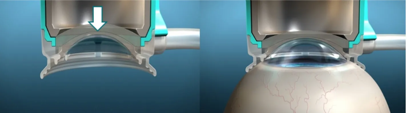
Figure 1. Advantages of SoftFit
TM patient interface. In contrast to solid direct patient interface, the design of the new hydrogel SoftFit™ interface (white arrow) has an extended suction ring skirt that allows the natural curvature of the cornea and delivers a gen- tle, secure fit with minimal corneal distortion. With the soft contact lens in place, the intraocular pressure remains much lower dur- ing the procedure and corneal compression and corneal folds are minimized.탈이 적고14, 고식적 백내장수술과 비교하여 측정된 수차 값이 낮고 시력의 질이 뛰어나며15, 내피세포의 손상과 전 방의 염증반응이 적고 창상치유가 빨라 안내염의 예방효과 도 있다고 알려져 있다.16-19 2009년 이후 해외의 여러 연구 에서 보고된 바에 따르면 펨토초레이저를 이용한 백내장수 술은 안전성이 높고 고식적 백내장수술에 비해 합병증 발 생률이 비슷하거나 적은 것으로 알려졌으나,20-23 국내에서 는 아직 이와 관련된 보고가 없다.
이에 저자들은 본 연구에서 펨토초레이저를 이용한 각막 절개, 수정체낭원형절개, 수정체 분할 시의 합병증의 발생 률을 조사하였고, 각 합병증에 관련된 위험인자에 대한 보 고가 없는 점에 착안하여 그와 관련된 위험인자가 무엇인 지 의무기록을 통해 후향적으로 분석해 보았다.
펨토초레이저 백내장 수술 시 사용되는 인터페이스는 펨 토초레이저의 기종에 따라 접촉과 비접촉 방식이 있으며, 본 연구에서 사용된 접촉 방식의 인터페이스는 작은 직경 을 가지고 있어 적은 용적을 가진 안와에 적합한 장점이 있 으나, 안압 상승과 결막하 출혈, 각막 주름 등의 문제가 자 주 발생하는 단점이 있었다.24
이러한 기존의 direct한 인터페이스와 비교하여 흡착 시 각막에 압박을 최소화시켜 안압상승을 줄이고 시술 중 합 병증을 감소시키는 것으로 알려진24 SoftFitTM 인터페이스 (Fig. 1)의 합병증 발생률에 대해서는 국내에서 보고된 바 가 없어 SoftFitTM 인터페이스 전후의 합병증 발생률을 비 교해 보았으며, 시술하는 술자의 숙련도가 수술 결과에 영 향을 미칠 것으로 생각되어 펨토초레이저 시행 초기 200안 과 후기 398안을 두 그룹으로 나누어 각 그룹 간의 합병증 발생률을 비교하였다.
대상과 방법
2012년 7월부터 2017년 1월까지 1명의 술자에 의해 펨 토초레이저를 이용하여 백내장수술을 시행 받은 337명 598 안을 대상으로 하였다. 펨토초레이저를 이용한 백내장수술 은 LenSx (Alcon LenSx, Fort Worth , TX, USA)를 이용하 였으며 투명각막절개, 수정체전낭절개, 수정체분할단계에 서 사용되었고 수정체 분할 패턴은 격자 방식을 채택하였 으며, 필요시 윤부이완절개술을 추가하였다. 투명각막절개 창 길이는 2.2 mm, 수정체전낭절개 직경은 5.0 mm, 수정 체분할 시 직경 길이는 5.2 mm로 설정하였다.
모든 수술은 0.5% proparacaine hydrochlorid로 점안 마 취 후 시행하였다. 펨토초레이저 시술 시 환자 인터페이스 를 각막에 흡착시킨 다음 시행하였고, 공기방울의 존재로 인한 흡착 실패 시 인터페이스를 완전히 분리한 후 재시도 하여 시행하였다. 수정체전낭절개는 산동된 동공 중심으로 설정하였다.
펨토초레이저를 이용하여 투명각막절개, 수정체전낭절 개, 수정체분할이 이루어진 이후 절개 부위를 blunt spatula 로 확인하였고, 불완전한 각막절개 시 각막절개도를 이용 하여 추가 시술하였으며, 불완전한 수정체 전낭절개의 경 우 낭집게를 이용하여 수정체낭의 방사상 파열 없이 전낭 절개가 불완전한 부분을 조심스럽게 제거하였다. 수정체분 할이 불완전한 경우 prechopper를 이용하여 재분할하였다.
수정체유화술과 잔여 수정체피질의 흡인은 Infinity (Alcon Laboratories Inc., Fort Worth, TX, USA)를 이용하여 시행 하였으며, 점탄물질로 전방을 채우고 인공수정체를 삽입한 다음 관류흡인으로 점탄물질을 제거하고 기질수화로 창상 을 폐쇄하였다. 술 후 2개월간 항생제, 스테로이드제제, 비 스테로이드성 항염증제제를 점안하였다
수술 중 발생한 합병증은 녹화된 영상을 단일 관찰자를 통
Table 3. Risk factors of intraoperative complications (n = 598)
Risk factor Incomplete corneal
incision
Incomplete capsulotomy
Incomplete nucleus
fragmentation Total
Corneal central opacity 7 (1.17) 5 (0.84) 3 (0.50) 15 (2.51)
Corneal peripehreal degeneration 3 (0.50) 0 (0) 0 (0) 3 (0.50)
Pterygium 2 (0.33) 0 (0) 0 (0) 2 (0.33)
Conjunctival chalasis 1 (0.17) 0 (0) 0 (0) 1 (0.17)
Mature cataract 0 (0) 17 (2.84) 13 (2.17) 30 (5.01)
Anterior polar or subcapsular cataract 0 (0) 7 (1.17) 2 (0.33) 9 (1.50)
Pupillary abnormality 0 (0) 3 (0.50) 1 (0.17) 4 (0.67)
Lens subluxation 0 (0) 3 (0.50) 3 (0.50) 6 (1.00)
Poor pupil dilation 0 (0) 1 (0.17) 0 (0) 1 (0.17)
Idiopathic 5 (0.84) 7 (1.17) 0 (0) 12 (2.01)
Total 18 (3.01) 43 (7.19) 22 (3.67) 83 (13.87)
Values are presented as n (%).
Table 1. Baseline demographic characteristics of all patients
Characteristic Value
Number of eyes (patients) 598 (337)
Age (years) 62.1 ± 11.9 (24–83)
Sex (male:female) 151:186
Eyes (right:left) 317:281
SoftFit™ interface (pre:post) 365:233 Values are presented as mean ± SD (range) or n (%) unless other- wise indicated.
Table 2. List of intraoperative complications encountered in
femtosecond laser cataract surgery cases (N = 598)Intraoperative complication Number of eyes
Incomplete corneal incision 18 (3.01)
Incomplete capsulotomy 43 (7.19)
Incomplete nucleus fragmentation 22 (3.67) Values are presented as n (%). Iris hemorrhage, endothelial in- cision, posterior capsule rupture, nucleus dislocation were not observed.
해 확인하였으며, 합병증에 해당하는 환자들의 의무기록을 후향적으로 확인하여 관련 인자를 분석하였다. 위의 연구는 성모안과병원 기관윤리심의위원회의 승인(2017-BM-01-1) 후 진행되었다.
2014년 4월에 도입된 SoftFit™ 인터페이스 사용 전후의 각 합병증의 발생률을 비교하고 유의한 차이가 있는지 검정 을 통해 비교하였다. 비교를 위하여 Pearson Chi-square test 를 시행하였고, 통계적인 분석은 SPSS® v22.0 for Windows (SPSS Inc., Chicago, IL, USA)를 사용하였다. p값이 0.05 미만일 경우 통계적으로 유의한 차이가 있다고 분석하였다.
결 과
총 337명 중 남자가 151명, 여자가 186명이었고 평균 나 이는 62.1 ± 11.9세였으며, 수술 받은 598안 중 우안이 317
안, 좌안이 281안, SoftFit™ 인터페이스 사용 전이 365안, 사용 후가 233안이었다(Table 1). 녹화된 비디오를 확인한 결과 펨토초레이저 수술 시 불완전한 투명각막절개가 18안 (3.01%), 수정체전낭절개가 43안(7.19%), 수정체분할이 22 안(3.67%)으로 나타났다(Table 2). 수술 중 합병증이 일어 난 환자의 의무기록에서 관련 인자를 후향적으로 조사한 결과 불완전한 투명각막절개의 경우 전체 18안 중 선천적 원인 또는 과거 감염, 외상 등으로 인한 각막 중심부 혼탁 이 7안, 노인환이나 테리엔각막가장자리변성, 잘쯔만결절 변성 등의 각막 주변부 변성이 3안, 익상편이 2안, 결막이 완이 1안, 특별한 원인이 없는 특발성이 5안 있었다.
불완전한 수정체전낭절개의 경우 전체 43안 중 관련 인 자로 팽대 백내장이나 모르가니 백내장 등의 과숙 백내장 이 17안, 전극 혹은 전낭하 백내장이 7안, 각막 중심부 혼 탁이 5안, 동공막 잔존이나 동공편위 등의 동공 형태 비정 상이 3안, 섬모체소대약화로 인한 수정체떨림이나 수정체 탈구가 3안, 당뇨나 거짓비늘증후군으로 인한 산동 불량이 1안, 특별한 원인이 없는 경우가 7안 있었다. 불완전한 수 정체분할의 경우 전체 22안 중 관련 인자로 과숙 백내장이 13안, 수정체떨림이나 수정체이탈이 3안, 각막 중심부 혼탁 이 3안, 전극 혹은 전낭하 백내장이 2안, 비정상적인 동공 이 1안 있었다.
모든 합병증과 관련된 위험인자를 발생 빈도순으로 정리 해보면 과숙 백내장이 30안, 중심성 각막혼탁이 15안, 특발 성이 12안, 전극 혹은 전낭하 백내장이 9안, 수정체탈구가 6안, 동공 형태 비정상이 4안, 각막 주변부 변성이 3안, 익 상편이 2안, 결막이완과 산동불량이 각각 1안 있었다(Table 3). 펨토초레이저 시술 시 홍채의 출혈, 불필요한 각막 절개 및 후낭 파열이나 수정체 이탈은 발생하지 않았다.
SoftFit™ 인터페이스 사용 전후의 합병증 발생률을 비 교해 보면, 사용 전 365안과 사용 후 233안을 대상으로 조
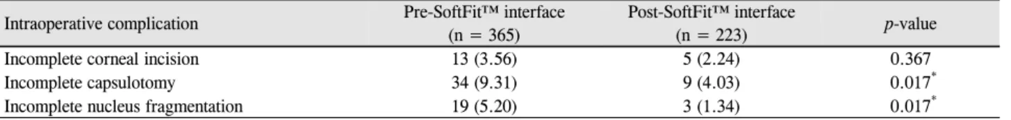
Table 4. Intraoperative complications rate before and after SoftFit™ interface
Intraoperative complication Pre-SoftFit™ interface
(n = 365)
Post-SoftFit™ interface
(n = 223) p-value
Incomplete corneal incision 13 (3.56) 5 (2.24) 0.367
Incomplete capsulotomy 34 (9.31) 9 (4.03) 0.017*
Incomplete nucleus fragmentation 19 (5.20) 3 (1.34) 0.017*
Values are presented as n (%) unless otherwise indicated.
*p-value < 0.05 between groups using Pearson Chi-square test.
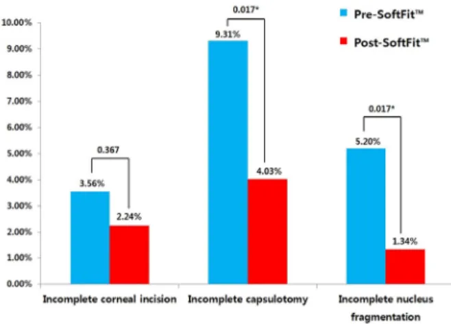
Table 5. Comparison of intraoperative complications between novice group (200 cases) and experienced group (398 cases)
Intraoperative complication Novice group (n = 200) Experienced group (n = 398) p-value
Incomplete corneal incision 11 (5.50) 7 (1.75) 0.012*
Incomplete capsulotomy 25 (12.50) 18 (4.52) <0.001*
Incomplete nucleus fragmentation 16 (8.00) 6 (1.50) <0.001*
Values are presented as n (%) unless otherwise indicated.
*p-value < 0.05 between groups using Pearson Chi-square test.
사한 결과 불완전한 투명각막절개의 경우 사용 전 13안 (3.56%)에서 사용 후 5안(2.24%), 불완전한 수정체전낭절 개의 경우 사용 전 34안(9.31%)에서 사용 후 9안(4.03%), 불완전한 수정체분할의 경우 사용 전 19안(5.20%)에서 사 용 후 3안(1.34%)으로 SoftFit™ 인터페이스 사용 후 모든 합병증의 발생률이 감소한 것을 알 수 있었다(Table 4).
술자의 숙련도에 따른 수술의 결과를 비교하기 위하여 펨토초레이저 수술 초기 200안과 후기 398안에서 발생한 합병증을 살펴보면, 불완전한 투명각막절개는 초기 11안 (5.50%)에서 후기 7안(1.75%), 불완전한 수정체전낭절개는 초기 25안(12.50%)에서 후기 18안(4.52%), 불완전한 수정 체분할은 초기 16안(8.00%)에서 후기 6안(1.50%)으로 술자 의 경험이 증가할수록 합병증이 감소하는 것을 확인하였다 (Table 5).
고 찰
2009년부터 백내장수술에 도입된 펨토초레이저 백내장 수술은 고식적 백내장수술법에 비하여 시술의 결과와 안전 성이 우수하다고 알려져 있다.20-23 수술 중 발생한 합병증 발생률에 대한 보고는 Roberts et al21이 1,500안을 대상으 로 조사한 불완전한 투명각막절개가 51안(3.40%), Chee et al25이 1,105안을 대상으로 조사한 불완전한 수정체전낭절 개 97안(8.78%) 및 불완전한 수정체분할 52안(4.71%)이 보 고되었으며, 발생 빈도는 불완전한 수정체전낭절개가 가장 많고 불완전한 수정체분할, 투명각막절개 순으로 나타났다.
국내의 경우 2012년부터 펨토초레이저 백내장수술이 백 내장수술에 도입되어 사용되고 있으나, 아직까지 국내에서 펨토초레이저 백내장수술 중 발생한 합병증에 대하여 보고 된 바는 없었다. 본 연구에서 1,129안을 대상으로 조사한
결과는 불완전한 수정체전낭절개 85안(7.53%), 불완전한 수정체분할 36안(3.19%), 불완전한 투명각막절개가 33안 (2.92%)으로 이전 연구에서 보고한 수술 중 합병증과 발생 빈도순이 일치하나, 각각의 발생률은 Roberts et al21이나 Chee et al25이 보고한 합병증의 비율보다 감소되어 있는 것 으로 나타났다. 합병증의 비율이 감소한 이유는 기존의 연구 에서 사용하지 않았던 SoftFit™ 인터페이스를 본 연구에서 2014년 4월부터 도입하여 시술하였기 때문으로 생각된다.
SoftFit™ 인터페이스는 기존의 단단한 고체 형태의 direct 인터페이스에 비하여 상대적으로 부드러운 하이드로젤 소 재를 사용하고 흡착 고리의 주변부 길이를 연장하여 보다 자연스럽게 각막 흡착을 가능하게 하며, 각막 표면의 압박 을 최소화시켜 안구내 안압의 상승을 낮추고 각막의 주름 을 줄임으로써 펨토초레이저가 목표점에 보다 정확히 도달 하게 하는 효과를 가지고 있다.24 Asena and Kaskaloglu24가 100안을 대상으로 발표한 보고에 따르면 SoftFit™ 인터페 이스 사용 전 50안과 사용 후 50안의 합병증 발생률은 불 완전한 각막절개의 경우 사용 전 10%에서 사용 후 2%, 불 완전한 수정체전낭절개의 경우 사용 전 12%에서 사용 후 0%, 불완전한 수정체분할의 경우 사용 전 10%에서 사용 후 4%로 모두 감소하였다. 본 연구에서도 발생률의 차이는 있으나 불완전한 투명각막절개, 불완전한 수정체전낭절개, 불완전한 수정체분할 등 모든 시술에서 SoftFit™ 인터페이 스 사용 후 합병증이 감소한 결과를 보였으며, 불완전한 수 정체전낭절개와 불완전한 수정체 분할의 발생률은 SoftFit™
인터페이스 사용 전후 군 간에 유의한 차이가 있음을 알 수 있었다(Table 4, Fig. 2). 반면 불완전한 각막절개 발생률은 유 의미한 감소를 보이지 않았는데 이는 Asena and Kaskaloglu24 가 분석한 바와 같이 인터페이스를 이용하여 각막 흡착 시 나타나는 각막 후면의 주름은 각막절개에 필요한 펨토초레
Figure 2. Rates of intraoperative complications before and af-
ter SoftFitTM interface. Comparison (%) of incomplete corneal incision, incomplete capsulotomy, incomplete nucleus frag- mentation between femtosecond laser refractive cataract sur- gery with a direct contact interface (Pre-SoftFit™) and SoftFit™interface (Post-SoftFit™). *p-value<0.05 between groups us- ing Pearson Chi-square test.
Figure 3. Rates of intraoperative complications between nov-
ice group and experienced group. Comparison (%) of in- complete corneal incision, incomplete capsulotomy, in- complete nucleus fragmentation between novice group (200 cases) and experienced group (398 cases). *p-value < 0.05 be- tween groups using Pearson Chi-square test.이저의 에너지가 각막 기질에 도달하는 데 영향을 주지 않 으며, 따라서 기존 인터페이스보다 각막 후면 주름이 적게 발생하는 SoftFit™ 인터페이스를 사용하여도 두 군 간의 불 완전한 각막절개의 발생에는 차이가 없을 것으로 추정된다.
또한 펨토초레이저 시술 시 술자의 숙련도와 합병증의 연관성을 확인하기 위하여 수술 초기 200안과 후기 398안 으로 나누어 합병증 발생률을 비교한 결과, 불완전한 투명 각막절개, 불완전한 수정체전낭절개, 불완전한 수정체분할 모두 초기보다 후기 398안의 합병증 발생률이 유의하게 낮 음을 확인할 수 있었다(Table 5, Fig. 3). 따라서 Bali et al26
이 보고한 바와 같이 술자의 숙련도가 증가할수록 수술 중 합병증 발생은 감소할 것으로 생각된다.
수술 중 발생한 합병증의 위험인자를 발생빈도가 높은 순으로 살펴보면 불완전한 투명각막절개의 경우 각막 중심 부 혼탁(1.17%), 특발성(0.84%), 각막 주변부 변성(0.50%), 익상편(0.33%), 결막이완(0.17%) 순이었다. 특발성을 제외 하고는 각막 혼탁이나 각막 주변부의 변성과 관련된 각막 병변(1.67%)이 불완전한 투명각막절개와 가장 많은 연관성 이 있음을 알 수 있었으며, 이는 투명각막절개 시 펨토초레 이저가 전달되는 과정에서 각막의 병변으로 인하여 조직에 도달하는 에너지에 상실이 일어났기 때문인 것으로 추측된 다. 익상편과 결막이완이 있는 경우는 인터페이스를 이용 하여 흡입하는 과정에서 계측 값과의 오차가 발생할 수 있 을 것으로 보인다.
불완전한 수정체전낭절개의 경우 관련인자는 과숙 백내장 (2.84%), 특발성(1.17%), 전극 혹은 전낭하 백내장(1.17%), 각막 중심부 혼탁(0.84%), 동공 형태 비정상(0.50%), 수 정체탈구(0.50%), 당뇨나 거짓비늘증후군으로 인한 산동 불량(0.17%) 순이었으며, 불완전한 수정체분할의 경우 과숙 백내장(2.17%), 각막 중심부 혼탁(0.50%), 수정체 이탈(0.50%), 전극 혹은 전낭하 백내장(0.33%), 비정상적 인 동공(0.17%) 순으로 나타났다. 과숙 백내장이나 전극 혹 은 전낭하 백내장의 경우 수정체전낭절개나 수정체 분할 시 펨토초레이저의 에너지가 일반 백내장 시술보다 더 많 은 에너지가 필요했을 가능성이 있으며, 동공 형태의 비정 상이나 산동 불량, 수정체의 탈구 등은 수정체전낭절개나 수정체 분할 시 해당 시술 부위를 동공 중심으로 판단하므 로 펨토초레이저의 정확성이 낮을 가능성이 있다. 각막 중 심부에 혼탁이 있는 경우는 시술 부위에 도달하는 에너지 가 부족했을 것으로 판단된다.
전체적으로 수술 중 합병증과 관련된 위험인자는 과숙 백내장(5.01%), 중심성 각막혼탁(2.51%), 특발성(2.01%), 전극 혹은 전낭하 백내장(1.50%), 수정체탈구(1.00%), 동공 형태의 비정상(0.67%), 각막 주변부 변성(0.50%), 익상편 (0.33%), 결막이완(0.17%)과 산동불량(0.17%) 순으로 조사 되었다(Table 3). 이는 펨토초레이저를 이용한 백내장수술 시 술 전 검사에서 해당 위험인자가 관찰되는 경우 관련된 불완전한 시술의 가능성을 인지하고 조심스럽게 접근해야 하며, 특별한 원인을 찾을 수 없는 특발성인 경우도 세 번 째로 많은 비율을 차지하고 있으므로 술 전 검사 시 관련 위험인자가 없고 펨토초레이저 시술 시 수술 시야상에서 합병증이 관찰되지 않더라도 수동적 투명각막절개 및 연속 곡선수정체낭원형절개, 수정체분할 등 추가적인 시술의 필 요성을 염두에 두고 수술에 임해야 할 것으로 사료된다.
결론적으로 저자들은 펨토초레이저를 이용한 백내장수 술이 기존의 고식적 백내장수술에서 발생하는 수술 중 합 병증을 완벽히 해결해 주지는 않으나, 술 전 검사에서 합병 증과 관련된 인자가 관찰된 경우에 해당 합병증에 유의하 여 시술한다면 백내장수술의 성공률을 향상시킬 수 있는 안전하고 효율적인 백내장수술법이라고 생각하며, 각막 흡 착 시 기존의 direct한 인터페이스보다 SoftFit™ 인터페이 스를 사용하고, 술자의 숙련도가 높아질수록 펨토초레이저 백내장수술 중 합병증의 발생을 더욱 감소시킬 수 있을 것 으로 기대하는 바이다.
REFERENCES
1) Krasnov MM. Laser-phakopuncture in the treatment of soft cataracts. Br J Ophthalmol 1975;59:96-8.
2) Ratkay-Traub I, Juhasz T, Horvath C, et al. Ultra-short pulse (femtosecond) laser surgery: initial use in LASIK flap creation.
Ophthalmol Clin North Am 2001;14:347-55. viii-ix.
3) Sugar A. Ultrafast (femtosecond) laser refractive surgery. Curr Opin Ophthalmol 2002;13:246-9.
4) Nagy Z, Takacs A, Filkorn T, Sarayba M. Initial clinical evaluation of an intraocular femtosecond laser in cataract surgery. J Refract Surg 2009;25:1053-60.
5) Masket S, Sarayba M, Ignacio T, Fram N. Femtosecond laser-as- sisted cataract incisions: architectural stability and reproducibility.
J Cataract Refract Surg 2010;36:1048-9.
6) Chang JS, Chen IN, Chan WM, et al. Initial evaluation of a femto- second laser system in cataract surgery. J Cataract Refract Surg 2014;40:29-36.
7) Park JH, Lee KH, Lee DJ. Comparison of continuous curvilinear capsulorhexis parameters between femtosecond laser and conven- tional cataract surgery. J Korean Ophthalmol Soc 2014;55:1800-7.
8) Kránitz K, Takacs A, Miháltz K, et al. Femtosecond laser capsu- lotomy and manual continuous curvilinear capsulorrhexis parame- ters and their effects on intraocular lens centration. J Refract Surg 2011;27:558-63.
9) Friedman NJ, Palanker DV, Schuele G, et al. Femtosecond laser capsulotomy. J Cataract Refract Surg 2011;37:1189-98.
10) Auffarth GU, Reddy KP, Ritter R, et al. Comparison of the max- imum applicable stretch force after femtosecond laser-assisted and manual anterior capsulotomy. J Cataract Refract Surg 2013;39:105-9.
11) Conrad-Hengerer I, Hengerer FH, Schultz T, Dick HB. Effect of femtosecond laser fragmentation on effective phacoemulsification time in cataract surgery. J Refract Surg 2012;28:879-83.
12) Conrad-Hengerer I, Hengerer FH, Schultz T, Dick HB. Effect of
femtosecond laser fragmentation of the nucleus with different soft- ening grid sizes on effective phaco time in cataract surgery. J Cataract Refract Surg 2012;38:1888-94.
13) Abell RG, Kerr NM, Vote BJ. Toward zero effective phacoemulsi- fication time using femtosecond laser pretreatment. Ophthalmology 2013;120:942-8.
14) Kránitz K, Miháltz K, Sándor GL, et al. Intraocular lens tilt and de- centration measured by Scheimpflug camera following manual or femtosecond laser-created continuous circular capsulotomy. J Refract Surg 2014;28:259-63.
15) Miháltz K, Knorz MC, Alió JL, et al. Internal aberrations and opti- cal quality after femtosecond laser anterior capsulotomy in cataract surgery. J Refract Surg 2012;27:711-6.
16) Takács AI, Kovács I, Miháltz K, et al. Central corneal volume and endothelial cell count following femtosecond laser-assisted re- fractive cataract surgery compared to conventional phacoe- mulsification. J Refract Surg 2012;28:387-91.
17) Conrad-Hengerer I, Al Juburi M, Schultz T, et al. Corneal endothe- lial cell loss and corneal thickness in conventional compared with femtosecond laser-assisted cataract surgery: three-month fol- low-up. J Cataract Refract Surg 2013;39:1307-13.
18) Abell RG, Allen PL, Vote BJ. Anterior chamber flare after femto- second laser-assisted cataract surgery. J Cataract Refract Surg 2013;39:1321-6.
19) Chen M. A review of femtosecond laser assisted cataract surgery for Hawaii. Hawaii J Med Public Health 2013;72:152-5.
20) Filkorn T, Kovács I, Takács A, et al. Comparison of IOL power cal- culation and refractive outcome after laser refractive cataract sur- gery with a femtosecond laser versus conventional phacoemulsification.
J Refract Surg 2012;28:540-4.
21) Roberts TV, Lawless M, Bali SJ, et al. Surgical outcomes and safe- ty of femtosecond laser cataract surgery: a prospective study of 1500 consecutive cases. Ophthalmology 2013;120:227-33.
22) Abell RG, Darian-Smith E, Kan JB, et al. Femtosecond laser-as- sisted cataract surgery versus standard phacoemulsification cata- ract surgery: outcomes and safety in more than 4000 cases at a sin- gle center. J Cataract Refract Surg 2015;41:47-52.
23) Chen M, Swinney C, Chen M. Comparing the intraoperative com- plication rate of femtosecond laser-assisted cataract surgery to tra- ditional phacoemulsification. Int J Ophthalmol 2015;8:201-3.
24) Asena BS, Kaskaloglu M. Laser-assisted cataract surgery: soft lens assisted interface (SoftFit) versus direct contact interface. Eur J Ophthalmol 2016;26:242-7.
25) Chee SP, Yang Y, Ti SE. Clinical outcomes in the first two years of femtosecond laser-assisted cataract surgery. Am J Ophthalmol 2015;159:714-9.
26) Bali SJ, Hodge C, Lawless M, et al. Early experience with the fem- tosecond laser for cataract surgery. Ophthalmology 2012;119:891-9.
= 국문초록 =
성공적인 펨토초레이저 백내장수술을 위한 수술 중 합병증 위험인자 분석
목적: 펨토초레이저 백내장수술 시 발생하는 합병증의 위험인자를 알아보고자 하였다.
대상과 방법: 2012년 7월부터 2017년 1월까지 본원에서 펨토초레이저 백내장수술을 시행한 337명 598안을 대상으로 연구를 실시하였 다. 모든 안에 투명각막절개, 수정체전낭절개, 수정체분할을 시행하였으며, 필요시 윤부이완절개술을 추가하였다. 수술 중 합병증은 녹화된 영상을 이용하여 결과를 확인하였고, 합병증 관련 위험인자는 의무기록을 후향적으로 분석하였다.
결과: 대상 환자군의 나이는 평균 62.1 ± 11.9세였으며, 불완전한 투명각막절개 18안 중 위험인자는 각막중심혼탁, 각막주변부변성, 익상편, 결막이완이 있었고, 불완전한 수정체전낭절개 43안 중 위험인자는 과숙백내장, 전극백내장, 각막중심혼탁, 비정상동공, 수정 체탈구, 산동불량이 있었으며, 불완전한 수정체분할 22안 중 위험인자는 과숙백내장, 수정체이탈, 각막중심혼탁, 전극 백내장, 비정상 동공이 있었다. SoftFit™ 인터페이스 사용 후 합병증 발생률은 투명각막절개(3.56%에서 2.24%, p=0.367), 수정체전낭절개(9.31%에서 4.03%, p<0.05), 수정체분할(5.20%에서 1.34%, p<0.05) 모두 감소하였으며, 초기 사례와 후기 사례의 합병증 발생률 비교 시 투명각 막절개, 수정체전낭절개, 수정체분할 모두 초기보다 후기에 발생률이 유의하게 감소하였다(p<0.05).
결론: 펨토초레이저는 백내장수술을 안전하고 효과적으로 할 수 있는 장점이 있다. 그러나 관련된 위험인자가 존재하는 경우 합병증 의 가능성을 고려하여 접근해야 할 것으로 보이며, SoftFit™ 인터페이스를 사용하고, 술자의 숙련도가 높을수록 합병증 발생을 감소시 킬 수 있을 것으로 생각된다.
<대한안과학회지 2017;58(5):539-545>