pISSN: 0378-6471 eISSN: 2092-9374 DOI : 10.3341/jkos.2011.52.6.671
= 증례보고 =
원자현미경을 이용한 마이토마이신 노출 시간에 따른 공막 표면 콜라젠의 변화 관찰
이희재1⋅최삼진2⋅정유진2⋅정경복2⋅진경현3⋅박헌국2,4⋅이승준1
강원대학교 의학전문대학원 안과학교실1, 경희대학교 의과대학 의공학교실2, 안과학교실3, 경희대학교 생체의과학 협동과정4
목적: 마이토마이신 노출 시간에 따른 공막 콜라젠의 변화를 원자현미경을 이용하여 보고자 하였다.
대상과 방법: 원자현미경을 이용하여 0.02% 마이토마이신을 공여 공막기질에 1, 3, 5분 동안 노출 전후의 표면변화를 관찰하였다. 공막 의 원자현미경 표면영상과 편향영상(deflection image)으로부터 콜라젠 피브릴의 구조 변화, 직경과 디-밴딩을 관찰하였다.
결과: 0.02% 마이토마이신을 1분간 노출한 군(155.04 ± 17.46 nm 직경과 70.02 ± 3.33 nm 디-밴딩)은 대조군과 콜라젠의 형태학적 변화를 보이지 않았지만 3분(182.33 ± 16.33 nm 직경과 70.27 ± 13.66 nm 디-밴딩, p<0.005)과 5분(199.20 ± 12.40 nm 직경과 72.75 ± 19.32 nm 디-밴딩, p<0.0001)간 노출한 군에서는 통계학적으로 유의하게 공막 콜라젠 피브릴의 증가가 관찰되었다.
결론: 0.02% 마이토마이신을 3, 5분 공막에 노출 시 공막 콜라젠의 형태학적 변화가 관찰되었으며 이는 마이토마이신을 3분 이상 공막에 노출 시 공막 콜라젠의 형태학적 변화가 일어남을 알 수 있었다.
<대한안과학회지 2011;52(6):671-678>
■ 접 수 일: 2010년 9월 15일 ■ 심사통과일: 2010년 12월 15일
■ 게재허가일: 2011년 3월 22일
■ 책 임 저 자: 이 승 준
강원도 춘천시 백령로 156 강원대학교병원 안과
Tel: 033-258-2443, Fax: 033-258-2191 E-mail: [email protected]
* 본 논문은 2010년도 강원대학교 학술연구조성비로 연구하였음.
마이토마이신(Mitomycin C)은 1955년 streptomyces caespitosus에서 추출한 대사억제제로 산소라디칼을 생성 하고 DNA와 RNA의 합성을 저해하며 섬유모세포의 증식 을 억제하는 것으로 알려져 있다.1-4 현재까지 마이토마이 신은 식도암, 유방암, 방광암 등의 여러 종양의 치료 약제로 널리 사용되고 있으며 식도 협착증과 기도 협착증의 치료 보조약제로도 사용되고 있다.5 안과 영역에서는 익상편 수 술, 굴절교정수술, 섬유주절제술, 코눈물관부분폐쇄, 미용 적 결막절제술 등에 사용되고 있으나 마이토마이신의 사용 후 공막궤양, 괴사성 공막염, 각막부종, 각막천공, 공막연 화, 포도막염, 녹내장 등의 부작용을 유발하는 것으로 알려 져 있다.6-14 공막은 대부분이 콜라젠으로 구성되어 있으며 이 중에서 I형 콜라젠이 50-70%로 가장 많고 III, V, VI형 콜라젠도 공막기질에서 발견된다.15 지금까지 공막 콜라젠 에 대한 연구는 전자현미경(scanning electron microscopy) 을이용하여 주로 조직을 검사해왔다.16-20그러나 전자현미
경으로 관찰하기 위해서는 탈수, 얇은 표본처리, 표면 코팅 처리와 같은 여러 단계의 복잡한 처리 과정으로 인해 조직 의 변성이 자주 유발된다. 이에 반해, 원자현미경(atomic forcemicroscopy)은 최소한의 조직 처리과정과 진공상태 에서만 영상획득이 가능한 전자현미경에 반해 제약을 받지 않고 영상을 얻을 수 있다. 또한, 3차원 영상 및 나노스케일 상에서 영상을 얻을 수 있어 보다 정확한 조직 검사가 가능 한 것으로 알려져 있다.21-24 현재까지 마이토마이신 합병증 에 대한 임상학적 보고는 많지만 마이토마이신이 공막에 미치는 영향을 조직학적 단계에서 접근한 연구보고는 제한 되어 있다. 따라서, 본 연구에서는 원자현미경을 이용하여 0.02% 마이토마이신의 노출 시간에 따른 공막 콜라젠 피브 릴의 구조적 변화를 통하여 마이토마이신의 효과를 관찰하 였다.
대상과 방법
조직준비과정
두 사람의 공막표본을 경희대학교 의과대학교 안과학교 실 안은행으로부터 얻었으며 기증자들은 간염, 매독, 후천 성면역결핍바이러스 감염 등이 음성으로 나왔으며 공막표 본은 안구 적출 후 90% 알코올에 3개월 동안 보관하였다.
공막조직을 평형용액(balanced salt solution)으로 세척한
A B
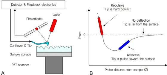
Figure 1. (A) Atomic force microscopy (AFM) block diagram, and (B) force vs. distance and relative zone modes.
후 30분간 평형용액에 담가 둔 후 각 그룹에 대해 5개 (n=20)의 시편으로 자른 후 총 영상 n=156개의 AFM 영 상을 촬영하였다. 0.02% 마이토마이신을 1분, 3분, 5분간 노출시킨 후 평형용액으로 세척 후 2분간 건조시켰다.
원자현미경 검사
원자현미경은 Figure 1A와 같이 스프링 캔틸레버(spring cantilever) 끝에 길이가 100-200 µm과 100 Å 이하의 직 경을 가진 팁(SiN)이 부착되어 있는 스프링 휨(deflection) 시스템이고 이 팁이 시편을 주사할 때 팁과 표면 사이에 반 데리발스(Vander Walls) 힘이 작용하여 캔틸레버를 굴곡 시킨다. 이것은 캔틸레버 뒷면에서 입사하는 레이저의 굴곡 으로 광검출기(photodetector)에서 감지되어 원자단위의 수준으로 물질의 표면구조를 3차원으로 형상화할 수 있고 표면의 변화양상을 실시간으로 관찰할 수 있다. 원자현미경 은 Figure 1B와 같이 크게 접촉식 모드(contact mode)와 비접촉식 모드(noncontact mode)로 구분된다. 표면과 팁 사이에 척력(repulsive force)이 작용하는 영역에서 작동되 는 모드가 접촉식 모드이며 표면과 팁 사이에 인력 (attractive force)이 작용하는 영역에서 작동되는 모드가 비접촉식 모드이다. 접촉식 모드에서는 원자현미경 팁과 시 편이 부드러운 “물리적 접촉”을 이룬다. 비접촉식 모드의 경우는 부드럽고 유연한 표면을 가진 시료측정에 유리하며 팁의 표면에 직접 닿지 않기 때문에 오염이 적다.
공막표면 영상은 42.5×42.5×4 μm3 XYZ 스캐너와 광학 현미경(Epiplan 200×/500×)이 장착된 NANOS N8 NEOS 원자현미경(Bruker, Herzogenrath, Germany)과 92.5×
92.5×6 μm3 XYZ 스캐너와 광학현미경(Epiplan 100×/
500×)이 장착된 NANOstation II 원자현미경(Surface
Imaging Systems, Herzogenrath) 두 대를 이용하여 비접 촉식 모드에서 촬영하였다. 외부에서 유입되는 노이즈 제거 를 위하여 원자현미경은 수동방진테이블(Passive vibration isolation table, Pucotech, Seoul, Korea) 안에 TS-150 능 동방진테이블(Active vibration isolation table, S.I.S., Herzogenrath)위에 설치하였다. 공막표면은 512×512 pixels 해상도, 0.8 line/sec 스캔 속도, 3개의 스캔영역(20
×20 µm2, 5×5 µm2, 1×1 µm2)에서 측정하였다. 각 샘플은 인테그럴 피라미드 모양 팁(Integral pyramidal shaped tip, SICONG, Santa Clara, CA, USA)을 가진 실리콘 캔틸레버 를 이용하여 35% 상대습도에서 10분 간격으로 검사하였 다. <10 nm 직경과 12-16 μm 높이를 가진 노미널 팁 (nominal tip)이 사용되었다. 샘플의 변형을 고려하여 모든 검사는 48시간 이내에 수행하였다.
분석과 통계방법
영상수집과 분석은 상용프로그램 SPIP (Scanning Probe Image Processor Version 4.8, Image Metrology, Denmark) 를 이용하여 수행하였다. 마이토마이신 노출시간에 따른 공 막표면의 콜라젠 피브릴의 직경(diameter)과 디-벤딩 (D-banding)의 변화를 통하여 간접적으로 마이토마이신 효과를 분석하였다. 통계처리는 SPSS version 16의 two- tailed Student’s t-test를 이용하여 분석하였고, p<0.05를 통계학적으로 유의한 것으로 간주하였다.
결 과
Figure 2에 서로 다른 3개의 스캔영역에서 건조된 공막 표면(대조군)의 원자현미경 표면(topography) 영상을 도
A B C
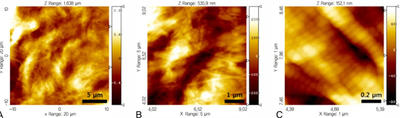
Figure 2. Representative atomic force microscopy topography images of the dehydrated human sclera in various scan sizes such as
(A) 20 × 20 µm2, (B) 5 × 5 µm2, and (C) 1 × 1 µm2.A B
C
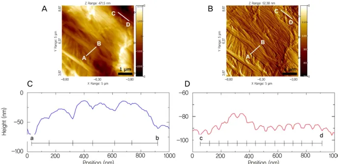
Figure 3. Representative three-dimensional images of the dehydrated human sclera in various scan sizes such as (A) 20 × 20 µm2, (B) 5 × 5 µm2, and (C) 1 × 1 µm2.
시하였다. Figure 2A, 20×20 µm2스캔영역에서 병렬구조 의 공막 피브릴의 교차된 다발(interlocked bundles) 형태 의 네트워크 구조를 볼 수 있다. 공막의 피브릴 구조의 자 세한 관찰을 위하여 동일 표면의 보다 작은 영역, 5×5 µm2 과 1×1 µm2영역 또한 검사되었다. Figure 2B와 C에서 공 막 콜라젠 피브릴의 규칙적인 병렬 배열은 물론 샘플 전처 리 과정에서 발생한 결함 혹은 콜라젠 분자 사이의 크로스 링크(cross-links)의 파괴에 의하여 변형된 직경과 디-밴 딩을 가진 콜라젠 피브릴 또한 관찰할 수 있었다.25 또한, 콜라젠 피브릴의 규칙적인 병렬배열과는 다르게 피브릴 다
발 위에 중첩된 형태의 독특한 배열과 상호교차 등의 구조 를 보였다.22Figure 3의 공막 콜라젠의 표면영상(Fig. 2)에 서 만들어진 3차원 영상으로부터 보다 정교한 공막 콜라젠 3차원 구조를 확인할 수 있었다. Figure 2C와 Figure 3과 같은 고분해능의 피브릴 영상으로부터 옥수수 구조와 유사 한 디-밴딩 구조를 확인할 수 있었다.21
Figure 4에서 보여진 것과 같이 표면영상(topography image, Fig. 4A)과 편향영상(deflection image, Fig. 4B)에 서 라인 프로파일 방법을 이용하여 직경(Fig. 4C)과 디-밴 딩(Fig. 4D)을 측정하였다. Figure 5와 6에 대표적인 대조
X X
A B
C D
Figure 4. Representative examples of the diameter (C) and D-banding (D) measurements using line profiling plots on AFM top-
ography (A) and deflection images (B) of the dehydrated scleral collagen fibrils in a scan size of 5 × 5 μm2.A B
C
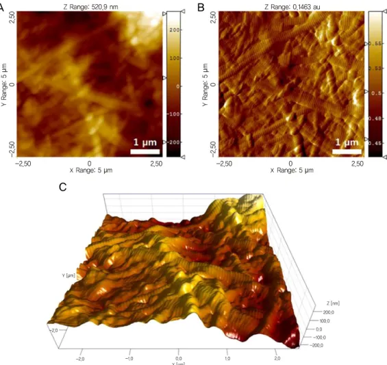
Figure 5. Representative AFM topography (A), deflection (B), and three-dimensional images (C) of the dehydrated
scleral collagen fibrils in a scan size of 5 × 5 μm2.A B
C
Figure 6. Representative AFM topography (A), deflection (B), and three-dimensional images (C) of the dehydrated
scleral collagen fibrils with 0.02% mitomycin C application for 5 minutes in a scan size of 5 × 5 μm2.Table 1. Morphological changes in the sclera fibrils for control and MMC-treated groups
Parameter Control MMC 1 min MMC 3 min MMC 5 min
Diameter (mean ± SD, nm) 145.22 ± 17.78 155.04 ± 17.46 182.33 ± 16.33* 199.20 ± 12.40†
D-banding (mean ± SD, nm) 69.14 ± 14.15 70.02 ± 3.33 70.27 ± 13.66 72.75 ± 19.32
MMC = Mitomycin C.
*p-value < 0.005 vs. Control group; †p-value < 0.0001 vs. Control group.
군과 0.02% 마이토마이신을 5분간 노출한 콜라젠 피브릴 원자현미경 영상을 도시하였다. 대조군(Fig. 5)에 비하여 5 분간 노출시킨 그룹(Fig. 6)에서 팽창된 콜라젠 피브릴 구 조를 확인할 수 있었다. 특히, 대조군에서는 콜라젠 피브릴 직경이 145.22 ± 17.78 nm이었고 디-밴딩이 69.14 ± 14.15 nm이었다. 0.02% 마이토마이신을 1분간 노출한 군 (MMC1min)에서는 콜라젠 피브릴 직경이 155.04 ±17.46 nm, 디-밴딩이 70.02 ±3.33 nm로 대조군과 유의성을 보 이지 않았다. 3분간 노출한 군(MMC3min)에서는 직경이 182.33 ±16.33 nm (p<0.005)의 증가함을 보였고 디-밴 딩은 70.27 ± 13.66 nm를 보였다. 5분간 노출한 군
(MMC5min)은 콜라젠 피브릴 직경이 199.20 ±12.40 nm (p<0.0001), 디-밴딩은 72.75 ± 19.32 nm를 보였다.
0.02% 마이토마이신을 1분간 군은 대조군과 콜라젠의 형 태학적 변화를 보이지 않았지만 3분과 5분간 노출한 군에 서는 통계학적으로 유의하게 공막 콜라젠 피브릴의 직경이 126% (3분)와 137% (5분) 증가함을 보였다(Table 1).
고 찰
마이토마이신은 안과 영역에서 익상편 수술 후 재발 방 지, 굴절교정수술후, 섬유주절제술, 코눈물관부분폐쇄 등에
X X
사용되고 있으며 그 농도는 0.02%와 0.04%가 주로 사용되 고 있다.6,7본 실험에서는 안과 영역에서 요즘 주로 사용되 는 농도인 0.02% 마이토마이신의 노출 시간에 따른 공막 표면 변화를 관찰하였다. 현재까지 마이토마이신 사용으로 인한 공막궤양, 괴사성 공막염, 각막부종, 각막천공, 공막연 화, 포도막염, 녹내장 등과 같은 많은 합병증이 보고되고 있
다.26-31 특히, 익상편 수술, 미용적 결막 절제술후 공막궤
양, 공막연화증 등의 합병증이 발생하는 이유로 마이토마이 신 사용에 따른 노출된 공막에 존재하는 섬유모세포의 증 식 억제, 세포자멸사 및 이에 따른 세포 이동과 증식의 억 제로 콜라겐 생성 저하 등이 병인으로 생각될 수 있으며 혈 관이 발달해 있는 상공막의 혈관을 수술 중 과도하게 소작 하는 경우, 상공막을 포함하여 제거한 경우, 방사선 치료를 받은 경우가 또한 그 원인으로 생각되고 있다.1-4
마이토마이신의 사용에 따른 합병증을 줄이기 위한 방법 들이 지금까지 많이 논의되고 있다.9,10,12-14Norliza et al32 은 익상편 수술 후 일주일간 하루에 4번 0.1% 마이토마이 신 점안 후 16년 후 공막연화증이 발생한 증례를 보고하였 고 발생 원인으로는 마이토마이신 자체의 섬유모세포의 증 식 억제 및 섬유모세포의 콜라젠 합성을 억제시켜 공막연 화증이 발생했을 것으로 보고하였다. 마이토마이신 사용에 따른 합병증을 줄이기 위해 낮은 농도의 점안제를 사용할 것과 짧은 기간 사용할 것을 권장하고 있다. Solomon et al33 은 익상편 수술 중 0.02% 마이토마이신을 5분간 노출 후 평형용액으로 세척을 시행하여 7년간 경과 관찰로부터 합병증이 발생하지 않았으며 익상편 수술 중 마이토마이신 을 사용한 경우에는 상피화가 완성될 때까지 지속적인 외 래 추적관찰을 시행할 것을 권장하였다.
본 연구에서는 원자현미경을 이용하여 가능한 조직 준비 과정을 간편화하고 전자현미경과 같이 조직 준비과정에서 올 수 있는 조직 손상을 최소화 하고자 하였다. 두 대의 원 자현미경을 가지고 대조군과 실험군을 동시에 검사하여 시 간차에 따른 오차를 최소화 하였다. 대조군의 공막 콜라젠 피브릴은 규칙적인 병렬배열은 물론 피브릴 다발 위에 중 첩된 형태의 독특한 배열과 상호교차 등의 구조를 보였다.
특히 Fullwood et al22는 공막 콜라젠 피브릴의 상호교차된 구조는 밀집된 단백당(proteoglycans), 즉 VI형 콜라젠과 관련이 있다고 발표하였다. 그리고 3차원 영상으로부터 보 다 정교한 공막 콜라젠 구조(옥수수 구조)를 확인할 수 있 었다.213, 5분간 0.02% 마이토마이신을 공막에 노출 시 공 막 콜라젠 피브릴 직경과 같은 형태학적 변화가 발생하였 다. 이는 수술 후 장기간에 걸친 마이토마이신 노출 시 공 막 콜라젠 변성이 발생할 수 있는 것을 암시하며 특히, 공 막이 넓게 노출되는 미용적 결막절제술 등의 시술에서는
장기간의 마이토마이신 점안은 여러 합병증의 발생을 증가 시킬 수 있을 것으로 생각한다. 향후 조직학적 변화를 유발 하지 않으면서 수술결과를 좋게 할 수 있는 마이토마이신 의 농도 및 노출 시간에 대한 보다 많은 연구가 필요할 것 으로 생각한다.
참고문헌
1) Danshiitsoodol N, de Pinho CA, Matoba Y, et al. The mitomycin C (MMC)-binding protein from MMC-producing microorganisms protects from the lethal effect of bleomycin: crystallographic anal- ysis to elucidate the binding mode of the antibiotic to the protein. J Mol Biol 2006;360:398-408.
2) Mao Y, Varoglu M, Sherman DH. Molecular characterization and analysis of the biosynthetic gene cluster for the antitumor antibiotic mitomycin C from Streptomyces lavendulae NRRL 2564. Chem Biol 1999;6:251-63.
3) Oum BS, Lee JS. The effect of Mitomycin C (MMC) on inhibition of cellular proliferation and type_I collagen, laminin synthesis of pterygial mesenchymal cell. J Korean Ophthalmol Soc 1999;40:
712-20.
4) Willems EW, Nooter K, Verweij J. Antitumor antibiotics. In:
Chabner BA, Longo DL, eds. Cancer Chemotherapy & Biotherapy:
Principles and Practice, 4th ed. Philadelphia: Lippincott Williams
& Wilkins, 2006; v. 1. chap. 16.
5) Ross P, Nicolson M, Cunningham D, et al. Prospective randomized trial comparing mitomycin, cisplatin, and protracted venous-in- fusion fluorouracil (PVI 5-FU) with epirubicin, cisplatin, and PVI 5-FU in advanced esophagogastric cancer. J Clin Oncol 2002;20:
1996-2004.
6) Lee TS, Rhee K. The effect of Mitomycin C eyedrops on prevention of intermal ostium obstruction after endonasal dacryocystorhinostomy.
J Korean Ophthalmol Soc 1998;39:1915-20.
7) Park DJ, Kwak MS. The effect of Mitomycin C on the success rate of endoscopic dacryo cystorhinostomy. J Korean Ophthalmol Soc 2000;41:1674-9.
8) Anderson RL, Edwards JJ. Indications, complications and results with silicone stents. Ophthalmology 1979;86:1474-87.
9) Tarr KH, Constable IJ. Late complications of pterygium treatment.
Br J Ophthalmol 1980;64:496-505.
10) Rubinfeld RS, Pfister RR, Stein RM, et al. Serious complications of topical mitomycin-C after pterygium surgery. Ophthalmology 1992;99:1647-54.
11) Wan Norliza WM, Raihan IS, Azwa JA, Ibrahim M. Scleral melt- ing 16 years after pterygium excision with topical Mitomycin C adjuvant therapy. Cont Lens Anterior Eye 2006;29:165-7.
12) Song HY, Im JS, Kwak JY. Acellular dermal allograft trans- plantation in patients with scleromalacia after pterygium excision.
J Korean Ophthalmol Soc 2008;49:1685-9.
13) Na YS, Joo MJ, Kim JH. Results of scleral allografting on scleral necrosis following pterygium excision. J Korean Ophthalmol Soc 2005;46:402-9.
14) Anduze AL, Burnett JM. Indications for and complications of mi- tomycin-C in pterygium surgery. Ophthalmic Surg Lasers 1996;
27:667-73.
15) Keeley FW, Morin JD, Vesely S. Characterization of collagen from normal human sclera. Exp Eye Res 1984;39:533-42.
16) Spitznas M, Luciano L, Reale E. Fine structure of rabbit scleral collagen. Am J Ophthalmol 1970;69:414-8.
17) Spitznas M. The fine structure of human scleral collagen. Am J Ophthalmol 1971;71:68.
18) Komai Y, Ushiki T. The three-dimensional organization of colla- gen fibrils in the human cornea and sclera. Invest Ophthalmol Vis Sci 1991;32:2244-58.
19) Marshall GE, Konstas AG, Lee WR. Collagens in the aged human macular sclera. Curr Eye Res 1993;12:143-53.
20) Lin Z, Chen X, Ge J, et al. Effects of direct intravitreal dopamine injection on sclera and retina in form-deprived myopic rabbits. J Ocul Pharmacol Ther 2008;24:543-50.
21) Meek KM, Fullwood NJ. Corneal and scleral collagens--a micro- scopist's perspective. Micron 2001;32:261-72.
22) Fullwood NJ, Hammiche A, Pollock HM, et al. Atomic force microscopy of the cornea and sclera. Curr Eye Res 1995;14:529-35.
23) Meller D, Peters K, Meller K. Human cornea and sclera studied by atomic force microscopy. Cell Tissue Res 1997;288:111-8.
24) Yamamoto S, Hitomi J, Sawaguchi S, et al. Observation of human corneal and scleral collagen fibrils by atomic force microscopy.
Jpn J Ophthalmol 2002;46:496-501.
25) Kadler KE, Holmes DF, Trotter JA, Chapman JA. Collagen fibril formation. Biochem J 1996;316:1-11.
26) Kwak JJ, Lee DH, Lew HM. Endoscopic dacryocystorhinostomy with Mitomycin-C application. J Korean Ophthalmol Soc 1998;
39:2211-7.
27) Lee KS, Byun YJ. Dacryocystorhinostomy with intraoperative Mitomycin C. J Korean Ophthalmol Soc 1998;39:1909-14.
28) Kim JH, Lee HB, Yoon DK. Scleral grafts fer the cases of scleral perforation, scleral ectasia and scleral necrosis. J Korean Ophthalmol Soc 1978;19:55-64.
29) Manning CA, Kloess PM, Diaz MD, Yee RW. Intraoperative mito- mycin in primary pterygium excision. A prospective, randomized trial. Ophthalmology 1997;104:844-8.
30) Khaw PT, Sherwood MB, Doyle JW, et al. Intraoperative and post operative treatment with 5-fluorouracil and mitomycin-c: long term effects in vivo on subconjunctival and scleral fibroblasts. Int Ophthalmol 1992;16:381-5.
31) Rubinfeld RS, Pfister RR, Stein RM, et al. Serious complications of topical mitomycin-C after pterygium surgery. Ophthalmology 1992;99:1647-54.
32) Wan Norliza WM, Raihan IS, Azwa JA, Ibrahim M. Scleral melt- ing 16 years after pterygium excision with topical Mitomycin C adjuvant therapy. Cont Lens Anterior Eye 2006;29:165-7.
33) Solomon A, Kaiserman I, Raiskup FD, et al. Long-term effects of mitomycin C in pterygium surgery on scleral thickness and the conjunctival epithelium. Ophthalmology 2004;111:1522-7.
=ABSTRACT=
Effects of Mitomycin C on Scleral Collagen Fibrils According to Atomic Force Microscopy
Hui-Jae Lee, MD1, Samjin Choi, PhD2, Youjin Cheong, MD, PhD Candidate2, Gyeong Bok Jung, PhD2, Kyung-Hyun Jin, MD3, Hun-Kuk Park, MD, PhD2,4, Seung Jun Lee, MD1
Department of Ophthalmology, School of Medicine, Kangwon National University1, Chuncheon, Korea Departments of Biomedical Engineering2, Ophthalmology, College of Medicine3, Program of Medical Engineering,
Kyung Hee University4, Seoul, Korea
Purpose: To investigate the effects of mitomycin C on the scleral collagen surfaces using atomic force microscopy (AFM).
Methods: Two non-contact mode AFM machines were used to observe changes in the morphological characteristics of human scleral surfaces before and after one, three, and five minutes of 0.02% mitomycin C application. Based on AFM topography and deflection images of the collagen fibril, the morphological characteristics of scleral fibrils including the fibril diameter and D-period were measured using the line profile.
Results: The sclera collagen fibril treated with 0.02% mitomycin C for one minute did not show any significant increases in mean fibril diameter (155.04 ± 17.46 nm) or mean D-periodicity (70.02 ± 3.33 nm), compared to those of the control group. However, the scleral collagen fibrils treated with 0.02% mitomycin C for three and five minutes showed significant increases in mean fibril diameter (182.33 ± 16.33 nm, 199.20 ± 12.40 nm, respectively) and mean D-periodicity (70.27 ± 13.66 nm, 72.75 ± 19.32 nm, respectively), compared to those of the control group.
Conclusions: The present study examined the structural changes in the scleral collagen fibrils before and after mitomycin C application according to atomic force microscopy. The results indirectly suggest that three or more minutes of 0.02%
mitomycin C application affects the morphology of scleral collagen.
J Korean Ophthalmol Soc 2011;52(6):671-678
Key Words: Atomic force microscopy, Collagen fibrils, Diameter, D-banding, Human sclera, Mitomycin C
Address reprint requests to Seung Jun Lee, MD
Department of Ophthalmology, Kangwon National University Hospital
#156 Baengryeong-ro, Chuncheon 200-722, Korea
Tel: 82-33-258-2443, Fax: 82-33-258-2191, E-mail: [email protected]