Biological markers around immediately placed titanium implant in the extraction socket of diabetic and insulin-treated rat maxilla
Suhyun Park, Hyun-A Heo, Won Lee, Sung-Woon Pyo
Department of Oral and Maxillofacial Surgery, College of Medicine, The Catholic University of Korea, Seoul, Korea
Abstract(J Korean Assoc Oral Maxillofac Surg 2012;38:204-11)
Objectives: Dental implants installation in patients with diabetes remains controversial as altered bone healing around implants has been reported.
And little is known about the biological factors involved in bone healing around implants. The present study aimed to investigate the biological markers around immediately placed implants in rats with controlled and uncontrolled diabetes.
Materials and Methods: Twenty rats (40 sites) were divided into the control, insulin-treated and diabetic groups. The rats received streptozotocin (60 mg/kg) to induce diabetes; animals in the insulin-treated group also received three units of subcutaneous slow-release insulin. Two threaded titanium alloy implant (1.2×3 mm) were placed in the extraction socket of the both maxillary first molars and allowed for healing. Bone blocks including implant were harvested at 3 days, 1, 2 and 4 weeks. The levels of bone morphogenetic protein (BMP)-4, transforming growth factor (TGF)-β1, osteocalcin (OC) and osteonectin (ON) were measured in the peri-implant osseous samples by RT-PCR.
Results: The BMP-4 level increased immediately in all groups by day 3, then decreased abruptly in the control and the insulin-treated groups.
However, by week 4, all groups showed mostly the same amount of BMP-4 expression. The level of TGF-β1 also instantly increased by day 3 in the insulin-treated group. This level elevated again reaching the same values as the control group by week 4, but was not as high as the diabetic group. In addition, the expression of OC and ON in the control and insulin-treated groups was higher than that of the diabetic group at 2 weeks and 4 weeks, indicating active bone formation in these groups.
Conclusion: The immediate placement of titanium implants in the maxilla of diabetic rat led to an unwanted bone healing response. Conclusively, the results of this study suggest that immediate implant insertion in patients with poorly controlled diabetes might be contraindicated.
Key words: Type 1 diabetes, Dental implant, Insulin, Biological markers
[paper submitted 2012. 3. 12 / revised 2012. 3. 13 / accepted 2012. 5. 18]
immediate implant placement.
The clinical studies on immediate placement of dental implants, which provided much of the evidence for their success, employed strict patient selection3. Patients with many systemic disorders were excluded. Such patients might not be suitable candidate even for delayed implant placement.
However, in recent years, the justification for some of these assumptions has been challenged. The evidence for an increased failure rate of implant treatment in medically compromised patients has not been confirmed4,5.
For a long time, diabetic patients have been denied implant therapy because of increased susceptibility to infection, delayed wound healing and microvascular complications6. Recently, a comprehensive and critical review of dental implant placement in diabetic subjects was performed7,8. Although it is generally accepted that patients with controlled diabetes have similar rates of success for dental implants as healthy individuals9,10, various reports have shown con tra- dictory results11,12.
I. Introduction
Traditionally, dental implant placement has been indicated for healed extraction sites. However, with the development of a better understanding of the biological principles of bone healing around dental implants, has allowed for placement in fresh extraction sockets with a high degree of success1. The immediate placement of implants provides significant advantages, including fewer surgical procedures, shorter treatment time, and improved esthetics2. However, clinical judgment is important for the appropriate case selection for
Sung-Woon Pyo
Department of Oral and Maxillofacial Surgery, Bucheon St. Mary’s Hospital, The Catholic University of Korea, 327, Sosa-ro, Wonmi-gu, Bucheon 420-717, Korea
TEL: +82-32-340-2134 FAX: +82-32-340-2135 E-mail: [email protected]
This is an open-access article distributed under the terms of the Creative Commons Attribution Non-Commercial License (http://creativecommons.org/licenses/by-nc/3.0/), which permits unrestricted non-commercial use, distribution, and reproduction in any medium, provided the original work is properly cited.
CC
II. Materials and Methods
All experiments were conducted under the guidelines and protocols of the ethics committee for maintenance and experimentation on laboratory animals of the Bucheon St.
Mary’s Hospital, The Catholic University of Korea (HFA 09- 001).
1. Animals
Twenty male 8 weeks old Sprague-Dawley rats of with an average weight of 350 g were obtained from the animal facility. The rats were divided into three groups: control (n=4), diabetic (n=8), and insulin-treated (n=8). Three animals were placed per cage and maintained in a day/night cycle of 12 h, while their weight and blood glucose were monitored at least once a week.
2. Diabetic induction and insulin treatment
The diabetic group received a peritoneal injection of strep- to zotocin (STZ [60 mg/kg, Sigma, St. Louis, MO, USA]) diluted in 0.2 M citrate buffer. The control animals received an injection of saline only. Blood glucose was monitored from tail-nicked blood samples prior to diabetic induction, at the time of implant surgery, throughout the treatment period, and at the time of sacrifice using the glucose-oxidase method (AccuCheck Advantage; Roche Diagnostics, Indianapolis, IN, USA). Blood glucose levels greater than 300 mg/dL were considered diabetic. Based on the glucose levels, a single daily dose (2-3 IU) of insulin glargine (Lantus; Sanofi- Aventis, Frankfurt, Germany) that was administered subcu- taneously to the insulin-controlled group was titrated to ensure normal regulation (300 mg/dL). The insulin treatment started on the second day after STZ injection and continued throughout the experiment.
3. Surgical implant procedure
The implant surgery was performed three days after inducing diabetes and following confirmation of diabetes condition. All surgical procedures were done under using general anesthesia with ketamine (44 mg/kg, Ketalar; Yuhan Co., Ltd., Seoul, Korea) and xylazine (5 mg/kg, Rompun;
Bayer Korea, Seoul, Korea) by intraperitoneal injection.
Briefly, the upper first molars on both sides of maxilla were extracted with a forceps and bone cavities for implantation The majority of animal studies addressing the issue of
diabetes mellitus and implants have reported that diabetic animals have lower rate of bone formation and osseointe- gration around titanium implants when compared to control animals13-15. Newly formed bone volume in these animals has been shown to decrease with time and their bone marrow underwent adipocyte differentiation14. Numerous studies have observed, on the other hand, that the injection of insulin partially improved the healing process and increased the bone-implant contact area as well as trabecular bone volume surrounding the implant. Diabetic animals treated with insulin were not statistically different from controls. It is asserted that induction of diabetes with streptozotocin has been associated with increased bone response compared to controls, yet this response was mediated by the treatment with insulin.
Therefore, bone repair around endosseous implants appears to be regulated, at least in part, by insulin15-17.
The above mentioned experimental studies demonstrated that the detrimental effects of diabetes on osseointegration can be modified using insulin. This now raise the question of clinical application of the immediate implant placements in the diabetics. Besides, to our knowledge, little is known about the tissue response to titanium implantation in the rat maxilla18 and bone formation around immediately placed implants19. In addition, in most prior studies, the implants were placed in the long bones such as fibula and tibia of the rat13,15-17,20,21. These sites provide an accessible site for implant placement in the rat model. However, they have different anatomical and biological potentials from the ones in the jaw bone. Thus, assessment of tissue reactions should be performed while keeping in mind the nature of the recipient bone for implantation.
Furthermore, even though several biological factors involved in bone skeletal repair have been described22, most published studies have evaluated the histological and histo- morphological aspects of implants in diabetic and non-diabetic animals13-15. And, no study has examined the biological factors involved in the bone healing and remodeling processes that take place around the implants. Our research has been focused on the basic biomolecular events around implants in the streptozocin-induced diabetic rat model.
The present study investigated bone healing around tita- nium implants in the maxilla after immediate implantation in the type 1 diabetes rat model. This was studied by evalu ating the expression of biologic molecules associated with the bone response around the implants during osseointe gration.
and DEPC-D/W for a total volume of 10 μL was followed by 35 amplification cycles of 30 sec, 94oC denaturation, 30 sec, 55oC annealing, and 30 sec, 72oC extension, and a final extension at 72oC for 5 min.
The following forward (fw) and reverse (rv) primers were constructed: for TGF-β1 fw 5’-CAGGTGTTGAGCCC- TTTCCA-3’, rv 5’-GGCACCATCCATGACATGAA-3, for BMP-4 fw 5’-CCGGCCCCTCCTGGTCACTTTTG-3’
r v 5’-ATCCGCACCCCTCCACCACCATC-3’, for ON fw 5’-CTGCGTGTGAAGAAGATCCA-3 rv 5’
-GGTCTCAAAGAAGCGAGTGG-3’, for O C f w 5 ’ -CAAAGCCCAGCGACTCTG-3’ rv 5’-AAACGGT- G GT GC C ATAGATG-3’ and for GAPDH 5’-GTGAT- GCTGGTGCTGAGTA-3’, rv 5’-ACTGTGGT CATGA- GCCCT TC-3’.
The PCR products were resolved on 2% agarose gels that contained ethidium bromide, and the fluorescence generated by ultraviolet transillumination was acquired and analyzed with image analysis program (Quantity One software ver 4.4 BioRad Laboratories, Hercules, CA, USA). Human GAPDH was used as a normalization control for the semi-quantitative analysis. The ratio of the fluorescence of each PCR product to that of the GAPDH at the same day of experiment was calculated.
III. Results
The blood glucose levels confirmed the onset of diabetes in the diabetic group. On the day of diabetic induction, the mean blood glucose level for all groups of animals was were created in the interradicular septum of the extraction
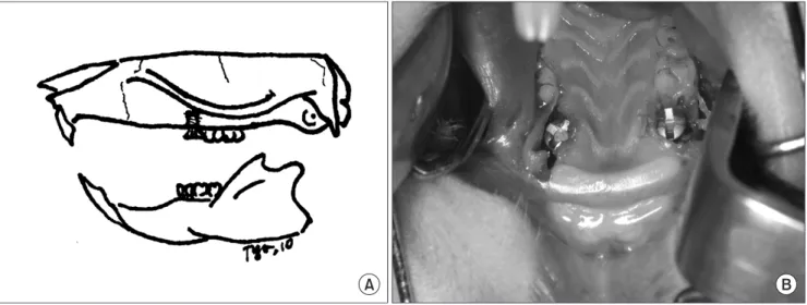
socket using a 1.0 mm twist drill. Two threaded titanium alloy implants (1.2×3 mm, Leibinger-Stryker, Freiburg, Germany) were inserted bilaterally into the prepared cavities by self-threading so that their tops were situated just above the alveolar crest.(Fig. 1) Profuse irrigation with sterilized physiological saline was maintained throughout this process.
The implants were allowed to healing for 3 days, 1, 2, and 4 weeks and bone block including implants were harvested.
4. Biomolecular analysis
Total RNA samples were extracted to evaluate the content of bone morphogenetic protein (BMP)-4, transforming growth factor (TGF)-β, osteocalcin (OC) and osteonectin (ON) in the bone surrounding the implants during the healing and remodeling processes. All the specimens were placed in RNA solution (TRIzol; Invitrogen Life Technologies, Rockville, IN, USA) and maintained at -80 until testing. The blocks were then ground under a liquid nitrogen stream. Bone powder (150 to 200 mg) was used to extract total RNA with an isolation kit (RNase Mini Kit;
Qiagen, Valencia, CA, USA). Reverse transcription reaction for cDNA was performed using 1.5 μg RNA, 1×RT buffer (Promega, Madison, WI, USA), 20 μM dNTP, 0.25 μg oligo (dT) 15 primer, 5 unit AMV reverse transcriptase (Pro emga) and DEPC-D/W for a total volume of 20 μL with a pre- dena turation step at 42oC for 65 min and 70oC for 15 min.
Then the PCR cycle using 4 μL of cDNA, 5×PCR pre-mix (Bioneer, Seoul, Korea), 10 pmol sense and antisense primer
Fig. 1. Schematic design of the experiment (A) and titanium implants placed in the extraction socket of both maxillary first molars (B).
Suhyun Park et al: Biological markers around immediately placed titanium implant in the extraction socket of diabetic and insulin-treated rat maxilla. J Korean Assoc Oral Maxillofac Surg 2012
A B
contrary to our expectations, the BMP-4 level peaked in the diabetes group by week 1. However, at week 4, there was a gradual increase in the control and the insulin-treated groups, then all groups showed mostly similar amounts of BMP-4 expression.(Fig. 2, 3)
2. Cytokine expression
The level of TGF-β1 increased instantly by day 3 in the bone that surrounded the implants of the insulin-treated and diabetic groups, compared to the control group. Control group level showed gradually increasement. Diabetes (DM) and diabetes controlled (DMC) level decreased at 1 and 2 121.9±14.6 mg/dL. The mean blood glucose level of the
insulin controlled diabetic animals was maintained at 160.2±
35.8 mg/dL at week 4. In contrast, the uncontrolled diabetic animals had a mean glucose level of 415.2±48.4 mg/dL at week 4. The onset of diabetes was further confirmed by a significant weight loss in the diabetic group compared to the control and insulin-treated groups.
1. Morphogenetic protein expression
The BMP-4 level increased immediately in the bone adjacent to the implants of all groups by day 3, then decreased abruptly in the control and the insulin-treated groups. On the
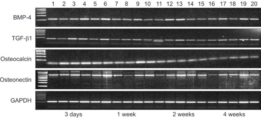
Fig. 2. Representative RT-PCR. RT-PCR products run on an agarose gel. The ratio of the fluorescence of each RT-PCR product to that of GAPDH was calculated. Lane 1-5: product of 3 days, lane 6-10: 1 week, lane 11-15: 2 week and lane 16-20: 4 week, respectively. (BMP:
bone morphogenetic protein, TGF: transforming growth factor, GAPDH: glyceraldehyde 3-phosphate dehydrogenase)
Suhyun Park et al: Biological markers around immediately placed titanium implant in the extraction socket of diabetic and insulin-treated rat maxilla. J Korean Assoc Oral Maxillofac Surg 2012
Fig. 3. BMP-4 expression pattern in the surrounding bone of immediately placed implant in the control, insulin-treated and diabetic rats at different healing phases by reverse transcription polymerase chain reaction analysis. (BMP: bone morphogenetic protein, DM: diabetes, DMC: diabetes controlled, C: controlled) Suhyun Park et al: Biological markers around immediately placed titanium implant in the extraction socket of diabetic and insulin-treated rat maxilla. J Korean Assoc Oral Maxil- lofac Surg 2012
Fig. 4. TGF-β1 expression pattern in the surrounding bone of immediately placed implant in the control, insulin-treated and diabetic rats at different healing phases by reverse transcription polymerase chain reaction analysis. (TGF: transforming growth factor, DM: diabetes, DMC: diabetes controlled, C: controlled) Suhyun Park et al: Biological markers around immediately placed titanium implant in the extraction socket of diabetic and insulin-treated rat maxilla. J Korean Assoc Oral Maxil- lofac Surg 2012
bone formation, mineralization, and resorption over a 10- day period using double-tetracycline labeling of bone in control, untreated diabetic and insulin-treated diabetic rats.
They found reduced bone formation and osteoid volume early in the course of healing in the animals with diabetes.
Verhaeghe et al.24 also studied bone morphology and function in spontaneously diabetic rats after 3 to 4 weeks of diabetes.
The number and function of osteoblasts were severely suppressed in the diabetic rats, resulting in decreased osteoid surfaces, mineral apposition rates, and plasma OC levels.
The osseointegration process occurs when the matrix is exposed to the extracellular fluid, causing the release of non-collagenous proteins and growth factors, which in turn activate the bone repair process. Osteoprogenitor cells of the bone marrow and the periosteum are attracted via chemotaxis to the site of the lesion. They proliferate and differentiate into osteoblasts and begin depositing bone25.
The present study was undertaken to identify the biological factors and their role in bone formation and repair during bone healing and remodeling processes around implants in diabetic compared to non-diabetic rats undergoing immediate implant surgery.
Important bone biological factors include the BMPs, which are members of the TGF-β family that play an essential role in osteogenesis. More than 15 BMP genes have been identified in vertebrates26 and they regulate osteoblast and chondrocyte differentiation during skeletal development.
BMPs have also been demonstrated to induce osteoblast weeks, but increased at 4 weeks. In addition, DMC becomes
similar to control at 4 weeks.(Fig. 2, 4)
3. Extracellular matrix protein expression
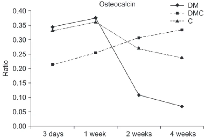
To compare the osteogenic potential of each group, the expression of OC, and ON was examined. On day 3, the expression of OC and ON was identified, suggesting the initiation of osteogenesis. The progressive increase in OC and ON expression from day 3 to week 1 were noticed in all three groups and then gradually decreased until 4 weeks. However, the OC, which initially demonstrated a lower expression than the other groups, exhibited a steady raising in expression.
Nevertheless, the OC in the control and the insulin-treated groups was higher than the diabetic group at 2 weeks and 4 weeks, indicating active bone formation in the control and the insulin-treated groups.(Figs. 2, 5, and 6)
IV. Discussion
This study reports on the effects of experimental diabetes on osseous healing around endosseous implants in the maxillary bone of rats. In addition, the effects of insulin on the expression of biological molecules in the peri-implant during bone healing processes were assessed.
Bone formation, osteoid formation, and bone mineral homeostasis have all been shown to be reduced in experi- mental models of diabetes. Goodman and Hori23 measured
Fig. 5. Osteocalcin expression pattern in the surrounding bone of immediately placed implant in the control, insulin-treated and diabetic rats at different healing phases by reverse transcription polymerase chain reaction analysis. (DM: diabetes, DMC: diabetes controlled, C: controlled)
Suhyun Park et al: Biological markers around immediately placed titanium implant in the extraction socket of diabetic and insulin-treated rat maxilla. J Korean Assoc Oral Maxil- lofac Surg 2012
Fig. 6. Osteonectin expression pattern in the surrounding bone of immediately placed implant in the control, insulin-treated and diabetic rats at different healing phases by reverse transcription polymerase chain reaction analysis. (DM: diabetes, DMC: diabetes controlled, C: controlled)
Suhyun Park et al: Biological markers around immediately placed titanium implant in the extraction socket of diabetic and insulin-treated rat maxilla. J Korean Assoc Oral Maxil- lofac Surg 2012
differentiation. Previous investigation of ovariectomized rats demonstrated the expression of OC mRNA was elevated when bone turnover was increased37. Therefore, the increase in the OC gene expression may be interpreted as the result of an increased number of differentiated osteoblasts or increased activity of osteoblasts as shown in the former studies.
On the other hand, ON is a major non-collagenous protein, it functions as a cell attachment factor for osteoblasts and osteoclasts, and is involve in the cell signaling pathway of osteoclasts and the modulation of osteoclast differentiation.
ON also shows a high affinity for mineral crystals and controls the shape of apatite crystals as they form and grow38. Our results clearly show that the levels of OC expression gradually increased during the later stage of bone healing in the insulin-treated group compared to the diabetic group.
Differently, the levels of ON decreased afterward in the bone around the implants in all groups.
It is not clear whether the osteoblastic gene expression was related to the activity of osteoblasts around the immediately placed implants. However, increased OC expression induced by the insulin treatment looked like to be associated with an increase in the bone formation activity in the insulin- treated group. Sharply declined expression of ON among all experiment group will remains to be explained in future studies.
Fiorellini et al.11 compared the course of osseous healing around endosseous implants in normal non-diabetic and insulin-controlled diabetic rats. The results indicated that the insulin therapy upregulated the formation of bone around implants inserted in the streptozotocin-induced diabetic rat model. However, the histometric parameters indicate that, although the total quantity of bone formation was greater in the insulin-controlled diabetic group, there was significantly less bone-to-implant contact in the insulin-controlled diabetic group when compared to the normal non-diabetic group4,11.
In the present study, the affects of insulin on the prevention of the detrimental effects of diabetes on osseointegration was evaluated by the expression of biologic molecules.
Comparison of the expression of the biological molecules of diabetic animals to those of diabetic animals treated with insulin suggests that that insulin enhances the bone response in the diabetic animals. And we concentrated on the effect of a hyperglycemic state that might show the inhibition of osteoblastic cell proliferation and collagen production during the early stages of callus development, resulting in reduced bone formation. In the future, it will be need to prove the diminished mechanical properties of the newly formed bone differentiation in some cultured cells27,28.
This study showed that BMPs, and in particular BMP-4, are involved in bone healing27,28. Most studies in the literature deal with the involvement of BMP-2 during this process29. Little attention has been given about the involvement of BMP-4 with regard to bone healing in vivo, especially around implant surfaces such as those analyzed in this study. BMP activity is stimulated by many well-known growth factors, such as the fibroblast growth factor, TGF-β, and platelet- derived growth factor, which can be found in bone30.
One of the best-known growth factors in skeletal tissues is TGF-β, the isoforms of which (TGF-β1, TGF-β2, and TGF-β3) exert multiple functions in bone metabolism31 including the promotion of bone for mation and the healing of fracture32,33.
The biomolecular analysis performed in this study reflected that the insulin-treated groups had relatively better results when the tests were carried out on the rat maxillary bone.
Thus, the levels of BMP-4 increased earlier in the insulin- treated group than in the diabetic group. Similarly, the levels of TGF-β1 increased earlier in the bone around the implants in the control and insulin-treated groups.
The administration of recombinant TGF-β1 has been shown to stimulate bone formation in vivo34, this cytokine may act in the early phases of bone healing just before the BMPs. Interestingly, the results of our study showed that TGF-β1 was present in higher levels in bone surrounding the implants in the insulin-treated and diabetic groups than in the control bone at 3 days and 1 week post-implantation. This increase was probably owing to the presence of inflammatory cells, which may in turn be responsible for the stimulation of BMP-4 synthesis. We observed that the levels of TGF-β 1 and BMP-4 increased only at 1 week post-implantation, at which time bone ossification was not evident around the implants. However, the TGF-β1 levels, which were higher in the control and insulin-treated groups compared to the diabetic group, appeared to modulate and complete the bone formation process. In addition, it showed little difference at 4 weeks between the groups in terms of the upregulation of BMP in the surrounding bone.
Because it has been believed that the gene expression of matrix protein is closely associated with bone turnover activity in vivo35,36, the analysis of the bone matrix protein may be a good indication of bone healing and remodeling in the present study.
OC has a high specificity for bone, and the expression of OC mRNA is detected at a later stage of osteoblast
JP. Wound healing around endosseous implants in experimental diabetes. Int J Oral Maxillofac Implants 1998;13:620-9.
14. Fiorellini JP, Nevins ML, Norkin A, Weber HP, Karimbux NY.
The effect of insulin therapy on osseointegration in a diabetic rat model. Clin Oral Implants Res 1999;10:362-8.
15. Mccracken MS, Aponte-wesson R, Chavali R, Lemons JE. Bone associated with implants in diabetic and insulin-treated rats. Clin Oral Implants Res 2006;17:495-500.
16. Takeshita F, Iyama S, Ayukawa Y, Kido MA, Murai K, Suetsugu T. The effects of diabetes on the interface between hydroxyapatite implants and bone in rat tibia. J Periodontol 1997;68:180-5.
17. Siqueira JT, Cavalher-Machado SC, Arana-Chavez VE, Sannomiya P. Bone formation around titanium implants in the rat tibia: role of insulin. Implant Dent 2003;12:242-51.
18. Shirakura M, Fujii N, Ohnishi H, Taguchi Y, Ohshima H, Nomura S, et al. Tissue response to titanium implantation in the rat maxilla, with special reference to the effects of surface conditions on bone formation. Clin Oral Implants Res 2003;14:687-96.
19. Shyng YC, Devlin H, Ou KL. Bone formation around imme diately placed oral implants in diabetic rats. Int J Prosthodont 2006;
19:513-4.
20. Hasegawa H, Ozawa S, Hashimoto K, Takeichi T, Ogawa T. Type 2 diabetes impairs implant osseointegration capacity in rats. Int J Oral Maxillofac Implants 2008;23:237-46.
21. Kwon PT, Rahman SS, Kim DM, Kopman JA, Karimbux NY, Fiorellini JP. Maintenance of osseointegration utilizing insulin therapy in a diabetic rat model. J Periodontol 2005;76:621-6.
22. Matin K, Senpuku H, Hanada N, Ozawa H, Ejiri S. Bone regeneration by recombinant human bone morphogenetic protein-2 around immediate implants: a pilot study in rats. Int J Oral Maxillofac Implants 2003;18:211-7.
23. Goodman WG, Hori MT. Diminished bone formation in experimental diabetes. Relationship to osteoid maturation and mineralization. Diabetes 1984;33:825-31.
24. Verhaeghe J, Suiker AM, Nyomba BL, Visser WJ, Einhorn TA, Dequeker J, et al. Bone mineral homeostasis in spontaneously diabetic BB rats. II. Impaired bone turnover and decreased osteo- calcin synthesis. Endocrinology 1989;124:573-82.
25. Schenk RK, Buser D. Osseointegration: a reality. Periodontol 2000 1998;17:22-35.
26. Reddi AH. Bone morphogenetic proteins: from basic science to clinical applications. J Bone Joint Surg Am 2001;83-A Suppl 1:S1- 6.
27. Katagiri T, Takahashi N. Regulatory mechanisms of osteoblast and osteoclast differentiation. Oral Dis 2002;8:147-59.
28. Ahrens M, Ankenbauer T, Schröder D, Hollnagel A, Mayer H, Gross G. Expression of human bone morphogenetic proteins-2 or -4 in murine mesenchymal progenitor C3H10T1/2 cells induces differentiation into distinct mesenchymal cell lineages. DNA Cell Biol 1993;12:871-80.
29. Ogawa T, Sukotjo C, Nishimura I. Modulated bone matrix-related gene expression is associated with differences in interfacial strength of different implant surface roughness. J Prosthodont 2002;11:241- 7.
30. Carrington JL, Roberts AB, Flanders KC, Roche NS, Reddi AH.
Accumulation, localization, and compartmentation of transforming growth factor beta during endochondral bone development. J Cell Biol 1988;107:1969-75.
31. Horner A, Kemp P, Summers C, Bord S, Bishop NJ, Kelsall AW, et al. Expression and distribution of transforming growth factor- beta isoforms and their signaling receptors in growing human bone.
Bone 1998;23:95-102.
32. Noda M, Camilliere JJ. In vivo stimulation of bone formation by transforming growth factor-beta. Endocrinology 1989;124:2991-4.
33. Rosier RN, O'keefe RJ, Hicks DG. The potential role of transforming growth factor beta in fracture healing. Clin Orthop Relat Res 1998;355 Suppl:S294-300.
as well as difference between the integrated implant and the disintegrated implant.
Regarding clinical application of these data, STZ-induced diabetes in a small animal model may or may not have a direct correlation with the clinical course of patients with diabetes. Still, the findings of this study will hopefully add to our knowledge regarding host, implant, and diabetic inter- actions, especially in the situation of immediate implant installation.
V. Conclusion
In conclusion, despite the low number of samples used in the present study, we can affirm that insulin stimulate bone formation earlier in the DM model and to a greater degree.
Further studies are needed to consider these findings to the application to patients in the clinical setting.
References
1. Lazzara RJ. Immediate implant placement into extraction sites:
surgical and restorative advantages. Int J Periodontics Restorative Dent 1989;9:332-43.
2. Wagenberg BD, Ginsburg TR. Immediate implant placement on removal of the natural tooth: retrospective analysis of 1,081 implants. Compend Contin Educ Dent 2001;22:399-404.
3. Becker W, Goldstein M. Immediate implant placement: treatment planning and surgical steps for successful outcome. Periodontol 2000 2008;47:79-89.
4. Marder MZ. Medical conditions affecting the success of dental implants. Compend Contin Educ Dent 2004;25:739-42.
5. Oczakir C, Balmer S, Mericske-stern R. Implant-prosthodontic treat ment for special care patients: a case series study. Int J Prosthodont 2005;18:383-9.
6. McMahon MM, Bistrian BR. Host defenses and susceptibility to infection in patients with diabetes mellitus. Infect Dis Clin North Am 1995;9:1-9.
7. Kotsovilis S, Karoussis IK, Fourmousis I. A comprehensive and critical review of dental implant placement in diabetic animals and patients. Clin Oral Implants Res 2006;17:587-99.
8. Mombelli A, Cionca N. Systemic diseases affecting osseointegration therapy. Clin Oral Implants Res 2006;17 Suppl 2:
97-103.
9. Olson JW, Shernoff AF, Tarlow JL, Colwell JA, Scheetz JP, Bingham SF. Dental endosseous implant assessments in a type 2 diabetic population: a prospective study. Int J Oral Maxillofac Implants 2000;
15:811-8.
10. van Steenberghe D, Jacobs R, Desnyder M, Maffei G, Quirynen M.
The relative impact of local and endogenous patient-related factors on implant failure up to the abutment stage. Clin Oral Implants Res 2002;13:617-22.
11. Fiorellini JP, Chen PK, Nevins M, Nevins ML. A retrospective study of dental implants in diabetic patients. Int J Periodontics Restorative Dent 2000;20:366-73.
12. Morris HF, Ochi S, Winkler S. Implant survival in patients with type 2 diabetes: placement to 36 months. Ann Periodontol 2000;5:157-65.
13. Nevins ML, Karimbux NY, Weber HP, Giannobile WV, Fiorellini
37. Westerlind KC, Wronski TJ, Evans GL, Turner RT. The effect of long-term ovarian hormone deficiency on transforming growth factor-beta and bone matrix protein mRNA expression in rat femora. Biochem Biophys Res Commun 1994;200:283-9.
38. Boskey AL, Maresca M, Ullrich W, Doty SB, Butler WT, Prince CW. Osteopontin-hydroxyapatite interactions in vitro: inhibition of hydroxyapatite formation and growth in a gelatin-gel. Bone Miner 1993;22:147-59.
34. Marcelli C, Yates AJ, Mundy GR. In vivo effects of human recom- binant transforming growth factor beta on bone turnover in normal mice. J Bone Miner Res 1990;5:1087-96.
35. Seto H, Aoki K, Kasugai S, Ohya K. Trabecular bone turnover, bone marrow cell development, and gene expression of bone matrix proteins after low calcium feeding in rats. Bone 1999;25:687-95.
36. Ogawa T, Nishimura I. Genes differentially expressed in titanium implant healing. J Dent Res 2006;85:566-70.