http://dx.doi.org/10.12671/jkfs.2015.28.1.8
8
Copyright ⓒ 2015 The Korean Fracture Society. All rights reserved.
This is an Open Access article distributed under the terms of the Creative Commons Attribution Non-Commercial License (http://creativecommons.org/licenses/
by-nc/3.0) which permits unrestricted non-commercial use, distribution, and reproduction in any medium, provided the original work is properly cited.
Received June 9, 2014 Revised August 18, 2014 Accepted September 25, 2014
Address reprint requests to: Hong Je Kang, M.D.
Department of Orthopedic Surgery, Wonkwang University College of Medicine, 460 Iksan-daero, Iksan 570-749, Korea
Tel: 82-63-859-1360ㆍFax: 82-63-852-9329 E-mail: [email protected]
Financial support: None. Conflict of interest: None.
골 결손을 동반한 고령의 원위 요골 분쇄골절에서 동종 해면골 이식과 수장측 잠김 금속판을 이용한 치료
강홍제 ⋅신창현
원광대학교 의과대학 정형외과교실, 원광의과학연구소
Treatment of the Communited Distal Radius Fracture Using Volar Locking Plate Fixation with Allogenic Cancellous Bone Graft in the Elderly
Hong Je Kang, M.D. , Chang Hyun Shin, M.D.
Department of Orthopedic Surgery, Institute of Wonkwang Medical Science, Wonkwang University College of Medicine, Iksan, Korea
Purpose: We studied results of the communited distal radius fracture treated with allogenic cancellous bone graft and volar locking plate in the elderly.
Materials and Methods: We studied 29 cases of communited distal radius fracture treated with allogenic cancellous bone graft and volar locking plate from April 2009 to April 2013. Fracture was classified according to AO/OTA classification.
Postoperative clinical evaluation was performed with measurement of wrist range of motion (ROM) at last follow-up, modified Mayo wrist scoring system (MMWS), and visual analogue pain scale (VAS). Radiologic evaluation was performed with measure- ment of radial length on immediate postoperation and last follow-up, radial inclination, volar tilt and ulnar variance checked at the last follow-up using Sarmiento criteria.
Results: Using the MMWS, 13 cases were classified as ‘good’, 10 ‘fair’, and 5 ‘normal’. The average wrist ROM was 88.5%
for flexion, 92.2% for extension, 90.5% for adduction, and 94.0% for abduction. The average VAS was 1.7. On the last fol- low-up, average radius length, radial inclination and volar tilt did not show statistically significant improvement (p>0.05) compared to immediate post operation measurements, and according to Sarmiento criteria, 5 cases were classified as ‘good’, 14 ‘fair’, and 7 ‘normal’.
Conclusion: Treatment of severe communited distal radius fracture accompanied by bone defect with volar locking plate and allogenic cancellous bone graft is a satisfying and effective treatment method in the elderly.
Key Words: Distal radius fracture, Allogenic Cancellous Bone Graft, Volar Locking Plate
서 론
원위 요골 분쇄골절에서 보존적 치료만을 시행했을 경우 정상적 해부학적 구조를 회복하지 못하여 외상 후 관절염 이 발생하거나 정복 소실, 불안정성, 부정 유합, 신경 손상 등의 많은 합병증이 발생할 수 있어 수술적 치료가 선호되 고 있다.1,2)
Table 1. Demographic Data of the Patients (n=29)
Patient data Value
Gender (male : female) Age (yr)
Follow-up duration (mo) Fracture type (AO/OTA)
4 (13.7) : 25 (86.2) 73.1 (65-81) 22.4 (12-36) A3.1: 2, A3.2: 4, A3.3: 5 C2.2: 3, C2.3: 5, C3.2: 6, C3.3: 4 Values are presented as number (%), medians (range), or number only.
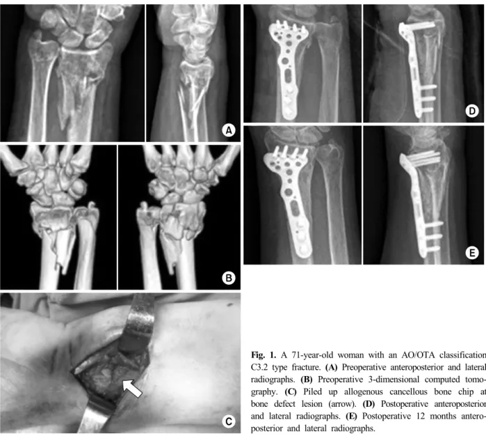
Fig. 1. A 71-year-old woman with an AO/OTA classification C3.2 type fracture. (A) Preoperative anteroposterior and lateral radiographs. (B) Preoperative 3-dimensional computed tomo- graphy. (C) Piled up allogenous cancellous bone chip at bone defect lesion (arrow). (D) Postoperative anteroposterior and lateral radiographs. (E) Postoperative 12 months antero- posterior and lateral radiographs.
원위 요골 분쇄골절의 수술적 치료 방법으로는 외고정 장치를 이용한 고정술, 도수 정복 및 경피적 K-강선을 이 용한 내고정술, 관혈적 정복 및 금속판을 이용한 내고정술 등이 있으며,3,4) 그 중 최근 고안된 수장측 잠김 금속판은 잠김 나사와 금속판의 기계적인 연결으로 안정성을 얻고 후방 고정 시 발생하는 신전건의 파열이나 자극 증상, 건 막염 등의 합병증을 피할 수 있다는 장점이 있어 점차 흔 하게 사용되고 있다.3)
하지만 심한 골 결손을 동반한 고령의 원위 요골 분쇄골 절에서 금속판만을 이용한 내고정술을 시행하면 수술 후 골간단부 골 결손과 피질골의 약화로 이차적인 전위나 관 절면의 붕괴가 일어날 수 있다. 따라서 이러한 문제점들을 최소화하기 위해 골 결손 부위에 골 이식 또는 골 대치물 삽입 등이 고려될 수 있다.5-8)
본 연구는 심한 골 결손을 동반한 고령의 원위 요골 분 쇄골절에서 동종 해면골 이식술과 수장측 잠김 금속판을 이용한 내고정술의 임상적, 방사선적 치료 결과에 대해 알
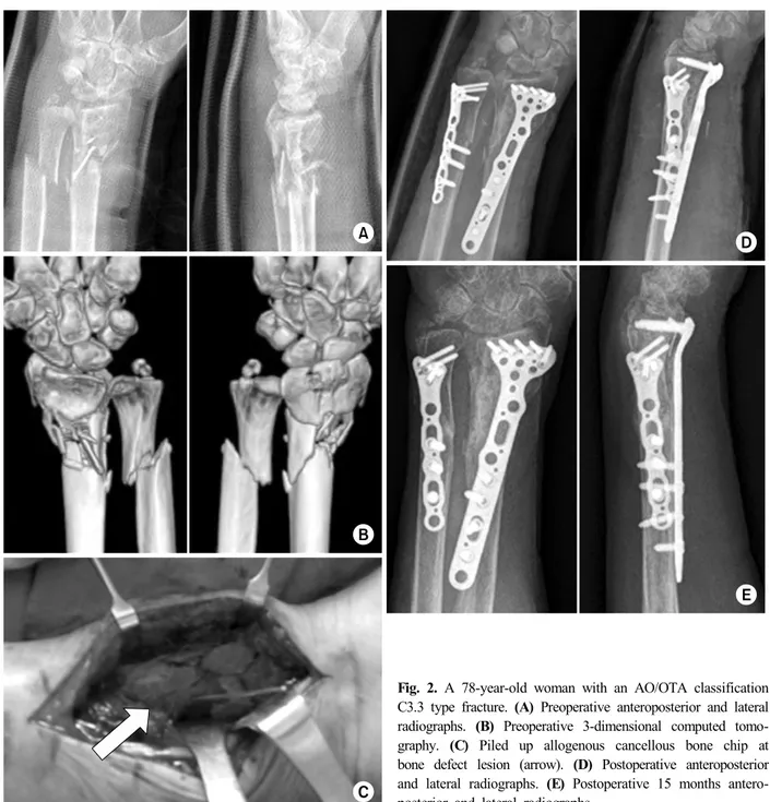
Fig. 2. A 78-year-old woman with an AO/OTA classification C3.3 type fracture. (A) Preoperative anteroposterior and lateral radiographs. (B) Preoperative 3-dimensional computed tomo- graphy. (C) Piled up allogenous cancellous bone chip at bone defect lesion (arrow). (D) Postoperative anteroposterior and lateral radiographs. (E) Postoperative 15 months antero- posterior and lateral radiographs.
아보고자 하였다.
대상 및 방법
1. 대상
2009년 4월부터 2013년 4월까지 원광대학교 의과대학병 원에서 원위 요골 골절에 대하여 수장측 잠김 금속판을 이
용하여 187예를 치료하였다. 수술적 치료는 10도 이상의 후방 굴곡, 5 mm 이상의 요골 단축 혹은 2 mm 이상의 관절 내 층형성을 보이는 불안정성 골절에서 시행하였다.
이 중 나이가 65세 이상이며 골 결손과 함께 분쇄골절이 동반하여 동종 해면골 이식술을 동시에 시행하고 12개월 이상 추시 관찰이 가능하였던 29예를 대상으로 하였다. 동 종 해면골 이식술은 전방과 후방 피질골의 심한 분쇄와 골 결손으로 수술 중 골정복의 유지가 어려운 환자에게 시행
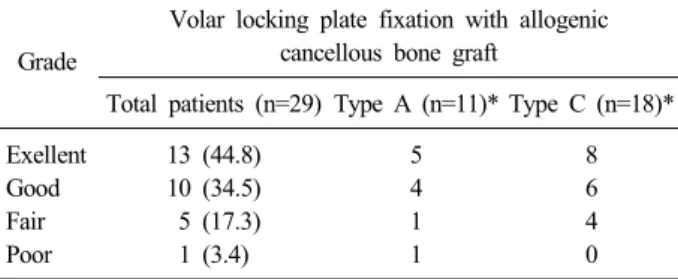
Table 2. Clinical Outcome of Communited Distal Radius Fracture according to Modified Mayo Wrist Scoring System
Grade
Volar locking plate fixation with allogenic cancellous bone graft
Total patients (n=29) Type A (n=11)* Type C (n=18)*
Exellent Good Fair Poor
13 (44.8) 10 (34.5) 5 (17.3) 1 (3.4)
5 4 1 1
8 6 4 0 Values are presented as number (%) or number only. *Classifi- cation according to the standard of AO/OTA fracture type.
하였다. 후방 피질골에만 분쇄가 있거나 수장측 또는 수배 측 Barton 골절, 추가적으로 후방 접근법을 이용하여 수술 을 시행한 경우는 제외하였으며 관절면의 분쇄가 심하여 추가적으로 관절 내시경을 이용한 정복술을 시행하거나 외 고정술을 시행한 경우도 제외하였다.
남자는 4명, 여자는 25명이었고, 평균 연령은 73.1세 (65-81세)였으며, 평균 추시기간은 22.4개월(12-36개월)이었 다. 모든 환자에서 수술 전, 대퇴 경부와 제1요추에서 제4 요추까지 에너지 방사선 흡수 계측 방법을 이용한 골밀도 검사(bone mineral density)를 시행하였으며, t-score는 평균
−4.3 (−5.2-−2.9)이었다. 골절의 분류는 단순 방사선 전 후면 및 측면 촬영과 컴퓨터 단층 촬영에 근거하여 AO/OTA 분류 이용하였고 A형 골절이 11예(A3.1: 2, A3.2:
4, A3.3: 5), C형 골절이 18예(C2.2: 3, C2.3: 5, C3.2: 6, C3.3: 4)였다(Table 1).
2. 수술 방법 및 재활
수술 방법은 전신 마취하에 상완부에 지혈대를 시행하였 으며 수술적 접근은 전방 도달법을 이용하였다. 요수근 굴 건의 건을 촉지한 후 피부절개를 시행하여 요수근 굴건을 척측으로 견인하였다. 방형 회내근 노출시킨 후, 요골 부착 부 1/3 지점인 안전 지대에서 박리하여 골절부를 노출시켰 다. 그리고 골절 부위에서 골의 손실이 추가적으로 이루어 지는 것에 조심하고, 분쇄된 피질골에서 연부 조직이 떨어지 지 않도록 조심하며 세척술을 시행하여 혈종을 제거하였다.
골 결손부를 확인 후 골절에 대한 정복을 시행하였고, 골 결손에 의해 정복 유지가 안되는 경우 먼저 동종 해면골 (Cancellus, Coarse, Freeze dried; CommunityTissueServices, Dayton, OH, USA) 이식을 시행하여 골 결손부를 충진한 이후 골절에 대한 정복을 시행하였으며 K-강선을 삽입하여 추가적인 고정을 시행하였다. 관절면을 침범한 골절의 경 우 요골 경상돌기에서 관절면과 평행하게 K-강선을 먼저 삽입하여 관절면 골절을 먼저 정복하였다. 이후 수장측 잠 김 금속판(2.3 mm Acu-LocⓇ Volar Distal Radius Plate;
ACUMED, Hillsboro, OR, USA)을 고정하였고, C-arm fluo- roscopy을 이용한 정복의 적절성을 평가한 이후 골 결손부 가 남아 있는 경우 추가적으로 골 이식을 시행하였다(Fig. 1).
요척 관절의 불안정성을 유발하는 척골 경상 돌기 기저 부 골절 혹은 척골두 골절이 동반된 경우 K-강선이나 척측 잠김 금속판(Acu-LocⓇ VDU Plate; ACUMED)을 이용하여 고정하였으며(Fig. 2), 이 경우에는 장상지 부목을 시행하 였고, 그렇지 않은 경우 수술 후 2주간 단상지 부목을 시 행하였다. 수술 직후부터 모든 수지의 능동적 관절 운동을 시행하였으며 수술 후 2주부터 제거 가능한 손목 보호대를
착용하였으며 능동적인 손목 관절 운동을 시행하였다.
3. 임상적 결과 및 방사선적 결과
술 후 임상적 결과는 최종 추시 시 modified Mayo wrist scoring system (MMWS), 손목의 관절 운동 범위, 그리고 visual analogue pain scale (VAS)를 이용하여 평가하였다.
손목의 관절 운동 범위는(굴곡, 신전, 회외전, 회내전) Goniometer를 이용하여 측정하고 건측의 값과 비교해 백 분율 값을 산출하였다.
술 후 방사선적 결과는 골절의 유합 시기와 수술 직후와 최종 추시에서 Goldfarb 등9)이 보고한 평가방법을 적용하 여 요골 길이, 요골 경사각, 수장측 경사각을 측정하여 비 교하였으며, Sarmiento criteria10)의 평가 방법을 적용하여 요골 원위부의 변형, 요골 단축, 요골 경사의 소실, 배측 경사의 변화 정도를 측정하여 우수, 양호, 보통, 그리고 불 량의 4단계로 나누어 평가하였다.
손목 관절 운동, 방사선적 지표는 Student t-test를 시행 하여 분석하였다. p값은 0.05 미만인 경우를 통계적인 유 의성이 있는 것으로 간주하였다. 분석은 PASW Statistics ver 18.0 (IBM Co., Armonk, NY, USA)으로 검정하였다.
결 과
MMWS에서 우수 13예, 양호 10예, 보통 5예, 불량 1예 로 양호 이상이 79.3%였다(Table 2). 평균 손목 관절 운동 범위 평균값은 굴곡 62o (70o, 88.5%), 신전 71o (77o, 92.2%), 회내전 79o (85o, 90.5%), 회외전 82o (87o, 94.0%) 였으며 VAS는 평균 1.5점이었다(Table 3).
방사선적 결과에서 평균 유합기간은 12.5주(9-22주) 전체 예의 환자에서 방사선적 유합을 얻을 수 있었다. 수술 직 후 요골 길이는 평균 13.5 mm (9.3-13.8 mm), 요골 경사각
Table 5. Results according to Sarmiento Criteria Grade
Volar locking plate fixation with allogenic cancellous bone graft
Total patients (n=29) Type A (n=11)* Type C (n=18)*
Excellent Good Fair Poor
5 (17.3) 14 (48.3) 7 (24.1) 3 (10.3)
3 5 2 1
2 9 5 2 Values are presented as number (%) or number only. *Classifi- cation according to the standard of AO/OTA fracture type.
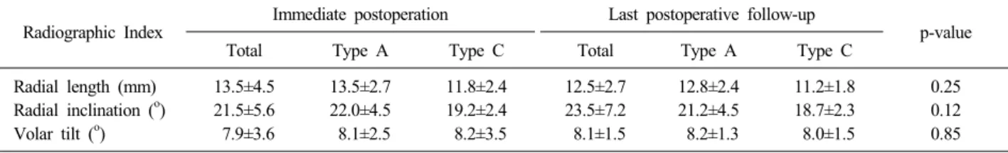
Table 4. Radiological Outcomes Immediate Postoperative Date and Last Postoperative Follow-Up Radiographic Index Immediate postoperation Last postoperative follow-up
p-value
Total Type A Type C Total Type A Type C
Radial length (mm) Radial inclination (o) Volar tilt (o)
13.5±4.5 21.5±5.6 7.9±3.6
13.5±2.7 22.0±4.5 8.1±2.5
11.8±2.4 19.2±2.4 8.2±3.5
12.5±2.7 23.5±7.2 8.1±1.5
12.8±2.4 21.2±4.5 8.2±1.3
11.2±1.8 18.7±2.3 8.0±1.5
0.25 0.12 0.85 Values are presented as mean±standard deviation.
Table 3. Wrist Functional Outcomes at Last Follow-Up Affected side
Contralateral side Affected side/contralateral side
p-value
Total Type A* Type C* Total Type A* Type C*
Flexion (o) Extension (o) Pronation (o) Supination (o) VAS (score)
62.3 71.6 79.4 82.2 1.5
68.4 75.3 83.2 86.4 1.0
58.2 69.2 77.4 80.6 1.7
70.3 77.1 85.6 87.9
88.5 92.2 90.5 94.0
97.1 97.4 97.6 98.8
82.8 89.6 90.5 91.9
0.03 0.64 0.12 0.76
The angle of the joint is shown in percentile compared to normal side. *Classification according to the standard of AO/OTA fracture type. VAS: Visual analogue pain scale.
은 평균 21.5o (18.5o-26.3o), 수장측 경사는 7.9o (4.8o-7.9o) 였으며, 최종 추시 시 각각 평균 12.5 mm (9.8-14.2 mm), 23.5o (16.2o-27.3o), 8.1o (6.6o-9.3o)로 수술 직후와 비교에 서는 통계적으로 의미있는 변화는 없었다(p=0.62) (Table 4). Sarmiento criteria상 우수 5예, 양호 14예, 보통 7예, 불량 3예로 양호 이상이 65.6%였다(Table 5).
술 후 합병증으로 1예에서 정중 신경 압박 증상이 있어 수술 후 3개월에 수근관 감압술을 시행하였으며 이후 증상 이 소실되었다.
고 찰
원위 요골 분쇄골절은 골 결손이 심하여 해부학적 정복
및 유지에 어려움이 있어 수술적인 치료가 요구된다. 원위 요골 골절의 수술적 치료 방법에는 관혈적 정복 및 금속판 내고정술, 도수정복 후 경피적 K-강선 내고정술, 외고정 장 치를 이용한 고정술 등 매우 다양하다.1-3)
Mah과 Atkinson11)은 도수 정복 및 금속 강선 고정술은 상대적으로 비침습적이고 시술 시간이 짧으며 비용적인 측 면에서도 장점이 있으나 상대적으로 고정력에서 떨어지고 골유합이 이루어지는 동안 정복의 소실과 그에 따른 기능 감소의 단점이 있다고 보고하였다.
Zhang 등12)은 26명의 원위 요골의 관절 내 분쇄 골절 환자를 대상으로 외고정 장치를 이용한 수술적 치료를 시 행하였고, 임상적 및 방사선적으로 좋은 결과를 얻었다고 보고하였다. 또한 Kofoed13)는 분쇄골절과 전위된 관절 내 골절에서 외고정 장치 및 골 시멘트를 이용한 보강을 시행 하였으며, 합병증 없이 양호한 결과를 보고하였다. 반면 Arora와 Malik14)은 외고정 장치의 축성 신연력만으로는 정 상적인 수근 관절면의 수장측 경사를 회복하기 어렵고, 관 절 내 골절편을 충분히 정복할 수 없을 뿐 아니라 해면골 의 소실로 인한 정복 소실이 발생할 수 있음을 보고하였으 며, Mudgal과 Jupiter15)는 과신연으로 인해 수근부 및 수지 의 구축이 올 수 있음을 보고하였다.
Na 등16)은 후방 분쇄 및 전위가 있는 경우 후방 금속판 을 이용한 고정술로 좋은 결과를 얻었다고 보고하였다. 그 러나 후방 접근법은 신근 지대의 절개가 필요하고, 신전건
의 손상이 발생하기 쉬우며, 금속판의 고정을 위해 리스터 결절의 절제가 필요하다는 단점을 가지고 있다.
여러 저자들은 불안정한 원위 요골 골절에서 수장측 잠 김 금속판을 이용하여 내고정술을 시행하여 좋은 결과를 보고하였다. 또한 수장측 잠김 금속판은 신전건의 손상이 적고, 배측에 비해 연부 조직이 풍부하여 내고정물이 만져 지는 합병증이 적으며, 기술적으로 손쉽다는 장점이 있다 고 하였다.17-19) 또한 T형 잠금 압박 금속판은 후방 피질골 분쇄를 동반한 불안정성 골절에서도 안정성을 얻을 수 있 어 정확한 해부학적 정복 및 견고한 고정을 얻을 수 있다 고 하였다.20) Kim 등21)은 고령의 골 간단부에 골 결손이 있는 불안정한 원위 요골 골절 환자에서 골 결손부 대체물 로 calcium phosphate bone cement (CPC)를 사용하고, 수 장측 잠김 금속판을 이용하여 내고정하였을 때 추시 결과 와 수장측 잠김 금속판만을 사용한 후 추시 결과의 차이가 없다고 보고하였다. 또한 Rhee 등22)은 고령 여성의 불안정 한 원위 요골 골절에서 수장측 잠김 금속판만을 이용하여 내고정하였을 때, 추시상 골편 전위 없이 만족할 만한 치 료 결과를 보고하였다.
한편, 여러 저자들은 불안정한 원위 요골 골절에서 수장 측 잠김 금속판만을 이용하여 내고정하였을 때, 금속판 손 상, 나사못 빠짐 현상 및 골편 정복 소실을 보고하였
다.23,24) 또한 다른 저자들은 후방 분쇄골절과 함께 수장측
피질골의 분쇄 및 골 결손이 동반된 경우 골간단부 골 결 손과 피질골의 약화로 이차적인 전위나 관절면의 붕괴가 일어나기 쉬울 뿐더러 지연 유합 혹은 불유합이 발생할 수 도 있다고 하였다.5-8) 따라서 이러한 문제점들을 최소화하 기 위해 골 결손부위에 골 이식 또는 대체물 삽입 등의 부 가적인 시술을 통한 구조적인 지지대가 필요하다고 보고하
였다.5-7,25,26) 현재 임상적으로 이용되는 골 결손부 대체물로
자가 골 이식, 동종 골 이식, 골 시멘트, 그리고 CPC 등을 삽입할 수 있다. Kainz 등25)은 요골 원위부의 골 결손을 동반한 분쇄골절 환자에서 분쇄골절 부분에 CPC를 사용한 뒤 수장측 잠김 금속판을 이용한 내고정술을 시행하였을 때 CPC를 사용하지 않은 대조군과 비교하여 최종 추시 결 과에서 안정성을 얻을 수 있었다고 보고하였다. 또한 Büyükkurt 등26)은 여성 37명의 원위 요골 골절 환자 중 골다공증이 있는 20명과 골다공증이 없는 17명의 환자에게 금속판만을 이용한 내고정술을 시행하였을 때, 추시 관찰 시 방사선학적 차이는 없지만 골다공증 환자에서 관절 운 동 제한과 일상 생활에 제한이 있음을 보고하였다. 이와 함께 Goto 등27)은 고령의 요골 원위부의 골 결손을 동반 한 분쇄골절 환자에서 분쇄골절 부분에 hydroxyapatite 골 이식을 시행한 뒤, 수장측 잠김 금속판을 이용한 내고정술 을 시행하였을 때, hydroxyapatite 골 이식을 하지 않은 대
조군과 비교하여 최종 방사선 추시상 만족할 만한 결과를 보고하였다.
자가 골 이식은 생체 적합성이 우수하면서 적당한 생역 학적 강도를 갖고 있으므로 골 결손이 동반된 불안정성 골 절에서 골절 치유를 촉진한다는 장점이 있어 전통적으로 장골 능에서 채취한 자가 골이 널리 이용되었다.28) 그러나 자가 골 이식은 공여부의 혈종과 감염, 동통, 장골 능 골 절, 신경 손상, 수술 시간 지연, 출혈량 증가 등의 합병증 등의 보고된 단점이 있다.29) 골 시멘트는 골실질의 재형성 을 기대할 수 없고, 자가골과 융합되지도 않으며, 시술 시 의 발열반응으로 인해 골절 치유를 방해할 뿐 아니라, 액 화상태에서는 세포독성이 있고, 골절부 외로 새어 나가면 조직손상의 위험이 있다.30) CPC는 생화학적 적합성과 골 전도 능력이 해면골과 유사하여 골 대치물로 많이 사용되 나 충진된 CPC가 관절 밖으로 흘러나와 동통과 관절증 등 합병증을 유발시킬 수 있다는 단점이 있어 제한적으로 사 용하고 있다.31)
본 연구에서는 고령의 골다공증이 있는 요골 원위부의 심한 골 결손을 동반한 분쇄골절 환자에게 동종 해면골 이 식을 시행하였다. 동종 해면골 이식의 장점으로는 첫째, 이 차적인 전위나 관절면의 붕괴를 막는 구조적인 지지대의 역할을 하기 때문 관절면에 기계적 안정성을 부여하고 정 복된 관절면을 유지하도록 하여 해부학적 정복을 유지시키 며, 둘째, 공여부의 이환이 없고 공급의 제한이 없다는 점, 셋째, 수술 중 출혈량을 줄일 수 있고, 넷째, 이식골 주위 는 수여자의 풍부한 해면골로 둘러싸인 부위이므로 이로부 터 이식된 동종골 내부로 가골이 신속하게 침투할 수 있다 는 점이 있다.32) 이후, 수장측 잠김 금속판을 사용하여 내 고정술을 시행하였고, 임상적으로 만족할 만한 결과를 보 였으며, 방사선학적으로 요골 원위부의 변형, 요골 단축, 요골 경사의 소실, 배측 경사의 의미있는 정복의 소실이 없이 골유합을 얻었다. 또한 좀 더 견고한 안정성을 얻을 수 있어 조기 관절운동이 가능하게 하여 일상 생활로의 빠 른 복귀가 가능하였다.
수장측 금속판을 삽입하는 경우 수지 굴곡건의 지연성 파열이 발생할 수 있으며 신경 손상을 유발하거나 방형 회 내근의 손상을 주며 전방 피질골의 혈액순환에 피해를 줄 수도 있으며 관절면을 침범한 골절의 경우 직접 관절면을 볼 수 없다는 단점이 있다.16,33) 본 논문에서도 정중 신경 압박 증상이 1예에서 발생하여 수근관 관압술을 시행하였 다. 또한 본 논문에서는 관절면 골절의 경우 먼저 C-arm fluoroscopy를 보며 관절면을 정복하여 금속 강선을 이용 하여 고정하였으며 심하게 골절되어 정복을 잘할 수 없는 경우에는 추가적으로 외고정 장치를 시행하거나 관절경하 정복술을 시행하였다.
본 연구의 한계점으로는 증례의 수가 29예로 적고, 골 이식을 하지 않은 환자와 비교 연구를 시행하지 않았다는 점이 있다. 또한 고령의 환자를 대상으로 하여 골 결손이 심한 젊은 환자의 경우 골 이식 시행하였을 때의 결과에 대하여는 알 수 없다. 이와 함께 외상 후 관절염의 발생 유무에 대해 장기 추시가 필요할 것으로 생각된다.
결 론
심한 골 결손을 동반한 고령의 원위 요골 분쇄골절 환자 에서 동종 해면골 이식과 수장측 잠김 금속판을 이용한 치 료는 임상적 및 방사선학적으로 만족할 만한 결과를 보이 는 효과적인 치료 방법이다.
References
1) Edwards GS Jr: Intra-articular fractures of the distal part of the radius treated with the small AO external fixator. J Bone Joint Surg Am, 73: 1241-1250, 1991.
2) Ring D: Treatment of the neglected distal radius fracture.
Clin Orthop Relat Res, (431): 85-92, 2005.
3) Leung F, Tu YK, Chew WY, Chow SP: Comparison of external and percutaneous pin fixation with plate fixation for intra-articular distal radial fractures. A randomized study. J Bone Joint Surg Am, 90: 16-22, 2008.
4) Ring D, Prommersberger K, Jupiter JB: Combined dor- sal and volar plate fixation of complex fractures of the distal part of the radius. J Bone Joint Surg Am, 86:
1646-1652, 2004.
5) Herrera M, Chapman CB, Roh M, Strauch RJ, Rosenwasser MP: Treatment of unstable distal radius fractures with cancellous allograft and external fixation. J Hand Surg Am, 24: 1269-1278, 1999.
6) Leung KS, Shen WY, Leung PC, Kinninmonth AW, Chang JC, Chan GP: Ligamentotaxis and bone grafting for comminuted fractures of the distal radius. J Bone Joint Surg Br, 71: 838-842, 1989.
7) McBirnie J, Court-Brown CM, McQueen MM: Early open reduction and bone grafting for unstable fractures of the distal radius. J Bone Joint Surg Br, 77: 571-575, 1995.
8) Wolfe SW, Pike L, Slade JF 3rd, Katz LD:
Augmentation of distal radius fracture fixation with coral- line hydroxyapatite bone graft substitute. J Hand Surg Am, 24: 816-827, 1999.
9) Goldfarb CA, Yin Y, Gilula LA, Fisher AJ, Boyer MI:
Wrist fractures: what the clinician wants to know.
Radiology, 219: 11-28, 2001.
10) Sarmiento A, Zagorski JB, Sinclair WF: Functional bracing of Colles' fractures: a prospective study of im- mobilization in supination vs. pronation. Clin Orthop Relat Res, (146): 175-183, 1980.
11) Mah ET, Atkinson RN: Percutaneous Kirschner wire sta- bilisation following closed reduction of Colles' fractures. J Hand Surg Br, 17: 55-62, 1992.
12) Zhang SX, Gu FR, Peng YL, et al: External fixation and bone grafting for collapsed and comminuted distal ra- dius fracture. Chin J Traumatol, 8: 156-159, 164, 2005.
13) Kofoed H: Comminuted displaced Colles' fractures.
Treatment with intramedullary methylmethacrylate stabi- lisation. Acta Orthop Scand, 54: 307-311, 1983.
14) Arora J, Malik AC: External fixation in comminuted, displaced intra-articular fractures of the distal radius: is it sufficient? Arch Orthop Trauma Surg, 125: 536-540, 2005.
15) Mudgal CS, Jupiter JB: Plate fixation of osteoporotic fractures of the distal radius. J Orthop Trauma, 22:
S106-S115, 2008.
16) Na KT, Song SW, Lee YM, Kang BM: Dorsal plate fix- ation for dorsally displaced distal radius fractures. J Korean Soc Surg Hand, 19: 44-51, 2014.
17) Orbay JL: The treatment of unstable distal radius frac- tures with volar fixation. Hand Surg, 5: 103-112, 2000.
18) Lozano-Calderón SA, Souer S, Mudgal C, Jupiter JB, Ring D: Wrist mobilization following volar plate fixation of fractures of the distal part of the radius. J Bone Joint Surg Am, 90: 1297-1304, 2008.
19) Murakami K, Abe Y, Takahashi K: Surgical treatment of unstable distal radius fractures with volar locking plates. J Orthop Sci, 12: 134-140, 2007.
20) Kim SJ, Cho CH: 2.4 mm volar locking compression plate for treatment of unstable distal radius fractures. J Korean Fract Soc, 24: 151-155, 2011.
21) Kim JK, Koh YD, Kook SH: Effect of calcium phos- phate bone cement augmentation on volar plate fixation of unstable distal radial fractures in the elderly. J Bone Joint Surg Am, 93: 609-614, 2011.
22) Rhee SH, Kim J, Lee YH, Gong HS, Lee HJ, Baek GH: Factors affecting late displacement following volar locking plate fixation for distal radial fractures in elderly female patients. Bone Joint J, 95: 396-400, 2013.
23) Foo TL, Gan AW, Soh T, Chew WY: Mechanical fail- ure of the distal radius volar locking plate. J Orthop Surg (Hong Kong), 21: 332-336, 2013.
24) Arora R, Lutz M, Hennerbichler A, Krappinger D, Espen D, Gabl M: Complications following internal fix- ation of unstable distal radius fracture with a palmar lock- ing-plate. J Orthop Trauma, 21: 316-322, 2007.
25) Kainz H, Dall'Ara E, Antoni A, Redl H, Zysset P, Weninger P: Calcium phosphate cement augmentation af- ter volar locking plating of distal radius fracture sig- nificantly increases stability. Eur J Orthop Surg Traumatol, 24: 869-875, 2014.
26) Büyükkurt CD, Bülbül M, Ayanoğlu S, Esenyel CZ, Oztürk K, Gürbüz H: The effects of osteoporosis on functional outcome in patients with distal radius fracture treated with plate osteosynthesis. Acta Orthop Traumatol Turc, 46: 89-95, 2012.
27) Goto A, Murase T, Oka K, Yoshikawa H: Use of the volar fixed angle plate for comminuted distal radius frac- tures and augmentation with a hydroxyapatite bone graft substitute. Hand Surg, 16: 29-37, 2011.
28) Geissler WB, Fernandez DL: Percutaneous and limited open reduction of the articular surface of the distal radius.
J Orthop Trauma, 5: 255-264, 1991.
29) Arrington ED, Smith WJ, Chambers HG, Bucknell AL, Davino NA: Complications of iliac crest bone graft harvesting. Clin Orthop Relat Res, (329): 300-309, 1996.
30) Mjöberg B, Pettersson H, Rosenqvist R, Rydholm A:
Bone cement, thermal injury and the radiolucent zone.
Acta Orthop Scand, 55: 597-600, 1984.
31) Zimmermann R, Gabl M, Lutz M, Angermann P, Gschwentner M, Pechlaner S: Injectable calcium phos- phate bone cement Norian SRS for the treatment of in- tra-articular compression fractures of the distal radius in osteoporotic women. Arch Orthop Trauma Surg, 123:
22-27, 2003.
32) Enneking WF, Mindell ER: Observations on massive re- trieved human allografts. J Bone Joint Surg Am, 73:
1123-1142, 1991.
33) Kim JY, Kang HJ, Yi Y: Multiple flexor tendon injuries after volar plate fixation for distal radius fracture: two cases report. J Korean Soc Surg Hand, 17: 47-51, 2012.
Copyright ⓒ 2015 The Korean Fracture Society. All rights reserved.
This is an Open Access article distributed under the terms of the Creative Commons Attribution Non-Commercial License (http://creativecommons.org/licenses/
by-nc/3.0) which permits unrestricted non-commercial use, distribution, and reproduction in any medium, provided the original work is properly cited.
http://dx.doi.org/10.12671/jkfs.2015.28.1.8
골 결손을 동반한 고령의 원위 요골 분쇄골절에서 동종 해면골 이식과 수장측 잠김 금속판을 이용한 치료
강홍제 ⋅신창현
원광대학교 의과대학 정형외과교실, 원광의과학연구소
목 적: 골 결손을 동반한 고령의 원위 요골 분쇄골절에서 동종 해면골 이식과 수장측 잠김 금속판을 이용한 치료의 결과에 대해 알아보고자 한다.
대상 및 방법: 2009년 4월부터 2013년 4월까지 동종 해면골 이식과 수장측 잠김 금속판으로 치료한 29예를 대상으로 하였다.
골절 형태는 AO/OTA 분류를 이용하였다. 임상적 결과는 최종 추시 시 관절 운동 범위, modified Mayo wrist scoring system (MMWS), visual analogue pain scale (VAS)를 이용하고, 방사선적 결과는 수술 직후와 최종 추시 시 요골 길이, 요골 경사각, 수장측 경사각, Sarmiento criteria를 이용하였다.
결 과: MMWS에서 우수 13예, 양호 10예, 보통 5예였다. 평균 관절 운동 범위는 굴곡 88.5%, 신전 92.2%, 회내전 90.5%, 회외전 94.0%였다. VAS는 평균 1.7점이었다. 최종 추시 시 평균 요골 길이, 요골 경사각, 수장측 경사각은 수술 후와 통계적 의미있는 변화는 없었으며(p>0.05), Sarmiento criteria는 우수 5예, 양호 14예, 보통 7예였다.
결 론: 골 결손을 동반한 고령의 원위 요골 분쇄골절에서 동종 해면골 이식과 수장측 잠김 금속판을 이용한 치료는 양호한 결과를 보이는 효과적인 치료 방법이다.
색인 단어: 원위 요골 골절, 동종 해면골 이식, 수장측 잠김 금속판
접수일 2014. 6. 9 수정일 2014. 8. 18 게재확정 2014. 9. 25 교신저자 강 홍 제
익산시 익산대로 460 원광대학교 의과대학 정형외과학교실
Tel 063-859-1360, Fax 063-852-9329, E-mail [email protected]
16