www.jpis.org
pISSN 2093-2278 eISSN 2093-2286 Copyright © 2013 Korean Academy of PeriodontologyThis is an Open Access article distributed under the terms of the Creative Commons Attribution Non-Commercial License (http://creativecommons.org/licenses/by-nc/3.0/).
Development of animal experimental periodontitis models
Min-Jae Do1,2, Kyuri Kim1, Haeshin Lee1, Seho Cha3, Taegun Seo3, Hee-Jung Park2, Jeong-Soon Lee2, Tae-Il Kim2,*
1Department of Chemistry, Graduate School of Nanoscience and Technology (WCU), Korea Advanced Institute of Science and Technology, Seoul, Korea
2Department of Periodontology, Dental Research Institute, Seoul National University School of Dentistry, Seoul, Korea
3Department of Life Science, Dongguk University, Seoul, Korea
Purpose: An animal periodontitis model is essential for research on the pathogenesis and treatment of periodontal disease.
In this study, we have introduced a lipopolysaccharide (LPS) of a periodontal pathogen to the alveolar bone defect of experi- mental animals and investigated its suitability as a periodontitis model.
Methods: Alveolar bone defects were made in both sides of the mandibular third premolar region of nine beagle dogs. Then, the animals were divided into the following groups: silk ligature tied on the cervical region of tooth group, Porphyromonas gingivalis LPS (P.g. LPS)-saturated collagen with silk ligature group, and no ligature or P.g. LPS application group as the con- trol. The plaque index and gingival index were measured at 0 and 4 weeks postoperatively. The animals were then euthanized and prepared for histologic evaluation.
Results: The silk ligature group and P.g. LPS with silk ligature group showed a significantly higher plaque index at 4 weeks compared to the control (P<0.05). No significant difference was found in the plaque index between the silk ligature group and P.g. LPS with silk ligature group. The P.g. LPS with silk ligature group showed a significantly higher gingival index com- pared to the silk ligature group or the control at 4 weeks (P<0.05). Histologic examination presented increased inflammatory cell infiltration in the gingival tissue and alveolar bone of the P.g. LPS with silk ligature group.
Conclusions: An additional P.g. LPS-saturated collagen with silk ligature ensured periodontal inflammation at 4 weeks.
Therefore, P.g. LPS with silk ligature application to surgically created alveolar bone defects may be a candidate model for ex- perimental periodontitis.
Keywords: Animal models, Lipopolysaccharides, Periodontitis, Porphyromonas gingivalis.
INTRODUCTION
Periodontitis is a prevalent inflammatory dental disease that causes the destruction of tooth-supporting structures and eventually results in the loss of the tooth [1]. To study the phenomenon of periodontal inflammation and the effect of periodontal treatment, several animal models have been ad- opted [2-4]. Among these animal models, dogs are one of the
commonly chosen models for periodontal study because of their high occurrence rate of periodontitis and the same eti- ologic factors as humans [5]. However, the natural periodon- titis of dogs produces inconsistent periodontal lesions with an uneven extent and localization of periodontal inflamma- tion [6]. Therefore, an experimental periodontitis model has been proposed.
It was previously reported that a surgically made critical- Received: Jan. 2, 2012; Accepted: Mar. 4, 2012
*Correspondence: Tae-Il Kim
Department of Periodontology, Dental Research Institute, Seoul National University Dental Hospital, Seoul National University School of Dentistry, 101 Daehak-ro, Jongno-gu, Seoul 110-744, Korea
E-mail: [email protected], Tel: +82-2-2072-2642, Fax: +82-2-744-1349
dogs is a reliable acute defect model for periodontal regener- ation procedures [7]. The surgical bone resection method does not lead to inflammatory bone loss, therefore, this sometimes makes it difficult to determine whether this model is practi- cally applicable to human periodontitis treatment. To repro- duce the periodontitis situation, a chronic defect model can be obtained by placing silk or cotton ligatures around tooth cervical areas, which takes over 4 months [8]. A combined model in which surgical defect formation after chronic in- flammation induction has been reported, but a several-month waiting period is also required to achieve chronic periodon- tal inflammation [9,10].
Porphyromonas gingivalis (P.g.), which resides in subgingival bacterial flora, has been reported to be the major pathogen in chronic periodontitis [11]. A previous study showed that li- popolysaccharide (LPS) of pathogenic bacteria application in- duced periodontal inflammation [12]. The purpose of this study was to evaluate the experimental periodontitis condi- tion in surgically created alveolar bone defects of dogs by us- ing P.g. LPS.
MATERIALS AND METHODS
Animals
Nine 18- to 24-month-old adult male beagle dogs around 10 kg each were used as study subjects. The animals were ac- climatized for 1 week prior to experimentation. The animals received dental prophylaxis for a healthy periodontal condi- tion. This study followed the protocols approved by the Insti- tutional Animal Care and Use Committee of Seoul National University.
P.g. LPS preparation
Ten miligram of P.g. LPS (InvivoGen, San Diego, CA, USA) was dissolved in 1 mL of Dulbecco’s phosphate-buffered sa- line (calcium/magnesium free, Gibco BRL, San Diego, CA, USA), and 20 µL of the solution was saturated into the colla- gen (ACE Surgical Supply Co., Brockton, MA, USA).
Surgical procedure
All of the surgical procedures were performed under gen- eral and local anesthesia induced by intravenous injection of atropine (0.04 mg/kg), intramuscular injection of 2% xylazine hydrochloride (Bayer Korea Ltd, Seoul, Korea), and ketamine hydrochloride (Yuhan, Seoul, Korea). Routine dental infiltra- tion anesthesia with 2% lidocaine hydrochloride/epineph- rine 1:100,000 (Kwangmyung Pharmaceutical, Seoul, Korea) was used at the surgical site. After mucoperiosteal flap eleva- tion on the mandibular third premolars of each dog, alveolar
ed on the buccal side. Then, the animals were divided into the following groups: 3-0 silk ligature (Ailee, Seoul, Korea) group, P.g. LPS-saturated collagen with 3-0 silk ligature tied on the cervical region of tooth group, and no ligature or P.g.
LPS application group. For postsurgical care, 20 mg/kg ce- fazolin sodium (Yuhan) was administered. Then, soft diet feeding was provided.
Periodontal examination
The plaque index [13] and gingival index [14] were measured before surgery and 4 weeks after surgery.
Specimen processing and histological evaluation
The animals were euthanized 4 weeks after surgery. The mandibular block specimens were rinsed in sterile saline and fixed in 10% formaldehyde solution at a volume 10 times that of the block section for 10 days. Paraffin blocks were made and sectioned in the mesio-distal vertical plane, with a thick- ness of 5 µm, at 80 µm intervals. Each section was stained with hematoxylin and eosin, then examined under a bright- field microscope (BX 51, Olympus Co., Tokyo, Japan).
Statistical analysis
Data are presented as the mean and standard error of means.
Statistical analyses were performed using one way analysis of variance with Tukey post hoc test. Differences were con- sidered significant if the P-value was <0.05.
RESULTS
Plaque index
Before the surgical procedure, there was no significant dif- ference in the plaque index among any of the groups (Fig. 1).
The plaque indices of the control group, silk ligature group, and P.g. LPS with silk ligature group were 0.125±0.069, 0.167±
0.078, and 0.167±0.078, respectively. Although the plaque in- dex of all of the groups had increased after 4 weeks, the con- trol group showed a significantly lower value (0.625±0.101) compared to the silk ligature group (1.79±0.134) or P.g. LPS with silk ligature group (1.75±0.138) (P<0.05). There was no significant difference between the silk ligature group and P.g.
LPS with silk ligature group at 4 weeks.
Gingival index
Fig. 2 shows that none of the baseline gingival index values of any of the groups were significantly different. The control group value was 0.167±0.078, while those of the silk ligature group and P.g. LPS with silk ligature group were 0.208±0.085 and 0.167±0.078, respectively. On the other hand, at 4 weeks
after surgery, significant differences were revealed among each of the groups. The P.g. LPS with silk ligature group showed a significantly higher value (2.208±0.159) compared to the silk ligature group (1.708±0.095) or the control (0.792±0.159) (P<
0.05). There was a significant difference between the silk lig- ature group and the control at 4 weeks (P<0.05).
Histologic examination
Fig. 3 demonstrates the severe inflammatory cell infiltra- tion in the periodontal tissue of the P.g. LPS with silk ligature group. A huge loss of attachment along the root surface and apically spread inflammatory cell infiltration were evident.
The silk ligature tied group showed gingival inflammation, which was milder than that of the P.g. LPS with silk ligature group. The control group showed minimal attachment loss and less inflammatory cell infiltration.
The alveolar bone of the P.g. LPS with silk ligature tied group showed severe inflammatory cell presence, while the silk ligature tied group showed reduced inflammation. As shown in Fig. 4, signs of inflammation in the alveolar bone can hardly be seen in the control group.
DISCUSSION
Periodontitis is one of the most prevalent diseases in hu- mans, so many studies have used experimental animals to investigate its pathogenesis [15-17]. Dog model is widely used for periodontal study because periodontal inflammation is common in dogs and its pathological characteristics are sim- ilar to those of periodontitis in humans [5]. Periodontitis of dogs arises naturally from gingivitis with the aging process, but the time of onset is unpredictable and the defects are in- consistent, which are considered to be the drawbacks of this model [6].
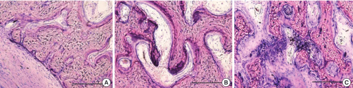
Therefore, ligature placement in the teeth of dogs has been proposed to obtain an experimental periodontitis condition more quickly than periodontitis naturally occurs [18]. This Figure 3. Histologic views of each group (H&E, bar=1 mm). (A) The control group showed minimal attachment loss and very little in- flammation infiltration. (B) The silk ligature group showed mild gin- gival inflammation. (C) The Porphyromonas gingivalis lipopolysaccha- ride with silk ligature group showed severe inflammatory cell infil- tration.
A B C
Figure 1. Plaque index for each group. There were no significant differences in any of the groups at baseline (week 0). After 4 weeks, the silk ligature group (Ligature) and silk ligature with Porphyromon- as gingivalis lipopolysaccharide (P.g. LPS) group (Ligature+LPS) showed marked differences compared to the control (P<0.05). There were no significant differences between the silk ligature with P.g. LPS group and silk ligature group. Different asterisks indicate statistically signif- icant differences. Data are presented as mean±standard error.
0 4
*
** **
Plaque index
2
1
0
Ligature Ligature + LPS
Weeks
Figure 2. Gingival index for each group. There were no significant differences among the groups at week 0. After 4 weeks, the silk liga- ture with Porphyromonas gingivalis lipopolysaccharide (LPS) group (Ligature+LPS) showed significant differences compared to the oth- er groups (P<0.05). There were also significant differences between the silk ligature group (Ligature) and the control. Different asterisks indicate statistically significant differences. Data are presented as mean±standard error.
0 4
*
**
***
Gingival index
3
2
1
0
Control Ligature Ligature + LPS
Weeks
chronic defect model takes over 4 months, and mainly de- stroys the interproximal bone, which is not suitable for peri- odontal treatment research [8]. In order to investigate the periodontal healing capacity of a therapeutic modality, surgi- cally created class III furcation defects in the mandibular pre- molar area were introduced [7]. This model provided critical- sized defects; however, they are acute defects, which are not regarded as periodontal inflammation. A combined model, a surgical defect with chronic inflammation, has also been pro- posed [10]. Despite the gingival inflammation that occurred in this model, the surgical defect was not directly influenced by experimental periodontitis. Herein, we modified the sur- gical bone defect to be influenced by periodontal inflamma- tion for a new combined experimental periodontitis model.
It was previously reported that P.g. comprises a major por- tion of bacteria in the dental plaque of dogs, and its quantity increases with age, plaque amount, and gingival inflamma- tion [19]. LPS comprises the outer surface of all subgingival gram-negative bacteria, and it can induce inflammation in periodontal tissue [20,21]. LPS injection induced periodontitis in a rat model, which has been regarded as a time-saving method for producing experimental periodontitis [12,22].
Because the gingival sulcus is absent in a periodontally healthy dog, a proper plaque accumulation method is re- quired to produce gingival inflammation [23-25]. Ligature placement on the cervical area of the tooth can collect dental plaque, and plaque bacteria provokes gingival inflammation [26]. Because it takes several months to produce periodontitis in this chronic model, additional measures are needed to achieve periodontal inflammation within a short period of time.
In our study, the P.g. LPS-saturated collagen with silk liga- ture group had developed periodontal inflammation that af- fected the gingiva and alveolar bone at 4 weeks after surgical defect creation. Although the plaque index was similar to that of the silk ligature group, the gingival index was significantly
higher and the alveolar bone was highly infiltrated by inflam- matory cells in the P.g. LPS-saturated collagen with silk liga- ture group.
Our findings are consistent with the results of previous studies that bone tissue may be influenced by LPS through inflammatory cytokines [27,28]. It has been proposed that LPS can affect circulating leukocytes directly and activate osteo- clasts [29]. Based on these findings, we can deduce that the P.g. LPS in our study plays an important role in alveolar bone defects and may prevent compensatory bone formation. In our model, P.g. LPS directly influenced periodontal tissue in- cluding alveolar bone, and accumulated plaque bacteria in the silk ligature also affected the gingival tissue.
It has not been reported that P.g. alone can colonize peri- odontal lesions in a dog model, which is possibly due to the variability of periodontitis development in a dog model [30].
In the present study, we utilized collagen material to be satu- rated with P.g. LPS and also added silk ligature placement.
Plaque bacteria might have been absorbed in P.g. LPS-satu- rated collagen and might have aggravated the periodontal inflammation. However, more evidence is needed. Our study has some limitations, in that the sample size is small and a P.g. LPS-deficient collagen with silk ligature group was not adopted, requiring further research.
Within the limits of this study, we were able to achieve periodontal inflammation at 4 weeks after surgical bone de- fect creation by using P.g. LPS-saturated collagen with silk ligature. This could be a possible candidate for an experimen- tal periodontitis model for surgically created alveolar bone defects.
CONFLICT OF INTEREST
The authors report no conflicts of interest related to this study.
Figure 4. Alveolar bone images of each group (H&E, bar=100 µm). (A) There was no sign of inflammation in the control. (B) The silk ligature group showed reduced inflammation. (C) The Porphyromonas gingivalis lipopolysaccharide with silk ligature group showed a severe inflam- matory cell presence in the alveolar bone tissue.
A B C
This study was supported by a grant of the Korean Health Technology R&D Project, Ministry for Health, Welfare &
Family Affairs, Republic of Korea (A101768).
REFERENCES
1. Pihlstrom BL, Michalowicz BS, Johnson NW. Periodontal diseases. Lancet 2005;366:1809-20.
2. Schou S, Holmstrup P, Kornman KS. Non-human primates used in studies of periodontal disease pathogenesis: a re- view of the literature. J Periodontol 1993;64:497-508.
3. Yamasaki A, Nikai H, Niitani K, Ijuhin N. Ultrastructure of the junctional epithelium of germfree rat gingiva. J Peri- odontol 1979;50:641-8.
4. Eggert FM, Germain JP, Cohen B. The gingival epithelium of rodent molars with limited eruption. Acta Anat (Basel) 1980;107:297-306.
5. Attström R, Graf-de Beer M, Schroeder HE. Clinical and histologic characteristics of normal gingiva in dogs. J Periodontal Res 1975;10:115-27.
6. Haney JM, Zimmerman GJ, Wikesjo UM. Periodontal re- pair in dogs: evaluation of the natural disease model. J Clin Periodontol 1995;22:208-13.
7. Wikesjo UM, Kean CJ, Zimmerman GJ. Periodontal repair in dogs: supraalveolar defect models for evaluation of safety and efficacy of periodontal reconstructive therapy. J Periodontol 1994;65:1151-7.
8. Holland M, Boring JG, Boyle CR, Pickrum HM, Jeffcoat MK. Radiographic bone loss correlations and technetium- 99m-MDP bone uptake in ligature-induced periodontal disease in the beagle. Vet Radiol Ultrasound 1998;39:366-74.
9. Clergeau LP, Danan M, Clergeau-Guerithault S, Brion M.
Healing response to anorganic bone implantation in periodontal intrabony defects in dogs. Part I. Bone regen- eration. A microradiographic study. J Periodontol 1996;67:
140-9.
10. Hayashi C, Kinoshita A, Oda S, Mizutani K, Shirakata Y, Ishikawa I. Injectable calcium phosphate bone cement provides favorable space and a scaffold for periodontal re- generation in dogs. J Periodontol 2006;77:940-6.
11. Andrian E, Grenier D, Rouabhia M. Porphyromonas gin- givalis-epithelial cell interactions in periodontitis. J Dent Res 2006;85:392-403.
12. Dumitrescu AL, Abd-El-Aleem S, Morales-Aza B, Donald- son LF. A model of periodontitis in the rat: effect of lipo- polysaccharide on bone resorption, osteoclast activity, and local peptidergic innervation. J Clin Periodontol 2004;31:
596-603.
relation between oral hygiene and periodontal condtion.
Acta Odontol Scand 1964;22:121-35.
14. Loe H, Silness J. Periodontal disease in pregnancy. I. Prev- alence and severity. Acta Odontol Scand 1963;21:533-51.
15. Page RC, Schroeder HE. Pathogenesis of inflammatory periodontal disease: a summary of current work. Lab In- vest 1976;34:235-49.
16. Lallam-Laroye C, Escartin Q, Zlowodzki AS, Barritault D, Caruelle JP, Baroukh B, et al. Periodontitis destructions are restored by synthetic glycosaminoglycan mimetic. J Biomed Mater Res A 2006;79:675-83.
17. Peruzzo DC, Benatti BB, Antunes IB, Andersen ML, Sal- lum EA, Casati MZ, et al. Chronic stress may modulate periodontal disease: a study in rats. J Periodontol 2008;79:
697-704.
18. Lindhe J, Hamp S, Loe H. Experimental periodontitis in the beagle dog. J Periodontal Res 1973;8:1-10.
19. Allaker RP, de Rosayro R, Young KA, Hardie JM. Prevalence of Porphyromonas and Prevotella species in the dental plaque of dogs. Vet Rec 1997;140:147-8.
20. Garrison SW, Nichols FC. LPS-elicited secretory responses in monocytes: altered release of PGE2 but not IL-1 beta in patients with adult periodontitis. J Periodontal Res 1989;
24:88-95.
21. Page RC. The role of inflammatory mediators in the pathogenesis of periodontal disease. J Periodontal Res 1991;26(3 Pt 2):230-42.
22. Rogers JE, Li F, Coatney DD, Rossa C, Bronson P, Krieder JM, et al. Actinobacillus actinomycetemcomitans lipo- polysaccharide-mediated experimental bone loss model for aggressive periodontitis. J Periodontol 2007;78:550-8.
23. Egelberg J. Local effect of diet on plaque formation and development of gingivitis in dogs: 3. Effect of frequency of meals and tube feeding. Odontol Revy 1965;16:50-60.
24. Hamp SE, Lindhe J, Loe H. Experimental periodontitis in the beagle dog. J Periodontal Res 1972;(10):13-4.
25. Lindhe J, Hamp SE, Loe H. Experimental periodontitis in the beagle dog. Int Dent J 1973;23:432-7.
26. Lindhe J, Ericsson I. Effect of ligature placement and den- tal plaque on periodontal tissue breakdown in the dog. J Periodontol 1978;49:343-50.
27. Lindemann RA, Economou JS, Rothermel H. Production of interleukin-1 and tumor necrosis factor by human pe- ripheral monocytes activated by periodontal bacteria and extracted lipopolysaccharides. J Dent Res 1988;67:1131-5.
28. Garrison SW, Holt SC, Nichols FC. Lipopolysaccharide- stimulated PGE2 release from human monocytes. Com- parison of lipopolysaccharides prepared from suspected periodontal pathogens. J Periodontol 1988;59:684-7.
osteoclastogenesis via an osteoblast-independent path-
way. Infect Immun 2002;70:3143-8. tis in adult dogs: a clinical and histopathological survey. J Periodontol 1981;52:60-73.