太 ~;~IjJ(~‘t~↓협쩡짤표‘ 찌'í 22 양 第 3 ~Æ pp. 384 - 391, 1986 Journal of Korean Radiological Society, Vo1.22, No.3, 1986
〈국문초록〉
Sonographic Changes of the Gallbladder Wall in Cholecystitis
- A Sonographic-Pathological Correlation -
Jae Hoon lim, M.D., Young Tae Ko, M.D., and 500n Yong Kim, M.D.*
Depar/Jηen/ 01 Diagηosl1‘c Radiology, Kyung Hee Universi/)’ Hosþita/
* Presen/ Adress: Deþartmeη t 01 R,αdiology, Han Yang Ulliversi/y Hosþila/
H홈킹훌싹에 있어서 擔훌훌의 超音波 所見 - 超音波所見과 病理組織所見의 .It較 -
g얄熙大쌓!校 짧科大學 放射線科웰敎室
林在勳·高永泰·金짧鋼
a홉않t~時 뼈察되는 超흡波所見의 病理學的 根題을 究明하기 위하여 수울로 切除한 急性體짧~ 17 例와 ↑성性廳짧~ 27 例의 廳짧慶을 組織學的으로 觀察하고 이를 超룹波所見파 各各 比較하여 마음 과 같은 結폐을 얻었마.
1) 超흡波所見上 廳製훨 自體內의 低에코혐은 急性觸짧~인 경우는 類陳下層의 F¥種, 出血 또는
~j[쩌flH댐용뼈等에 의하고
↑상性願짧췄인 경우는 觸肉혐의 nEJl길에 의한마.2) 不解明한 內測 廳짧훨 혹운 內떼의 低에코輪은 챔廳의 l뾰짧이나 *퍼願주릉의 消失에 의한다.
3) 觸製훨에코의 全般的인 減少는 以上의 두가지, 즉 짧l않下혐의 ~j[性 짧化와 *김願脫落 또는
*6 1l1윗주릉消失에 의한다.
著者들은 이 3 가지 超륨波檢효所見 즉, 1!1쩔웰t용훌 自體內의 低에코햄, 不解明한 內에 廳製용훌 또는 低에코輪, 그러고 m쩔짧툴풍에코의 全般的 減少等은 擔製찢 該斷에 매우 有用한 所見임을 彈調한마.
To assess the pathological basis of the sonographic changes of the gallbladder wall in cholecystitis, the sonographic appearances of the gallbladder wall were analysed in 17 patients with acute cholecystitis and 27 patients with chronic cholecystitis, and correlated with the pathological specirnens rernoved at surgery In aCllte cholecystitis, a thin sonolucent layer within the echogenic gallbladder wall corresponds to subserosal edell1a, hell10rrhage and inflall1rnatory cell infiltration; in chronic cholecystitis it corresponds to rnuscular hypertrophy. Indistinctness and/or a low echogenicity rind along the inner ll1argin reflects rnucosal sloughing
。 robliteration of the ll1ucosal folds. Uniforll1ly decreased echogenicity of the wall is caused by severe inflall1-
ll1atory cell infiltration with sloughing of the ll1ucosa or obliteration of the ll1ucosal folds. These sonographic signs are cOl1sidered 1'0 be valllable Sigl15 of cholecystitis
Receivecl April 19, 1986; acceptecl May 17, 1986
384 -
- Jae Hoon Lim, et al.: Sonographic Changes of the Gallbladder Wall in Cholecystitis-
Introduction Materials and Methods
The role of ultrasound in the diagnosis of gallstone The study populalion COnSiSled of 17 patients with is well estab!ished but its role in the diagnosis 01' acute cholecystitis and 27 patients with chronic cholecystitis remains controversial. Ultrasonic cholecystitis who were referred for abdominal demonstration of gallstones is a supportive sign of sonography before surgery. At the time of sonography cholecystitis in the appropriate clinical settings, 1) but each patient had appropriate clinical evidence of in- its mere existence does not necessarily indicate inflam- flammation of the gallbladder and there was no in- mation of the gallbladder. Gallbladder wall thicken- dication of other diseases which might cause ing has been described as a sign of cholecystitis,2-7 sonographic changes of the gallbladder wall. The pa- but it is also seen in diseases other than tients who had acute cholecystitis superimposed on cholecystitis.Ó-1
") chronc cholecystitis were classified as acute
There have been several papers concerned with the cholecystitis, as acute inflammatory changes sonographic changes 01' the gallbladder wall in pa- predominated in the pathological findings and clinical tients with cholecystitis, (for example, sonolucency signs and symptoms were those of acute cholecystitis.
surrounding the gallbladder waI1.31<,",) “halo signs"7) The interval between sonography and surgery was less and hazy clelineation of the wall17) but few 01' them than 5 clays in most cases but up to 2 weeks in some have clealt Wilh the pathological basis 01' the cases 01' chronic cholecystitis. Sonograms were review- sonographic changes.17-19
) To verify lhe palhological ecl relrospectively and comparecl with the histological basis 01' lhe sonographic changes, the aUlhors analysed finclings.
the sonographic appearances ancl pathological changes Sonography was performed using commercially
01' the gallblaclcler wall in patienls with cholecystitis available gray-scale static or real-time scanners with ancl a sonographic-pathological correlalion \vas at- a 3.5 MHz transclucer. During the examination, an
lemptecl. attempt was macle to lessen subjective observation
A I!lν ‘ 옛iøP.: 엉양싫강r ‘ B
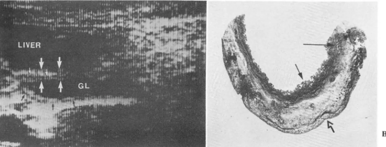
Fig. 1. A 4.5-year-old \\'oman with acute cholecystitis superimposed on chronic cholecystitis
A. Ultrasonogram of the gallblac1c1er wall shows an anechoic layer (smaller arrows) between two hyperechoic layer (bCl\、'een larger arro\Vs). GL Gallbladder lumen
ß, IJow-po\\'cr micrograph of thc gallblac1c1er wall (Masson ’s trichroe stain, 5X magnification) discloses severe cclcma. inllammatory cell infillration ancl some fibrosis in the subserosal (perimuscular) layer. Note intact
J11ucosal folcls (arrow), Infla11lmatory change, especially ec1ema, of subserosallayer explains the anechoic laycr on sonogram, Serosallayer (open arrow) explains the outer hyperechoic layer anc1 Jllucosal folc1s ex plain lhc c1islincl inner hypcrechoic layer. Long arrow points a Rokitansky-Aschoff sinus
- The Journal of the Korean Radiological Society, Vol. 22, No. 3, 1986
of the gaIlbladder waIl echoes. The sonographer tried
to maintain the incident beam perpendicular to the Results gallbladder waIl and longitudinal views were usuaIly
obtained in left lateral decubitus position. The seg- Sixteen of the 17 patients with acute cholecystitis ment of the gaIlbladder waIl in contact with the liver and 14 of the 27 patients with chronic cholecystitis was chosen for sonographic analysis. showed one or two sonographic abnormalities (Table
Sonograms were analysed for the presence of the 1).
foIlowing: The sonographic-pathological correlation may be
1. a sonolucent layer within the hyperechoic summerized as foIlows:
gaIlbladder waIl which divided it into two layers (Fig. 1. The sonolucent layer was proven to represent
l-A), edema, hemorrhage and inflmmatory ceIl infiltration
2. indistinctness and/or a low echogenicity rind in the subserosal (perimuscular) layer in acute along the inner aspect of the waIl (Fig. 2-A), and cholecystitis (Fig. l-A,B) and moderate to severe 3. alteration of the overaIl waIl echogenicity with muscular hypertrophy in chronic cholecystitis reference to the hepatic echogenicity (Fig. 3-A). (Fig.4-A,B).In 14 cases of acute cholecystitis superim-
On histological examination, the foIlowing were posed on chronic cholecystitis both of these
recorded: pathological findings were observed in the same
1. integrity of the mucosa, gallbladder.
2. presence of subserosal edema, hemorrhage and 2. GaIlbladders showing an indistinctness and/or inflammatory ceIl infiltration, and low echogenicity rind along the inner aspect of the 3. muscular hypertrophy. gaIlbladder on sonography had sloughed mucosa and A direct correlation between the sonographic and obliteration or loss of the mucoosal folds (Fig.2-A,B).
pathological findings of the gaIlbladder was then at- Gallbladders showing both a sonolucent layer with
tempted. indistinctness and/or a low echogenicity rind show-
A . 향쩔얀짧향협聊f"~웠 깅I흩흩~ ,톨톨l1li tf..-- B
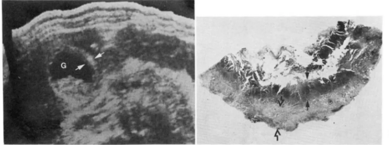
Fig. 2. A 67-year-old man with acute cholecystitis superimposed on chronic cholecystitis
A. Transverse ultrasonogram of the gallbladder wall (between arrows) shows a low echogenicity rind alol1g the il1ner side of the outer hyperechoic layer. NOl1shadowing echogenic foci occupy the clepenclant part of the gallbladder lumen. Note indistinct inner margin. G Gallblaclcler lumen
B. Low-power micrograph of the gallblaclcler wall (HE stain, 5X magnification) cliscloses slollghing of the
ITIlICOSa, cell infiltration, ancl necrosis (between arrows). Debris, tiny stones ancl epithelial cells fill the 11ll11en Fibrosis of subserosallayer (between open arrows) corresponcl to hyperechoic ollter layer ancl cell infiltra- tion and necrosis corresponcl to low echogenicity rincl of inner layer 011 50no15ral11. MlIcosal sloll15hing ex- plains inclistinctness of inner surface of the gallbladcler on sono15ral11
Jae Hoon Lim, et a1.: Sonographic Changes of the Gallbladder Wall in Cholecystitis-
ed both the subserosal and mucosal changes describ- to our parameters on sonography showed minimal ed above (Fig.5-A,B). inflammatory changes, fibromuscular thickening or
3. Gallbladders showing uniformly decreased wall fat cell infiltration.
echoes had a severe inflammatory cell infiltration in
the entire wall as well as sloughing of the mucosa Discussion or obliteration of the mucosal folds (Fig.3-A,B).
4. Gallbladders showing no abnormality according The pathological change of the gallbladder wall
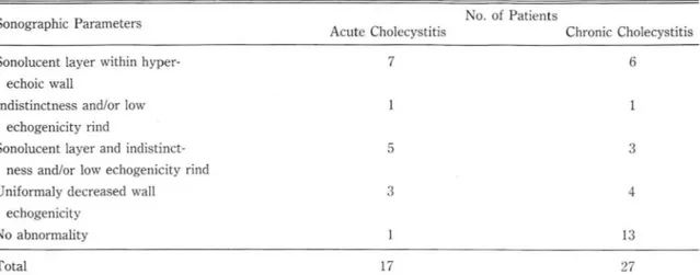
Table 1. Summary of Sonographic Findings
No. of Patients Sonographic Parameters
Acute Cholecystitis Chronic Cholecystitis
Sonolucent layer within hyper- echoic wall
Indistinctness and/or low echogenicity rind
Sonolucent layer and indistinct- ness and/or low echogenicity rind Uniformaly decreased wall
echogenicity No abnormality
Total
A
Fig. 3. A 74.year-old woman with chronic cholecystitis.
7 6
5 3
3 4
13
17 27
A. lJltrasonogrilm shows uniformly decreased gallbladder wall echo. Echo from the gallbladder wall is the same as that of the liver, and thus the gallbladder \Vall is not separable from adjacent liver
ß. L。이 power micrograph of the gallbladder wall (HE stain, 5X magnification) shows diffuse inflammatory cell infiltration as well as dense fibrosis in the subserosallayer ancl muscular hypertrophy. Arrows point patchy areas of mucosal folc\ obliteration. Diffuse inflaml11i1tory cell infiltration, fibrosis ancl ll1uscular hypenrophy explain uniform clecrease of the gallblaclc1er \\'all echo. anc1 piltchy ll1ucosal folcl obliteration explains the lack of sharp echogenic inner margin 01' the gallblaclc1er wall on sonogram. Note cholesterolosis :vlU :\1ucosal surface. SE Serosal surface
B
- The Journal of the Korean Radiological Society, Vol. 22, No. 3, 1986 -
MU
Fig. 4. A 65-year-old man with chronic cholecystitis
A. Ultrasonogram for the gallbladder wall (between arrows) shows a hypoechoic layer between two sharply defined hyperechoic layers. A large shadowing stone seen
B. Low-power micrograph of the gallbladclel ψall (HE stain, 5X Illagnification). There is severe subserosal edema with minimal inflalllll1atory cell infiltration and sOllle fibrosis. Note intact Illucosa ancl Illuscular hypertrophy. [nner echogenic layer on sonogram corresponcls to intact Illucosa and outer echogenic layer corresponcls to fibrosis of the subserosal layer. Subserosal eclerna ancl Illuscular hypertrophy between them are responsible for the hypoechoic layer between two hyperechoic wall echoes. SE Serosal Sllr- face. MU Mucosal surface
MU
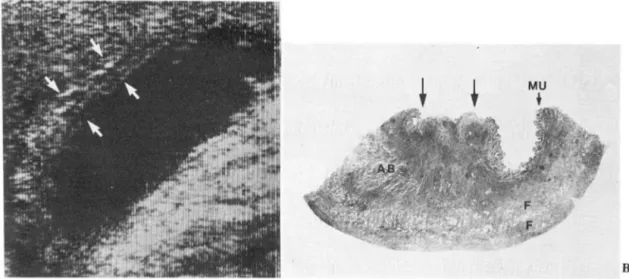
Fig. 5_ A 53-year-old man with aCllte cholecystitis slIperilllposecl on chronic cholecystitis
A. Ultrasonogram shows two hypoechoic layers in the thickenecl gallblac\c\er \Vall (between alTO\\’S). lnnel
Illargin is no1' sharply c1efinec\ ancl less echogenic
B. Low-power l11icrograph of I'he gallblaclclel 、\'all(HE stain, 5X Illagnification) discloses sc\'ere lhickenin 니 01' the \Vall up to 11 111111. Localizecl abscess (AB) ancl inflanllnatorv ccll infillral ion are re 、]lOllsihk for Ihe hypoechoic layers ancl fibrosis (F) of the 、\'allis responsible for the hyperechoic layers. i\il:cosal sloll미 ll J1g (arrows) e:ψlains the indistinct inner Illargin on sonogra111. MU ;Vlllcosal surface
B
B
- Jae Hoon Lim, et a1.: Sonographic Changes of the Gallhlaclclel 씨!all in Cholecystitis -
in acute cholecystitis is thickening 01‘ the gallbladder cholecyst it iSl7 15.'시 and pericholecystic abscess22) has wall which is caused by spreading of the tissue been documented. Marchal et al. described a sonolu- elements and filling of the these spaces by edema cent area within the wall as being a speciric sign for fluid, blood and inrIammatory cells.20) These inrIam- aCllte cholecystitis and attribllted it to sllbserosal matory changes are most severe in the sllbserosallayer edema.17) Raghavendra et al. described gallbladder as it is normally composed of loose connective wall anechoicity in 17 01' 24 patients with aCllte tissue.21) The mucosa may be either shed or missing cholecystitis and considered it represented inflam- in some areas and preserved in others. In chronic matory changes slIch as edema, blood and celllllar cholecystitis, the muscular layer Illay be greatly hyper- inriltration occurring predominently in the slIbserosal trophied with increased connective tisslle between the (pcrimllsclllar) loose connective tisslle spaces.'") muscJe bundles in the lamina propria and sllbserosal Joseph described the “dOllble wall sign'’ in acute layer. The epithelium is lIsllally intact bllt the mllcosal cholecystitis and confirmed that it was produced by folds may be obliterated 01' absent in advanced stages. ederna occllrring in the subserosal layer. He also At intervals, rnllcosal olltpollchings penetrate the observed the sign in chronic cholecystitis and thickened musclllar coat to varying depths towards postulated it was dlle to poor echoes arJSlllg from the external layer (Rokitansky-Aschoff sinllses). AII a thick, fibrolls layer laid down uniform and display- the connective tissue is infiltrated with IYll1phocytes, ing relative acoustic hOlllogenicity with few inter- plasll1a cells, large mononllclear cells and cosinphilic phases. '9)
granlllocytes. In chronic cholcιystitis with slIpcrill1- In this series, the allthors observecl this sonolu- posed aClIte cholecystitis, these inflall1l11atory changes cent layer in the gallblaclderwall in 1201' the 17 cases 01' aCllte and chronic cholecystitis occur at the sall1e (710/0) or aClIte cholecystitis and 9 01‘ the 27 cases time ancl grossly, sllch a gallblaclder differs little 1‘rom (33ψ'0) 01' chronic cholecystitis. These layers were one with aCllte cholecystitis except for the prcsencc anechoic 01' hypoechoic, continllolls 01' interrllpted
01' calcllli. 2띠 11‘ these pat
- 389-
- The Journal of the Korean Radiological Society, Vol. 22, No. 3, 1986 -
is contradiction with the observation of Joseph.19) are more or less subjective and one-to-one comparison However, several diseases that produce thicken- between the observed sonographic findings and ing of the gallbladder wall may also cause a sonolu- histologic findings is very difficult. Authors were cent layer. 14.19) We have seen this sonolucent layer aware of this fact and tried to lessen this effect at in patients with ascites, hypoalbuminemia, cirrhosis the time of sonography and review of pathology of the liver and hepatitis. Although there has been specimens.
no pathological confirmation, the mechanism of this In summary, it is concluded that 1) a thin sonolu- layer in these patients may be similar to that in cent layer within the echogenic gallbladder wall repre- cholecystitis, i.e., subserosal edema. sents subserosal edema, hemorrhage and inflam- Indistinctness of the inner margin of the gallblad- matory cell infiltration or muscu1ar hypertrophy,>) der wall has been regarded as sign of acute indistinctness and/or a low echogenicity rind along cholecystitis by several authors.17) Schneider et al. the inner aspect reflect sloughing of the mucosa or described a halo of low level echoes between the echo- obliteration of the mucosal folds and3) uniformly free gallbladder lumen and the confluent high level decreased wall echogenicity represents severe inflam echoes that surround it and called it the “halo sign".7) matory cell infiltration of the entire wall with This halo of low level echoes corresponds exactly to sIoughing of the mucosa or obliteration of the the low echogenicity rind in our series. However, there mucosal folds. Some diseases other than cholecystitis has been no pathological confirmation of these may results in a sonolucent layer within the gallblad sonographic signs. der wall and thus it is not. specific for cholecystitis
In our series, indistinctness and/or low but indistinctness and/or a low echogenicity rind of echogenicity rind along the inner aspect of the the inner aspect and uniformly decreased wall gallbladder wall was observed ll1 6 of 17 cases (35%) echogenicity may be considered specific fOI of acute cholecystitis and 4 of 27 cases (15070) of cholecystitis.
chronic cholecystitis. Pathology specimens showed a
partially or totally sloughed mucosa 01' obliteration Acknowledgement of the mucosal folds (Fig.2-a,b and Fig.5-a,b). Con
sidering the distinct hi
ed in 7 patients is considered to represent diffuse
severe inflammatory cell infiltration of the entire 、vall 1. Cintora 1, Ben.Ora A, MacNeil R, Gilsclorf RB as well as sloughing of mucosa or obliteration of the Cholecystosonography for the deísíon to operate when mucosal folds (Fig.3-A,B). Hypertrophied muscle is acute cholecystítís ís suspected. A/ll 1 SurglY79.
believed to be responsible for the decreased wall echo ηδ δ 18.820
on sonogram. 2. Hancller 51: Ullrasound of gallbladder wall thíckeníng and The authors admit that sonographic observations íts rclalíon 10 cholecystítís. Alk 7')7<); U2:S81.S8S
- 390-
Jae Hoon Lim. et al.: Sonographic Changes of the Gallbladder Wall in Cholecystitis-
3. Mindel Hj, Ring A: Callbladder wall thickening: u/trasonic findings. Radiology 1979; 133:699-701
4. Finberg Hj
,
Birnholz JC: Ultrasound evaluation of the gallbladder wall. Radiology 1979; 133:693-698 S. Engel jM, Deitch EA, Sikkerna W: Callbladder wallthickness: 50nographic accuracy and relation to disease AjR 198α 134:907-909
6. 5anders RC: The signiηcance of sonographic gallbladder wall thickening. jCU 198α 8:743-146
7. 5chneider M, Rubenstein 、VA, Auh YH, Kazam E Ultrasonography and computed tomography in the evalua- tion of biliary tract disease. In: Thorbjarnarson 8 ed. Surgery of the biliary tract. Philadelphia, Londoπ Toronto, ιlex ico city, Rio de janeiro, 5ydneκ Tokyo : ,ν B. 5aunders Company. 1982; 79-60
8. Fiske CE, Laing FC, Brown TW: Ultrasonographic evidence of a gallbladdel νV.δ11 rhickening in association wirh hypoalbuminemia. Radiology 1980; 13.5:713-716 9. 5hlaer W j, Leopold CR, 5cheible FW: 50nography of the
thickened gallbladder νV긴 11: a nonspecific finding. AjR 7981;136:337-339
10. Yum HY, Fink AH: 5ol1ographic findings in primary car- cinoma of the gallbladder‘ Radiology 1980; 134:693-696 11. Ralls I'W , Quinn tv\F, juttner H-U, Halls jM, Boswell WD Callbladder νvall thickening: Patients without intrinsic gallbladder disease. AjR 1981; 137:6.5-68
12. juttner H-U , Ralls I'W, Quinn MF, jenny jM: Thickening of the gallbladder wall in acute hepatitis: ultrasound demonstration. Radiology 1982; 1-+2:-+65-466
-13. I'atrigui HB, Dipietro M, Barber FE, Tcele RL: 50nogr따Jhy
of thickened gallbladder wall: Causes in children. AjR 1983, 141:.57-60.
14. Maudgal D, Wansbrough-jones MH, joseph AEA Callblad der abnormalities of acute infectious hepatitis. A prospec- tive study. Oig Ois 5ci 1984; 29:257-260.
15. Creenberg M , Kangarloo H, Cochran 5t, 5ample WF: The Ultrasonographic diagnosis of cholecystitis and cholelithiasis in children. Radiology 1980; 137’745-749 16. Kane RA: Ultrasonographic diagnosis of gangrenous cholecystitis and empyema of the gallbladder. Radiology 1980; 134:191-194
17. Marchal GjF, Casaer ι1, Baert AL, Goddeeris I'G, Kerremans R, Fevery j: Callbladder wall sonolucency in acute cholecystitis. Radiology 1979;133.429-433
18. Raghavendra BN, Feiner HD, 5ubramanyam BR, Ranson jHC, T oder 51', Horii 5C, Madamba MR: Acute cholecystitis:
sonographic-pathologic anaylysis. AjR 1981; 137:327-332 19. joseph AEA: The gallbladder. In: joseph AEA, Consgrove
00 eds. Ultrasound in inflammatory disease. New York, Edinburgh, London, ι1elbourne: Churchill Livingstone, 1983, 761-183
20. Halpert B: Callbladder and biliary ducts. In: Anderson WAO, Kissane j,ι1 eds. Pathology. 5t Louis: Mosby. 1977; 1439-14.56
21 Thomas CG, Wonack NA: Acute cholecystitis, its pathogenesis and repair. Arch 5urg 19.52; 64:590-600 22. Bergman AB, Neiman, HL, Kraut ß: U/trasonographic
evalual'ion of pericholecystic abscesses. AjR 1979;
132:201-203
- 391-