I. Introduction
Studies of the IgG subclass responses in early- onset periodontitis(EOP) have frequently demon- strated elevated levels of total IgG or IgG2 to whole cell antigens, outer membrane proteins, and/or lipopolysaccharides(LPS) of Actinobacillus actino- mycetemcomitans(Aa)1,2) or Porphyromonas gingi- valis(Pg)3 according to the disease type.
(Hammarstrom et al., 1986) IgG1, IgG3 and IgG4 antibodies are primarily reactive to protein antigens and IgG2 to bacterial carbohydrates, such as LPS or capsular polysaccharides(CPS). Moreover, each IgG subclass response has been studied in the context of the genetic control of an individual.
Immunodominant antigens of Aa in localized juve- nile periodontitis(LJP) were reported to include the 29-kd, 40-kd and 75-kd outer membrane proteins, and capsular carbohydrates(CPS) as well as LPS, while those of Pgin rapidly progressive periodonti- tis(RPP) were fimbriae, 75-kd protein, CPS, and LPS5). There have been controversies in regard to the bacterial antigens being recognized as immun- odominant especially in the case of outer rnem- brane proteins, presumably due to individual differ-
ences in IgG subclass responsiveness or to the com- plexities of the bacterial antigens. There has also been some debate regarding bacterial LPS or CPS in terms of their immunodominant nature in the patho- genesis of EOP.
A previous investigation from our laboratory has focused on the various patterns of elevated IgG sun- bclasses to Pg381 in LJP or RPP patients6). To better understand the immunodominant antigens recog- nized by a panel of IgG subclass antibodies, we per- formed dot immunoblot analysis of selected bacteri- al antigens of Pgthat are recognized by either a sin- gle or by various combinations of IgG subclasses in early-oneset periodontitis.
II. Material and Methods
1. Purification of Fimbriae of Pg 381The fimbriae of Pg381 was prepared as described previously7). Briefly, cells were harvested by cen- trifugation and were suspended in 20 mM Tris- HCI(pH7.4)-0.15M NaC1-10 mM MgC12 by repeat- ed pipetting. The suspension was agitated by mag- netic stirrer for 30 minutes and the supernatant
IgG subclass-dependent Recognition of Porphyromonas Gingivalis Antigens in the Early-onset Periodontitis
Jeom Il Choi
1, Fuminobu Yoshimura
2, Robert E. Schifferle
3, and Katsuji Okuda
4Department of Periodontology, School of Dentistry, Pusan National University, Korea,
1Depatment of Microbiology, School of Dentistry, Aichigakuin University, Japan
2,
Departments of Periodontics and Oral Biology, School of Dentistry, USA,
3and Department of Microbiology, Tokyo Dental College, Japan
4대한치주과학회지 : Vol. 29, No. 4, 1999
obtained after centrifugation at 8,000×g for 20 min.
Ammonium sulfate was added to 4% saturation, pre- cipitated proteins collected by centrifugation, the precipitate was dissolved in 20 mM Tris-HC1(pH 8.0), and dialyzed against 20 mM Tris-HC1(pH 8.0).
The dialysate was clarified by centrifugation at 8,000
× g for 20 min, and applied to a column of DEAE- Sepharose CL-6B(1.5 by 16 cm)(Pharmacia, Piscataway, NJ) equilibrated with the above buffer.
The column was washed with 20 mM Tris-HC1, pH 8.0 and eluted with a linear gradient of 0 to 0.3 M NaC1. No 43K protein band was detected in the fractions eluted after 0.17 M NaC1. Fractions con- taining the 43K protein were concentrated by ammonium sulfate precipitation and dialyzed againt 3 mM Tris-HC1(pH 8.0) or 3 mM sodium bicarbon- ate(pH 8.0).
2. Preparation of Capsular Polysaccha- rides of Pg A7A1-28(ATCC 53977)
The capsular polysaccharide of PgATA1-28 was prepared by a modification of the method previous- ly described8). Briefly, bacterial cells were suspend- ed in water(0.2 to 0.4 g wet weight/ml), extracted with an equal volume of 90% phenol for 20 rain at 65 to 68°C, and stored overnight at 4°C. The aque- ous phase was obtained by centrifugation at 4000×
g for 1h at 10°C and dialyzed at 4°C against distilled water using Spectrapor 1 tubing. The dialyzed solu- tion was brought to 0.15 M sodium chloride, 4 mM MgC121 mM CaC12, and pH 7.5 with Tris-HC1 and treated with ribonuclease A(0.04 mg/ml) and deoxyribonuclease I(0.01 mg/ml)(Sigma, St. Louis, MO) for 2 hr at 37°C and then with Proteinase K(0.04mg/ml) for 1 hr at 60°C. The solution was dialyzed against dH2O and lyophilized The lyophilized extract was dissolved in 0.05M Tris-HCI buffer, pH 9.5, containing 0.3% deoxycholate and
0.001 M trisodium EDTA. This solution was applied to a column of Sephacryl S-400 HR(1.0×
47cm)(Pharmacia, Piscataway, NJ), at room temper- ature and eluted with the deoxycholate containing buffer. Fractions were assessed for LPS and CPS by double immunodiffusion in agarose, for LPS by SDS- PAGE, and at 280 nm. Appropriate fractions con- taining either PS or LPS were pooled, medium chlo- ride was added to 0.15 M NaCl. and PS and LPS precipitated with 4 volumes of 95% ethanol. The precipitates were isolated by centrifugation, dis- solved, dialyzed, and lyophilized
3. Preparation of Lipopolysaccharides of Pg 381
LPSs were extracted by the hot phenol water method by Westphal and Jann9). Briefly, lyophilized cells were suspended in pyrogen-free distilled water. 90% phenol was added, and the suspensions were shaken vigorously at 67°C for 15 min and cen- trifuged at 7,000×g for 20 min. The aqueous phase was removed and diayzed thoroughly against dis- tilled water. After centrifugation at 7,000×g for 20 min, equal volume of ethanol with 0.15 M NaCl was added to the supernatant. After centrifugation at 7,000×g for 15min, the resulting pellet was dis- solved in 30 ml of distulled water and centrifuged at 105,000×g for 2 h. The procedure was repeated, and the resulting precipitate was suspended in water and lyophilized.
4. Serum Samples
Serum samples of 35 EOP patients(1 I/LJP and 24 RPP), whose IgG subclass antibody levels were measured in a previous study, were reclassified according to the patterns of elevated antibody either in a single or in combination of IgG subclasses to Pg
381 whole cells6). LJP patients were diagnosed as those who were between 12-25 years of age with typical pattern of molar/incisor pattern of involve- ment showing>5 mm of attachment loss. RPP patients were diagnosed as those who were between 18-35 years of age showing generalized extensive pattern of attachment loss with severe alveolar bone destruction. Serum samples from 21 patients consisting of 5 rapidly progressive peri- odontitis(RPP) patients with elevated IgG2 antibody levels to Pg 381(group 1), 6 patients(2 localized juvenile periodontitis(LJP) and 4 RPP) with elevated IgG4(group 2), 2 RPP patients with elevated IgG2+4(group 3), and 8 patients(2 LJP and 6 RPP) with elevated IgG1+2+4(group 4), were selected for dot immunoblot analysis. 20 clinically healthy sub- jects(aged between 20-25 years) whose antibody levels were not elevated to any of tested antigens were designated as the control group.
5. Enzyme-liked immunosorbent assay (ELISA)
IgG subclass antibody titers to Pg were deter- mined by ELISA using alkaline phosphate assay sys- tem. 96-well microtiter plates were coated with 0.1 of purified antigens diluted in 50 mM carbonate/bicarbonate buffer(pH 9.6). After overnight incubation at 4°C, the plates were washed 3 times with phosphate-buffered saline(PBS) con- taining 0.1% Tween 20. 0.1 ml of serum samples diluted in PBS containing 0.1% Tween 20 were added into each well and incubated for 2 hours at room temperature. The plate was washed 3 times with PBS containing 0.1% Tween 20, and then 0.1 ml of four mouse antihuman IgG subclasses(affinity- purified monoclonal antibody, γ-chain specific, IgG1; HP6012, IgG2;HP-6014, IgG3;HP-6050, IgG4;HP-6025; Sigma Chemicals, Ohio, USA) diluted
in PBS containing 0.1% Tween 20 were added into each well and incubated for 2 hours at room tem- perature. After washing 3 times with PBS containing 0.1% Tween 20, 0.1 ml of goat anti-mouse IgG(heavy/light chain specific, affinity purified, alka- line phosphatase-conjugated, Calbiochem. Basel, Switzerland) diluted in PBS containing 0.1% Tween 20 were added into each well and incubated for overnight at room temperature. After the washing plates, 0.2 ml of of nitrophenyl phosphat(1 mg/ml) were added into each well and incubated for 30 minutes and finally 0.1 ml of 1N NaOH were added to stop color reaction. Optical density was mea- sured using an ELISA plate leader with wavelength set at 492 nm. To determine the serum IgG antibody tiers, optical densities(O.D.) were plotted as a fun- ciotn of serum dilution facror. Regression analysis was performed and reciprocals of the serum dilution factrors at the X-axis intersection of O.D.=1.0 expressed as the ELISA unit for each sample.
Patients whose IgG Levels exceeded the twice the value of the control group were assigned as the ele- vated group. For a comparison between groups, total IgG titer was measured against Pgor Fn
6. Dot Immunoblot Analysis
Three kinds of the purified antigens were serially diluted by halves from 0.5 microgram/ml to 0.0312 microgram/ml of buffer(32 mM sodium carbonate, 68 mM sodium bicarbonate) containing 200 mM MgC12. Four microliters of each sample were spelled onto nitrocellulose membrane and left to dry at room temperature for 1 hour. The membrane was incubated with 5% skin milk in 25 mM tris-buffered saline(TBS) overnight at 4°C followed by incubation by orbital shaking for 1 hour at room temperature.
After washing the membrane once with phosphate buffered saline(PBS), it was incubated with human
serum at 1;1,000 dilution in buffer(85 mM sodium carbonate/0.5 mL Tween 20) for 1 hour at 31°C.
The membrane was washed three times with 1%
skim milk in TBS for 5 minutes and finally washed once with PBS. The membrane was then incubated with the goat anti-human IgG(affinity purified, y- chain specific) (Calbiochem, Basel, Swizerland) with 1:1,000 dilutions in buffer(2.7 g of sodium phos- phate dibasic, 0.28 g of sodium phosphate monoba- sic, 8.75 g of sodium chloride, 0.5ml of Tween 20, 0.2 g of sodium azide/L) for 1 hour at room temper- ature followed by washing three times with 1% skin milk in TBS an once with PBS. The membrane was finally incubated with rabbit anti-goat IgG(havy/light chain specific, affinity purified, alkaline-phosphatase conjugated)(Sigma, St. Louis, MO) with 1:2,000 dilu- tions in buffer(2.7 g of sodium phosphate dibasic, 0.28 g of sodium phosphate monbasic, 8.75 g of sodium chloride, 0.5 ml of Tween 20, 0.2 g of sodi- um azide/L) for 1 hour at room temperature. After
washing three times with 1% skin milk in TBS and once with PBS 5-bromo-4-chloro-3-indolyl-phos- phate/nitroblue tetrazdium(BCIP/NBT) solution (Kirkegaard & Perry, Gaithersburg MD) was added and incubated for 30 minutes at room temperature for color development. The staining intensities were evaluated by densitometric analysis for quantitative comparison for the tested antigens in each group.
Pooled serum samples from the control group were used for comparison.
7. Statistical Analysis
To compare the IgG levels to each tested antigen within the groups, ANOVA was performed.
III. Results
A total of 21 serum samples from the 35 patients(1l LJP and 24 RPP patients) were selected
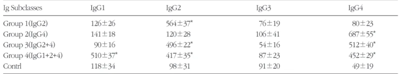
Table 1A. IgG subclass levels to whole Pg 381 cells in each experimental group and control group as determined by ELISA(mean±s.d.).
Ig Subclasses IgG1 IgG2 IgG3 IgG4
Group 1(IgG2) 126±26 564±37* 76±19 80±23
Group 2(IgG4) 141±18 120±28 106±41 687±55*
Group 3(IgG2+4) 90±16 496±22* 54±16 512±40*
Group 4(IgG1+2+4) 510±37* 417±35* 87±23 452±29*
Contrl 118±34 98±31 91±20 49±19
*higher than twice the value of the control group
Table 1B. IgG levels to each tested antigens in each experimental group and control group as determined by ELISA(mean
±s.d.).
Antigens fimbrillin LPS CPS
Group 1(IgG2) 88±27 49±16 246±18*
Group 2(IgG4) 468±34* 54±28 98±49
Group 3(IgG2+4) 350±41* 30±12 201±22*
Group 4(IgG1+2+4) 387±29* 61±15 225±19*
Contrl 68±16 38±16 89±40
*significantly higher than those to other antigen(s) by ANOVA(p<0.01) and greater than twice the value of the control group
based upon the pattern of elevated IgG subclass responses against Pg 381(see materials and method). Tables 1A and 1B summarize the IgG sub- class responses for either IgG2 or IgG4, or in combi- nations of elevated IgG2+4 or IgG1+2+4 antibodies against whole Pg 381 cells6), Group 1 pateints had significantly higher IgG levels to CPS, while group 2 to fimnrilin and CPS, group 3 to fimbrilin and finally group 4 to fimbrilin and CPS. Figure 1 demonstrates
the representative dot immunoblot pattern from each group against the 43-kd fimbrilin protein and LPS of Pg381, and the CPS of Pg A7A1-28, respec- tively. The intensity of each sample was determined by densitometric analysis(Figure 2). The IgG4 anti- body strongly reacted with the fimbrial antigen.
Some of the IgG4 antibodies that reacted strongly with fimbriae also demonstrated a positive reaction with the CPS antigen. In contrast, IgG2 antibody
2 1.5 1 0.5
01 2 3 4 5
dilution(log2)
geometric area
IgG2
6
4
2
0 CPS
43-kd Fim LPS Control
2 1.5 1 0.5 0
3.5 3 2.5 2 1.5 1 0.5 0
CPS 43-kd Fim LPS Control
CPS 43-kd Fim LPS Control
CPS 43-kd Fim LPS Control
1 2 3 4 5
dilution(log2)
geometric area
IgG2+4
1 2 3 4 5
dilution(log2)
geometric area
IgG4
1 2 3 4 5
dilution(log2)
geometric area
IgG1+2+4
Figure 1. Representative patterns of dot immunoblot from four different patient groups; patients with elevated antibody lev- els to Pg 381 in IgG2 only, IgG4 only, IgG2+4, and IgG1+2+4, respectively. The tested antigens were the 43-kd fimbrilin protein(Fim) and lipopolysaccharide(LPS) of Pg 381 and the capsular polysaccharide(CPS) of Pg A7A1- 28.
Figure 2. Diagrammatic representation of geometric area(mm2) of immunoblot described in Figure 1 determined by densit- ometric analysis. X-axis denotes serum dilution factors(log 2)
43-kd Fim LPS CPS
IgG2 IgG4 IgG2+4 IgG1+2+4 CONTROL
recognized primarily the CPS antigen. In most cases, if not all, the LPS antigen was not recognized by any of the IgG subclass groups. Minor individual varia- tions could be demonstrated for the staining intensi- ties within the groups(data not shown).
IV. Discussion
To better understand the immunopathogenesis of early-onset periodontitis, we initially evaluated the IgG subclass antibody levels to Pg381 in LJP or RPP patients6)to see how the d subclass in either a single class or in combinations selectively react with the three kinds of Pgantigens. Thus a dot immunoblot analysis was designed to elucidate the IgG subclass- dependent recognition of IgG known antigens, the 43-kd fimbrilin protein and LPS of Pg381, and the CPS of Pg A7A1-28. The dot immunoblot analysis clearly demonstrated that the fimbrial antigen was recognized primarily by IgG4(or IgG1 to a lesser extent) while the CPS was recognized primarily by IgG2. There was minimal evidence to suggest that any IgG subclass patterns reacted strongly or posi- tively with the LPS antigen from Pg 381. Since the combination of IgG1+2+4 was most frequently found one to be elevated, followed by IgG4 only, IgG2 only, and IgG2+46), it is likely that both fimbri- ae and the CPS of Pg are important bacterial anti- gens in the pathogenesis of EOP The study results clearly demonstrated the specific antibody binding patterns reflecting the magnitudes of IgG levels to the tested bacterial antigens determined by ELISA.
Our observation was consistent with that of other investigators10). However these results differed from reports of elevated levels of IgG2 against LPS3). As there are more than one specific protein antigen reported to be immunodominant and there exist dif- ferent opinions on the immunodcminant role of the bacterial LPS, further studies with individual bacterial
antigens are needed, using a study design which considers the various groupings of the elevated IgG subclasses. Only through this experimental design, may one obtain consistent rsults for the immun- odominant antigen(s) since the IgG subclass responses are highly antigen-dependent fimbrial proteins of Pg are important in bacterial survival against host defense mechanisms by contributing to bacterial adherence5)and are thought to be immun- odoninant antigens11). Moreover recent animal immunization studies with the fimbrial protein or its synthetic analogues have proven to be protective suggesting its possible use for the prevention of human periodontal diseases12). We used the purified fimbriae of Pg 381 in the dot immunoblot assay and it was strongly recognized by the IgG4 antibody and to some extent by IgG1(data not shown). This sug- gests that the fimbrial protein may be strongly immunogenic in EOf patients. Immunoblot staining intensity increased as the IgG4 antibody increased along with IgG1 or IgG2 it has widely been accept- ed that IgG1, IgG3 and to a lesser extent IgG4 are generated in response to bacterial protein antigens.
Ogawa13) also reported the antifimbrial antibody was primarily of the IgG3 subclass in adult peri- odontitis and RPP. They also reported a predomi- nance of IgG4-secreting cells over IgG1-secreting cells in periodontal lesions with severe destruction.
The dot immunoblot assay demonstrated that the IgG4 antibody reacted strongest against fimbriae.
The suggests that a subclass switch occurred in the immunoglobulin gene due to the prolonged anti- genic stimulus14). This may imply that the minimally- protective IgG4 antibody could contribute to the destructive process of EOP6). Evans et al.12)reported the protective effects of immunization with fimbrial protein and postulated a model for its use as an effective vaccine against periodontitis. Therefore, it is possible that antibody to the 43-kd may exert pro-
tective role. However if we consider the functional role funciotn. Rather it is probable that the failure to prevent bacterial adherence at the initial phase of the disease may adversely contribute to the destruc- tive process. Thus it seems reasonable to evaluate the data on the immunodominant antigen(s) in terms of the functional capacities of the reactive IgG subclass antibodies.
Bacterial CPS are structural components that help bacteria to evade phagocytosis by PMNL CPS are closely associated with bacterial invasiveness15). Several groups of investigators have reported that the carbohydrate moiety of either LPS or CPS of Aa may be the immunodominant antigen in localized juvenile periodontitis1). In the present study, the ele- vated IgG2 antibody exclusively recognized the CPS of Pg A7A1-28(strain 381 lacks K-antigen15)), and not the LPS. This finding was consistent with ether reports on Pg 10) and Aa1) Our results, however, were different from the findings of other for Pg11) and Aa16)in LJP, where an elevated IgG2 response was seen against LPS. Recently, Wilson and Hamilton17) reported elevated IgG2 levels against the 29-kd OMP of Aain the LJP. We also observed a similar pattern of IgG2 responses against fimbriae of Pg which seems to be worthy of further studios.
When IgG2 antibody again is viewed in terms of its functional properties, it is possible that the poor complement-fixation ability and the low opsonic properties of IgG2 antibody might have resulted in inadequate clearance of the infecting organism18), although IgG2 antibodies have been reported to promote neutrophil killing of Aa19). It is likely that the IgG2 antibody is reactive promarily with the car- bohydrate moiety of either LPS or CPS.
For all its immunobiological significance, most of the elevated IgG subclass antibodies did not react stongly against LPS in the dot immunoblot assay.
This differs from prerious findings where an IgG2
response to LPS was observed in ELISA studies11). The reaseon for the different results are unclear20). They may be due either to different methodologies or to the different racial origin of the patient sam- ples. In general, the group with elevated IgG4 anti- bodies had a stronger reaction with fimbriae than those with other elevated IgG subclasses(e.g.
IgG2+IgG4 or IgG1+IgG2+IgG4). Most of IgG4 anti- bodies, either singly or in combination with other IgG subclasses, demonstrated a strong reaction with the fimbrial protein and also showed a positive reac- tion with the CPS. IgG2 antibodies generally recog- nized the CPS with few exceptions although the blotting intensities were somewhat weaker than those noted for IgG4 against the fimbrial proteins.
There were a few IgG2 antibodies which also recog- nized the fimnbrial proteins although the reaction intensities were weak.
Schifferle et al.2)attempted to modify Pg infection in mice by immunization with a CPS-protein conju- gate. They were able to reduce the severity of infec- tion, but not prevent infection. Realizing that carbo- hydrate is a weak immunogen, many investigatiors have used a carbohydrate-outer membrane protein conjugate to elicit a thymus-dependent memory T- cll response22). Considering the frequencies and functional roles of the elevated IgG2 and IgG4 sub- classes in EOP, it may be a reasonable immunization strategy to employ a CPS-43 kd fimbrial protein con- jugate as a candidate for vaccine studies23). It is important to realize that IgG2 and other IgG sub- class responses against carbohydrate vaccines are under the control of immunogenetic make-up24). The Gm marker of an individual is highly race-spe- cific25). When employing an animal model for experimental immunization. consideration should be given to the immunoglobulin allotype marker of the animals to be used26).
The present experiment characterized IgG sub-
class reactivity in an immunoblot assay using three kinds of Pg antigens. The results do not exclude the possibility that each IgG2 subclass or subclasses in combination may recognize other immunodomi- nant antigens of Pg and/or other strains, especially protein antigens. We are currently performing addi- tional studies to further characterize the immun- odominant antigens of Pg recognized by IgG sub- classes in early-oneset periodontitis.
V. References
1. Califano, J.V., Schenkein, H.A, Tew J.G.
Immunodominant antigen of Actinobacillus actinomycetemoomitans serotypes a and c in high-responder patients. Infect. Immun.
57:1582-1589, 1989.
2. Califano, J.V., Schenkein, H.A., Tew, J.G.
Immunodominant antigens of Actinobacillus actinomycetemcomitans serotypes a and c in high-responder patients. Oral Microbiol.
Immunol. 6:228-235, 1991.
3. Ant. J., Whitney, C., Houstion, L. Serum IgG subclass response to Bacteroides gingivalisin rapidly progressive periodontitis. J. Dent.Res.
70:584,1991(Abst.#2546).
4. Hammarstom, L., Smith, C.I.E. IgG Subclasses in Bacterial Infections. In:Shakib F, ed. Basic and Clinical Aspects of IgG subclass. Monographs in Allergy vol.19, pp 122-133, Basel:Karger, 1986.
5. Genco, R.J., Sojar, H., Lee, J.Y., Sojar, A., Bedi, G., Cho, M.I., Dyer, D.W. Porphyromonas gin- givalis Fimbriae:Structure, Funciton, and Insertional Inactivation Mutants. In: Genco R, Hamada S, Lehner T, McGhee J, Mergenhagen S, eds. Molecular pathogenesis of Periodontal Disease. Weshington, DC; ASM Press, 13-24, 1994.
6. Choi, J.I., IgG subclass responses in the pheno-
typic subsets of early-onset periodontitis, J.
Korean Acad. Periodontal. 29:251-264, 1999.
7. Yoshimura, F., Takahashi, K., Nodasaka, Y., Suzuki, T. Purification and characterization of a novel type fimbrial from the oral anaerobe Bacteroides gingivalis.J. Bacteriol. 160:949-957, 1984.
8. Schifferle, R.E., Reddy, M.S., Zambon, J.J., Genco, R.J. Characterization of a polysaccharide antigen from Bacteroides gingivalis. J. Immunol.
143:3035-3042, 1989.
9. Westphal, O, Jahn, K. Bacterial lipopolysaccha- ride. Extraction with phenol-water and further applications of the procedure. Methods.
Carbohydr. Chem. 5:83-91, 1965.
10. Schifferle, R.E. Bacterial Polysaccharide as Microbial Virulence Factors. In: Genco R, Hamada S, Lehner T, McGhee J, Mergenhagen S, ed. Molecular Pathogenesis of Periodontal Disease. Washington DC: ASM Press, 83-92, 1944.
11. Chen, H.A., Weinberg, A., Daveau, R.P., Engel, D., Page, R,C. Immundominant antigens of Porphyromonas gingivalisin patients with rapid- ly progressive periodontitis. Oral Microbiol.
Immunol. 10:193-201, 1995.
12. Evans, R.T., Klausen, B., Sojar, H., Bedi, G.S., Sfintescu, C., Ramamurthy, N.S., Golub, L.M., Genco, R.J. Immunization with Porphyromonas (Bacteroides) gingivalisfimbriae protects against periodontal destruciton. Infect. Immun. 60:2926- 2935, 1992.
13. Ogawa, A., Kusumoto, Y., Hamada, S., McGhee, J.R., Kiyono, H. Bacteroides gingivalis- specific serum IgG and IgA subclass antibodies in periodontal diseases. Clin. Exp. Imnunol.
82:318-325, 1990.
14. Aalberse, R.C., van der Gaag, R, van Leeuwen, J. Serologic aspects of IgG4 antibodies. I.
Prolonged immunization results in an IgG4- restricted response. J. Immunol. 130:722-726, 1993.
15. Van Winkelhoff, A.J., Applemelk, B.J., Kippuw, N., de Graaff, J. K-antigens in Porphyromonas gingivalis are associated with virulence. Oral Microbiol. Immunol. 8:259-265, 1993.
16. Lu, H., Califano, J.V., Schenkein, H.A., Tew, J.G. Immunoglobulin class and subclass distrib- ution of antibodies reactive with the immun- odominant antigert of Actinobacillus actino- mycetemcomitans serotype b. Infect. Immun.
61:2400-2407, 1993.
17. Wilson, M.E. IgG antibody response of localized juvenile periodontitis patients to the 29 kilodal- ton outer membrane protein of Actinobacillus actinomycetemcomitans.J.Periodontol. 62:211- 218, 1991.
18. Page, R.C. The humoral response in patients with periodontitis; effects of treatment and prospects for a vaccine. Compend. Contin.
Educ. Dent. 15:S666-S671, 1994.
19. Wilson. M.E., Bronson, P.M., Hamilton, R.G.
Immunoglobulin G2 antibodies promote neu- trophilic killing of Actinobacillus actinomycetem- comitans.Infect. Immun. 63:1070-1075, 1995.
20. McArthur, W.P., Clark, W.B. Specific antibodies and their potential role in periodontal diseases.
J. Periodontol. 64:807-818, 1993.
21. Schifferle, R.E., Genco, R.J., Levine, M.J.
Modificaiton of experimental Porphyromonas
gingivalis murine infection by immunizaiton with a polysaccharide-protein conjugate. Oral Microbiol. Immunol. 8:266-271, 1993.
22. Jennings, H.J., Roy, R., Gamian, A. Induction of meningococcal group B polysaccharide-specific IgG antibodies in mice using an N-propionylated B polysaccharide-tetanus toxoid conjugate vac- cine. J. Immunol. 137:1708-1713, 1986.
23. Choi, J.L., Schifferle, R.E., Yoshimura, F., Kim, B.W. Capsular polysaccharide-fimbrial protein conjugate vaccine protects against Porphyromonas gingivalis infection in SCID mice reconstituted with human peripheral blood lymphocytes. Infect. Immun. 66:391-393. 1998.
24. Choi, J.I., Kim, J.H., Ha, M.H., Kim, S.J. An immunogenetic study on the IgG subclass responses in the localized juvenile and the rapid- ly progressive periodontitis. J. Korean Acad.
Periodontol.(in press). 1999.
25. Burtion, D.R., Gregory, L., Jefferis, R. Aspects of the Molecular Structure of IgG Subclasses In:
Shakib F, ed. Basic and Clinical Aspects of IgG Subclasses. Monographs in Allergy vol.19, pp 7- 35. Basel: Karger, 1986.
26. Granoff, D.M., Pandey, J.P., Boies, E., Squires, J., Munson, R.S., Squarez, B. Response to immunization with Haemophilus influenzatype b polysaccharide-pertusis vaccine and risk of haemophilus meningitisin children with the Km(I) immunoglobulin allotype. J. Clin. Invest.
74:1708-1714, 1984.
-국문초록-
조기발병형 치주염환자의 IgG subclass 별 Porphyromonas gingivalis 항원인지에 대한 연구
최점일1, Fuminobu Yoshimobu2, Robert E. Schifferle3, and Katsuji Okuda4
부산대학교 치과대학 치주과학교실1, Depatment of Microbiology, School of Dentistry, Aichigakuin University, Japan2, Departments of Periodontics and Oral Biology, School of Dentistry, USA,3and
Department of Microbiology, Tokyo Dental College, Japan4
본 연구는 세 종류의 Porphyromonas gingivalis(Pg) antigen의 IgG subclass associated recognition을 평가하 기 위해 수행했었다. 총 35명의 조기발병형치주질환자중, Pg381에 대한 IgG2항체의 증가를 보이는 5명이 급속 진행형 치주질환자, IgG4의 증가를 보이는 6명의 환자(국소유년형 치주질환자 2명과 급속진행형 치주질환자 4 명), IgG2+4의 증가를 보이는 2명의 급속진행형 치주질환자 그리고 IgG1+2+4의 증가를 보이는 8명의 환자(국 소유년형 치주질환자 2명과 급속진행형 치주질환자 6명)으로 구성된 21명의 환자를 dot immunoblot analysis 를 위해 선택했다. 실험에 사용된 정제된 항원은 Pg381에서 추출한 43-kd fimbrilin protein과 lipoplysaccha- ride(LPS), Pg A7A1-28(ATCC 53977)에서 추출한 capsular polysaccharide(CPS)였다. Immunoblotting pattern은 IgG4 antibody가 fimbrial antigen에 강력히 반응함을 보여주었다. Fimbriae에 잘 반응하는 몇몇의 IgG4 anti- body역시 antigen에 대해 양성반응을 보였다. 대조적으로 IgG2는 CPS antigen을 일차적으로 인식했다. 전부는 아니지만 대부분의 경우, single이나 group화된 IgG subclass는 모두 LPS antigen을 인식하지 못했다. 같은 group에서 염색강도의 개인적인 차이는 증명되었다. 이런 결과는 조기발병형 치주질환에서 Pg의 fimbriae와 CPS가 immunodominant antigen이 될 수 있음을 제시한다. 더욱이 IgG subclass antibody가 이런 Pg의 immunoglobulin antigen을 선택적으로 인식함을 알았고, 이는 조기발병형치주질환의 병리에 immunodomi- nant antigen과 함께 IgG의 기능적인 역할을 고려해야 함을 제시한다.