대한치주과학회지 : Vol. 37, No. 2(Suppl.), 2007
The Effect of Recombinant Human Bone Morphogenetic Protein-2/Macroporous Biphasic Calcium Phosphate Block
system on Bone Formation in Rat Calvarial Defects
Young-Jun Lee, Sung-Won Jung, Gyung-Joon Chae, Kyoo-Sung Cho, Chang-Sung Kim*
Department of Periodontology, Research Institute for Periodontal Regeneration, College of Dentistry, Yonsei University, Seoul, Korea.
I. INTRODUCTION
The use of autogenous bone grafts have been considered to be the gold standard for new bone formation. Currently, the use of block shaped bone grafts is a well-accepted procedure in oral and maxillofacial re- habilitation1,2). However, autogenous grafting has limitations including inadequate supply and surgical morbidity, as well as donor site pain and infection. Moreover, significant volumetric resorption of the graft poses a clinical problems in the case of block grafts from endochondral donor sites3). Therefore, an alternative biomaterial to autogenous bone is needed.
Since Urist discovered bone morphogenetic
proteins (BMPs)4), more than 20 BMPs have been identified with several BMPs, such as BMP-2, -4, -5, -6, and -7, being reported to have significant osteoinductive activity5-10). Accordingly, there have been many trials us- ing rhBMPs for bone tissue engineering. The implantation of BMPs alone does not induce bone formation because the protein rapidly diffuses from the site of implantation.
Therefore, many studies have been carried out with the aim of identifying an ideal car- rier system for rhBMPs.1)
For clinical success using rhBMPs, the carrier should be easy to manipulate and be made into a specific shape. It also needs to provide sufficient firmness against soft tissue pressure during the healing period. Therefore,
* This work was supported by a grant No. 10699 of the Seoul R&D Program.
* Correspondence: Chang-sung Kim, Department of Periodontology, Research Institute for Periodontal Regeneration, College of Dentistry, Yonsei University, Shinchondong 134, Seodaemungu, Seoul, 120-752, Korea (E-mail: [email protected])
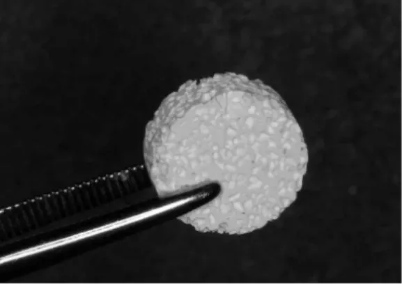
Figure 1. Block type MBCP implant used in this study.
the ability to predict the volume and shape of regenerated bone is essential.
To date, studies of such carriers include biological materials such as bone matrix10), absorbable collagen sponge (ACS)11-15), fibrin fibronectin sealing system(FFSS)16-18), syn- thetic polymers19,20), β-tricalcium phos- phate(TCP)11-13,18,21,22) and macroporous bi- phasic calcium phosphate (MBCP)23,24).
Although various carriers for rhBMPs have been investigated, there is as yet no ideal carrier system available. In many cas- es, volumetric reduction of the rhBMP-in- duced bone has been reported13,14).
MBCP is composed of 40% TCP and 60% hydroxyapatite (HA), which is consid- ered to be an ideal mixture ratio for bone substitutes26-28), and has been reported to have favorable osteoconductive and os- teoinductive properties26-31). Volumetric re- duction was reported to be considerably low due to its relatively low resorption rate27-28). Therefore, the block shape of the MBCP might be a suitable carrier for predictable bone formation in terms of the volumetric stability
The aim of this study was to determine the osteogenic effect of an MBCP block as a carrier system for rhBMP-2 using a rat calvarial defect model.
II. MATERIALS AND METHODS
1. Animals
Thirty-two male Sprague-Dawley rats (weight
250~300 g) were used. The rats were main- tained in plastic cages in a room with a 12 h-day/night cycle and an ambient temper- ature of 21°C, with access to water and standard laboratory pellets ad libitum.
Animal selection and management, surgical protocol, and preparation were in accordance with the routines approved by the Institutional Animal Care and Use Committee, Yonsei Medical Center, Seoul, Korea.
2. rhBMP-2 Implant Construction
Disc-shaped MBCP implants (Biomatlante, Vigneux de Bretagne, France) (3mm height and 8mm diameter) were manufactured.
rhBMP-2 (R&D Systems Inc., Minneapolis, MN ,USA) was reconstituted and diluted in a buffer to a concentration of 0.025 mg/ml.
For the rhBMP-2/MBCP implants, the MBCP implants were loaded with 0.2 ml of the rhBMP-2 solutions for one hour before surgery. For the MBCP implants, the MBCP implants were loaded with 0.2 ml of a buf- fer solution (Figure 1).
3. Surgical Procedures
The animals were anaesthetized by an in- tramuscular injection (5 mg/kg body wt.) of a 4:1 solution of ketamine hydrochloride Ketalar® (Yuhan Co.,Seoul, Korea):Xylazine, Rompun® (Bayer Korea, Seoul, Korea).
Routine infiltration anaesthesia, 2% lido- caine, 1:100,000 epinephrine (Kwangmyung Pharm., Seoul, Korea) was used at the surgi- cal site. An incision was made in the sag- ittal plane across the cranium and a full thickness flap was reflected to expose the calvarial bone. A standardized, circular, transosseous defect, 8 mm in diameter, was created on the cranium using a saline-cooled trephine drill (3i, Palm Beach Gardens, FL, USA). The animals were divided into 2 groups containing 16 animals each and al- lowed to heal for 2 (8 rats) or 8 weeks (8 rats). Each animal received 1 of the 2 ex- perimental treatments: MBCP carrier control and rhBMP-2/MBCP. The periosteum and skin were closed and sutured with an absorb- able monofilament suture, Monosyn® (Aesculap AG Co. KG., Tuttlingen, Germany) for primary intention healing.
4. Histology and Histometric Procedures
Two and 8 weeks after surgery, the ani- mals were sacrificed by CO2 asphyxiation . The block sections, including the ex- perimental sites, were removed and fixed in a 10% neutral buffered formalin solution for
10 days. The samples were decalcified using 5% formic acid for 14 days and embedded in paraffin. Serial sections, 7 μm thick, were prepared at 80 μm intervals, stained with hematoxylin/eosin (H-E), and examined by optical microscopy. The most central sec- tions from each block were selected for the histology and histometric evaluation.
The computer-assisted histometric meas- urements were obtained using an automated image analysis system coupled with a video camera attached to an optical microscope.
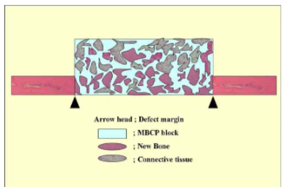
The sections were examined at magnifications of × 20 and × 100. The histometric parame- ters were defined as follows (Figure 2).
•Augmented area (mm2): all tissues with- in the boundaries of the MBCP carrier, i.e. new bone, fatty marrow, fi- brovascular tissue/marrow and residual biomaterial.
•New bone area (mm2): the area of new- ly formed bone within the total aug- mented area.
Figure 2. Schematic diagram of the calvarial osteotomy defect showing histometric analysis.
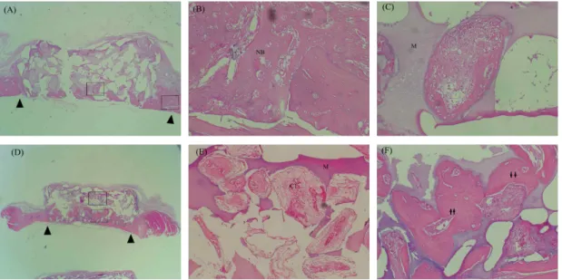
Figure 3. Representative photomicrographs of the MBCP control group at 2 (A,B and C) and 8 weeks (D,E and F) (arrow head: defect margin; arrow: cement lines, M;MBCP, NB: new bone, CT; connective tissue, H-E stain; original magnification A and D x20, B,C,E and F x100).
5. Statistical Analysis
The histometric recordings from the sam- ples were used to calculate the means and standard deviations (mean ± SD). The inter- actions between the healing interval and treatment condition were examined using a two-way analysis of variance (two-way ANOVA). A paired or unpaired t-test was used for the comparisons between the two groups. A p-value <0.05 was considered significant.
III. RESULTS
1. Histology Observations
1) MBCP control group (Figure 3).
At 2 weeks, there were generally no his- tological markers of inflammation or foreign
body reactions found. A small amount of new bone formation was observed adjacent to the margins of the defect and beneath the MBCP block. Evidence of osteogenic activ- ity, such as a dense osteoblast-like cell lin- ing, osteoid and bone apposition along the surface of the macropores in the lower part of the implant was observed. Macropores in the upper part were usually filled with loose fibrous connective tissue and the bone-form- ing activity was rare.
At 8 weeks, the majority of macropores in the lower part of the block were filled with newly formed bone. The specimens showed a more advanced stage of remodeling and consolidation. Loose or dense fibrous con- nective tissue was observed in the central and upper part of the block.
There was no significant resorption of the MBCP block observed during the healing time.
Figure 4. Representative photomicrographs of the rhBMP-2/MBCP group at 2 weeks (A,B and C) and 8 weeks (D,E and F) (arrow head: defect margin, arrow: cement lines, M;MBCP, NB:
new bone, CT; connective tissue, BM;bone marrow, H-E stain; original magnification A and D x20, B,C,E and F x100).
2) MBCP/ rhBMP-2 group (Figure 4).
At 2 weeks, the overall bone healing was similar to that of the MBCP control group.
The majority of macropores in the lower part of the block were filled with new bone.
Beneath the block, the defect sites were al- most completely bridged with new bone.
However, there was little bone forming ac- tivity in the central and upper part of the block. In the case of macropores with bone forming activity, new bone that was asso- ciated cement lines was in direct contact with the surface of the MBCP, and an os- teoblast-like cell lining was observed in the center area suggesting that bone formation and mineralization proceeded inwards from the implant surface.
At 8 weeks, the quantity of new bone be- neath the block and macropores in the lower part of the block was greater than that ob-
served at 2 weeks, and the specimens showed a more advanced stage of remodel- ing and consolidation. Even though some macropores with fibrous connective tissue was noted in the central and upper part of the block, macropores with newly formed bone could be also found.
The newly formed bone consisted of wo- ven and lamellar bone, and showed cement lines that were separated earlier from the more recently deposited bone. There was no evidence of cartilage formation. However, fatty marrow was observed in the central as- pect of the newly formed bone.
2. Histometric Analysis
Tables 1 and 2 show the results of histo- metric analysis.
The new bone area in the rhBMP-2/
2 weeks 8 weeks
MBCP 20.5 ± 3.5 20.3 ± 2.7
rhBMP-2/MBCP 22.4 ± 3.4 21.5 ± 1.8
No significant difference when compared to all groups (P>0.01)
2 weeks 8 weeks
MBCP 1.1 ± 0.6 3.0 ± 0.9*
rhBMP-2/MBCP 3.0 ± 0.9¶ 5.5 ± 2.2*
* : Statistically significant difference compared to 2 weeks (p < 0.01)
¶ : Statistically significant difference compared to MBCP group (p < 0.01)
Table-1. Total augmented area (group means ±SD, mm2, n=8)
MBCP group were significantly larger than in the MBCP group at each healing interval (p <0.01). In both groups, the quantity of the new bone was greater at 8 weeks than at 2 weeks (p <0.01). Two-way ANOVA re- vealed that the time had a significant effect on new bone formation in both groups (P<0.01). However, the pattern of the changes in bone healing over time was not significantly different.
IV. DISCUSSION
This study examined the osteogenic effect of a MBCP block as a carrier system for rhBMP-2 using a rat calvarial defect model.
The results after a 2- and 8-week of healing period were evaluated by the histology and histometric parameters . The rationale behind this was to utilize the favorable bone heal- ing capacity of MBCP block, which is known to have osteoconductive and os- teoinductive effect, and to have volumetric predictability over healing time because of low resorption rate and resistance of block
type against to soft tissue compression. It was expected that the rhBMP-2 using MBCP block would result in predictable new bone formation in terms of volume and shape.
rhBMPs have been shown to have potent osteoinductive activity4-6,13) and there have been many trials using rhBMPs in bone tis- sue engineering7-23). Hence, bone tissue en- gineering technology might be an alternative to autogenous bone graft. The results of bone tissue engineering using rhBMPs are fully dependent on the characteristics of the carrier systems as biomaterials. Therefore, many biomaterials have been evaluated as a carrier system for rhBMPs.
In this study, MBCP alone produced fa- vorable bone healing. Although limited bone formation was observed in the middle and upper part of the block, new bone was ob- served homogeneously in the lower part of the block. Almost complete defect closure beneath the block was observed and the ma- jority of the macropores in the lower part of the block were filled with newly formed
bone after 8 weeks in MBCP block alone group. These results are comparable to pre- vious studies using other biomaterials such as TCP granules11,12,21), FFSS17,18), and ACS9,11), where complete defect closure had not been achieved. Defect closure did not exceed 30-35% of the defect width after implanting these biomaterials. The favorable bone heal- ing effects of MBCP might explain the com- plete defect closure observed in this study .
The favorable osteoconductive effect of the MBCP in bone healing is well docu- mented25-28). It was suggested that higher lo- cal calcium and phosphate concentrations in MBCP might have had a stimulatory effect on bone formation. In addition to the osteo- conductive effect of MBCP, a there are many reports on the osteoinductive effect of MBCP when implanted in ectopic sites in various species29-32). Ectopic bone formation could be also observed using other type of synthetic bone substitute such as hydroxyapatite. However, MBCP was reported to have a higher osteoinductive potential29).
Synthetic biomaterials with a porous struc- ture such as MBCP have been reported to entrap rhBMPs within its pores, and intrinsi- cally diffusible rhBMPs can be retained and their activity prolonged22). It is believed that the porous structure of MBCP allows cells and newly formed tissues to migrate into the block. New bone formation in the macro- pores of the MBCP block in this study can explain this phenomenon. Ectopic bone for- mation around the MBCP was also reported when rhBMP were implanted with MBCP in
the rat subcutaneous model24). Therefore, the MBCP block could be recommended as a carrier for rhBMPs.
In a clinical point of view, a carrier for the delivery of BMPs should serve as a scaffold for bone forming cells while provid- ing a space in which bone formation can occur. In addition, the space should be maintained for a relatively long period in or- der to allow sufficient maturation of the newly formed bone. In terms of clinical bone tissue engineering, the provision of the volume and shape of the bone tissue is a key factor in treatment success.
When rhBMPs was impregnated with bio- materials as a carrier, some of which in- clude ACS11-13), ß-TCP11-13,18,21) and FFSS
17,18), they induced excellent new bone
formation. However, the amount of newly formed tissue was reduced at a later healing stage compared with at an earlier stage be- cause of soft tissue compression during the healing periods. When implanted at an ec- topic site such as a subcutaneous pouch, the new bone induced by rhBMP-2/ACS at two weeks could not be observed at eight weeks, while more expanded bone maturation was observed in the rhBMP-2/ β-TCP sites dur- ing that same period13). Moreover, ACS was not effective as a carrier for rhBMP-2 on ridge augmentation in chronic alveolar ridge defects in dogs, which was attributed to a non space-providing defect due to soft tissue compression during the healing period14). The lack of space-providing capacity of ACS was suggested to be a major factor re-
sponsible for its failure to maintain the new- ly induced bone. Although particle types such as ß-TCP can serve as an effective carrier for rhBMPs, there are questions as to whether clinical applications are possible be- cause the nature of the particulate form cre- ates difficulties regarding its manipulation for the intended shape of bone. Since pre- dictable bone tissue regeneration could be one of the major clinical therapeutic goals, a carrier system for rhBMPs needs to provide stability over the time in terms of volume and shape.
In this study, the total augmented area was stable during the healing periods in two groups. As reported earlier, MBCP is com- posed of 40% TCP and 60% HA26,27,30). The higher TCP content in MBCP suggests that it would be resorbed. However, there was no decrease in size or change in shape re- lated to resorption. Moreover, there was no active resorption phenomenon observed on histology findings at 8 weeks. It was re- ported that there was no significant re- sorption with decreases in size with a longer healing time using other species26,29). These results suggest that a MBCP block could serve as a carrier system for predictable bone tissue engineering using rhBMPs. In particular, the rhBMPs/MBCP block system is recommended for bone tissue reconstruction in oral and maxillofacial areas, in which a characteristic 3-dimensional topography and soft tissue compression during the healing periods are important consideration factors.
V. CONCLUSION
The block type of MBCP as a carrier for rhBMP-2 is effective in new bone formation.
The total augmented area induced by the rhBMPs/MBCP block system was stable dur- ing the observed healing period. These re- sults suggest that the rhBMPs/MBCP block system can be used for predictable bone tis- sue reconstruction
IV. REFERENCE
1. Thor A, Wannfors K, Sennerby L, Rasmusson L. Reconstruction of the se- verely resorbed maxilla with autogenous bone, platelet-rich plasma, and implants:
1-year results of a controlled prospective 5-year study. Clin Implant Dent Relat Res 2005;7:209-220.
2. Schwartz-Arad D, Levin L, Sigal L.
Surgical success of intraoral autogenous block onlay bone grafting for alveolar ridge augmentation. Implant Dent 2005;
14:131-138.
3. Cordaro L, Amade DS, Cordaro M.
Clinical results of alveolar ridge augmen- tation with mandibular block bone grafts in partially edentulous patients prior to implant placement. Clin Oral Implants Res 2002;13:103-111.
4. Urist MR. Bone Formation by autoinduc- tion. Science 1965;150:893-899.
5. Sampath TK, Maliakal JC, Hauschka PV.
Recombinant human osteogenic protein-1 (hOP-1) induces new bone formation in vivo with a specific activity comparable with natural bovine osteogenic protein and stimulates osteoblast proliferation and dif- ferentiation in vitro. J Biol Chem 1992;
267:20352-20362.
6. Wikesjo UM, Guglielmoni P, Promsudthi A, et al. Periodontal repair in dogs: effect of rhBMP-2 concentration on regeneration of alveolar bone and periodontal attachment. J Clin Periodontol 1999;26: 392-400.
7. Kim CS, Choi SH, Choi BK, et al. The effect of recombinant human bone mor- phogenetic protein-4 on the osteoblastic differentiation of mouse calvarial cells af- fected by Porphyromonas gingivalis. J Periodontol 2002;73:1126-1132.
8. Choi SH, Kim CK, Cho KS, et al. Effect of recombinant human bone morphoge- netic protein-2/absorbable collagen sponge (rhBMP-2/ACS) on healing in 3-wall in- trabony defects in dogs. J Periodontol 2002;73:63-72.
9. Hyun SJ, Choi SH, Chai JK, et al. The effect of recombinant human bone mor- phogenetic protein-2, 4 and 7 on bone formation in rat calvarial defects. J Periodontol 2005;76:1667-1674.
10. Yasko AW, Lane JM, Fellinger EJ, et al.
The healing of segmental bone defects, induced by recombinant human bone mor- phogenetic protein (rhBMP-2). J Bone Joint Surg 1992;74-A:659-671.
11. Ahn SH, Kim CS, Suk HJ, et al. Effect of Recombinant Human Bone Morphogenetic Protein- 4 with Carriers in Rat Calvarial Defects. J Periodontol 2003;74:787-797.
12. Pang EK, Im SU, Kim CS, et al. Effect of recombinant human bone morphoge- netic protein-4 dose on bone formation in rat calvarial defects. J Periodontol 2004;
75:1364-1370.
13. Kim CS, Kim JI, Kim J, et al. Ectopic bone formation associated with recombinant human bone morphogenetic protein-2 using absorbable collagen sponge and beta trical- cium phosphate as carriers. Biomaterials
2004;19:1-7.
14. Barboza EP, Duarte ME, Geolas L, et al.
Ridge augmentation following implantation of recombinant human bone morphoge- netic protein-2 in the dog. J Periodontol 2000;71:488-496.
15. Wikesjo UM, Guglielmoni P, Promsudthi A, et al. Periodontal repair in dogs: effect of rhBMP-2 concentration on regeneration of alveolar bone and periodontal attachment.
J Clin Periodontol 1999;26: 392-400.
16. Kawamura M and Urist MR. Human fi- brin is a physiologic delivery system for bone morphogenetic protein. Clin Orthop Rel Res 1988;235:302-310.
17. Han DK, Kim CS, Jung UW, et al.
Effect of a Fibrin-fibronectin sealing sys- tem as a carrier for recombinant human bone morphogenetic protein-4 on bone formation in rat calvarial defects. J Periodontol 2005 Dec;76:2216-2222.
18. Hong SJ, Kim CS, Han DK, et al. The Effect of a Fibrin-Fibronectin/ß-Tricalcium Phosphate/Recombinant Human Bone Morphogenetic Protein-2 System on Bone Formation in Rat Calvarial Defects.
Biomaterials 2006;27:3810-3816.
19. Miyamoto S, Takaoka K, Okada T, et al.
Polylactic acid-polyethylene glycol block copolymer: a new biodegradable synthetic carrier for bone morphogenetic proteins.
Clin Orthop Rel Res 1993;294:333-343.
20. Miki T, Imai Y. Osteoinductive potential of freeze-derived, biodegredable, poly(gly- colic acid-co-lactic acid) disks in- corporated with bone morphogenetic pro- tein in skull defects in rats. J Oral Maxillofac Surg 1996;25:402-426.
21. Jung UW, Choi SY, Pang EK, et al. The Effect of varying the Particle Size of Beta Tricalcium Phosphate carrier of Recombinant Human Bone Morphogenetic Protein-4 on
Bone Formation in Rat Calvarial Defects.
J Periodontol 2006;77: 765-772
22. Barnes B, Boden SD, Louis-Ugbo J, et al. Lower dose of rhBMP-2 achieves spine fusion when combined with an os- teoconductive bulking agent in non-human primates. Spine 2005;30:1127-1133.
23. Oda S, Kinoshita A, Higuchi T, et al.
Ectopic bone formation by biphasic cal- cium phosphate (BCP) combined with re- combinant human bone morphogenetic protein-2 (rhBMP-2). J Med Dent Sci 1997;44:53-62.
24. Nery EB, LeGeros RZ, Lynch KL, Lee K. Tissue response to biphasic calcium phosphate ceramic with different ratios of HA/beta TCP in periodontal osseous defects. J Periodontol 1992 ;63:729-735.
25. Schopper C, Ziya-Ghazvini F, Goriwoda W, et al. HA/TCP compounding of a po- rous CaP biomaterial improves bone for- mation and scaffold degradation--a long- term histological study. J Biomed Mater Res B Appl Biomater 2005;74: 458-467.
26. Cornell CN, Lane JM. Current understanding of osteoconduction in bone regeneration.
Clin Orthop Relat Res 1998;355 Suppl:
S267-273.
27. Gauthier O, Bouler JM, Aguado E, Pilet P, Daculsi G. Macroporous biphasic cal- cium phosphate ceramics: influence of macropore diameter and macroporosity percentage on bone ingrowth. Biomaterials 1998;19:133-139.
28. Yuan H, van Blitterswijk CA, de Groot K, de Bruijn JD. A comparison of bone formation in biphasic calcium phosphate (BCP) and hydroxyapatite (HA) implanted in muscle and bone of dogs at different time periods. J Biomed Mater Res A 2006;78:139-147.
29. Le Nihouannen D, Daculsi G, Saffarzadeh A, et al. Ectopic bone formation by mi- croporous calcium phosphate ceramic par- ticles in sheep muscles. Bone 2005;36:
1086-1093.
30. Habibovic P, Yuan H, van den Doel M, et al. Relevance of osteoinductive bio- materials in critical-sized orthotopic defect.
J Orthop Res 2006 ;24:867-876.
31. Manjubala I, Sastry TP, Kumar RV. Bone in-growth induced by biphasic calcium phosphate ceramic in femoral defect of dogs. J Biomater Appl 2005;19:341-360.
- 국문초록 -
백서 두개골 결손부에서 bone morphogenetic protein-2의 전달체로서 macroporous biphasic
calcium phosphate-block의 골재생효과
이용준, 정성원, 채경준, 조규성, 김창성* 연세대학교 치과대학 치주과학교실, 치주조직재생연구소
골형성 유도 단백질(bone morphogenetic protein, BMP)은 성장이나, 골형성과정에서 중용 한 역할을 한다고 입증되었고 그것의 운반체에 대한 연구가 이뤄져 왔다. 하지만 수직압 이 존재하는 곳에서 골증대술에 적용할 수 있을 만큼 강한 공간유지능력이 있는 운반체 에 대한 연구는 그리 많지 않았다. Macroporous biphasic calcium phosphate block (MBCP block)은 공간유지능력이 뛰어나며 강한 수직압을 견딜수 있는 골대체물질이다. 이 연구 의 목적은 MBCP block을 골형성유도 단백질(rhBMP-2)의 운반체로 사용하여 백서 두개골 결손부에 적용하였을 때, 골 형성 효과를 평가하는 것이다.
36마리의 웅성백서에서 8mm 지름을 갖는 임계크기의 두개부 결손을 형성하였다. 20마 리씩 2개의 군으로 나누어 MBCP block만 이식한 군, MBCP block을 운반체로 사용하여 농도 0.025mg/ml rhBMP-2를 이식한 군으로 나누어 술 후 2주와 8주에 치유결과를 조직학 적, 조직계측학적으로 비교 관찰 하였다.
조직계측학적 관찰 결과, rhBMP-2/MBCP block 군에서 MBCP block군에서 보다 2, 8주 모두 골밀도(bone density)가 유의성있게 증가하였다 (P<0.01). 각 군에서도 8주째가 2주째 보다 골밀도가 유의성있게 증가하였다(P<0.01). 총조직 형성량 (augmented area)에서는 변 화가 없었다. 이 연구 결과, 백서 두개골 결손부에서 MBCP block은 rhBMP의 운반체로 사용하였을 때 신생골 형성에 유의한 효과가 있을뿐 아니라 공간유지능력이 우수해서 수 직압이 존재하는 골증대술(bone augmentation)시 rhBMP의 운반체로 가능성이 있다고 사료 된다.2)
Key worlds: 골형성 유도 단백질, Macroporous biphasic calcium phosphate block, 골증대술, 백서두개골 결손부