대한치주과학회지 : Vol. 35, No. 2, 2005
무기인산염이 골유도재생에 미치는 영향
정종혁 ․ 권영혁 ․ 박준봉 ․ 허 익 경희대학교 치과대학 치주과학교실
I. 서론
40)임플란트를 이용한 치료가 보편화되고 심미적인 치료에 대한 환자의 요구가 증가하면서 치조골의 결 손은 치료의 결과를 결정하는 중요한 요소가 되었다.
상실된 치조골의 재생을 통해 임플란트의 성공률을 높이고 보다 심미적인 치료가 가능하게 하기위해 다 양한 재료와 수술방법들을 이용한 치조골재생술식이 널리 이용되고 있다.
이러한 술식들 중 골유도재생술은 차폐막이 상방 연조직세포의 골 결손부로의 이주를 차단함으로써 하부 잔존골로부터 골세포의 분화 및 증식을 유도하 여 골조직을 재생시키는 술식1)으로, 차폐막은 골 결 손부 내의 혈병을 보호하며 상부연조직으로부터의 외력을 차단함으로써 창상을 고정하여 골조직의 재 생을 촉진시킨다.2-6) 골유도재생술은 다양한 형태의 골결손부, 치조제증대술 및 임플란트의 식립과 연관 된 골결손부 등에 널리 사용되고 있다.7-16)
골유도재생술에 사용되는 차폐막이 갖추어야 할
조건은 인체에 무해하고 조직친화성이 있어야 하며 치은결합조직의 세포유입을 차단할 수 있는 세포차 단성이 있어야 하고 골재생에 관여하는 세포가 이주, 증식할 수 있는 재생공간의 확보와 유지를 할 수 있 는 물리적 성질이 있어야 한다.1,17-20)재생공간 확보 및 유지를 위하여 소형 스크류나 핀을 사용하여 차 폐막을 기저골에 고정시키거나,21) 기계적 강도가 부 족한 점을 보완하고자 티타늄을 보강한 티타늄강화 차폐막(titanium-reinforced expanded poly- tetrafluoroethylene; TR-ePTFE)막을 사용하기 도 하였다.22-24) Jovanovic 등(1995)22)은 5마리의 성견의 연구에서 티타늄강화차폐막 사용시 6개월의 치유기간후 수직적으로 4mm 정도의 골재생을 보고 하였는데, 부가적인 골이식재의 사용없이도 큰 공간 을 잘 유지할 수 있으며 이전의 ePTFE막과 비교시 더 많은 양의 골을 형성할 수 있다고 제안하였다.
차폐막 하방의 재생공간을 확보하고 유지하기 위 한 방법으로 여러 골이식재 등을 함께 사용하기도 하며 자가골, 탈회동결건조골, 동결건조골, 탈단백우 골 및 합성골 등이 이용되고 있다.10,17-20,25-32)이 중
*본 연구는 보건복지부 보건의료기술 진흥사업의 지원에 의하여 이루어진 것임(03-PJ1-PG1-CH08-0001).
교신저자 : 권영혁, 서울시 동대문구 회기동 1번지 경희대학교 치과대학 치주과학교실 우편번호: 130-702 E mail: [email protected]
대표적인 이종골이식재인 탈단백우골은 사람과 같은 칼슘/인 비율을 갖는 천연 인회석결정체로 이루어져 있고 비교적 낮은 온도인 300℃에서 화학적 과정을 거쳐 골의 유기질 성분을 제거하기 때문에 합성골과 달리 골의 본래의 다공성 구조를 그대로 유지하는 특징을 갖는다.33,34)또한 탈단백우골은 다른 골이식 재에 비해 5배 이상의 높은 표면적을 보이며 수 많 은 결정체 공간과 미세터널을 갖기 때문에 혈관과 골의 성장을 증가시켜 골유착을 일으키는 것으로 보 고되고 있으며35) 생체친화성이 있으며,36,37) 골형성 의 비계로 작용하는 골전도성이 있는 것으로 알려져 있다.38,39)
탈단백우골이 골형성에 미치는 영향에 대해서는 골조직의 재생을 증가시키지 못한다는 보고들도 있
고,40,41) 오히려 골재생을 방해한다는 부정적인 보고
42-45)들도 있는데, Slotte과 Lundgren(1999)45)은 쥐의 두개골에 탈단백우골을 이식한 결과, 탈단백우 골이 초기 골형성을 방해한다고 하였다.
이러한 골이식재들은 골채취를 위한 추가적인 수 술의 필요, 이식편의 빠른 흡수, 골형성능의 부족, 감염의 가능성 및 면역반응 등의 문제점을 나타내어 다른 물질들에 대한 연구가 진행되고 있다. 그 중 성 장인자들이 골형성과 재생에 관여한다는 연구들을 통해 인슐린유사성장인자(IGF), 변형성장인자(T- GF-β), 그리고 골형태형성단백질-2(BMP-2)와 같 은 성장인자들이 주목을 받아왔으며 골아세포의 분 화를 유도하고 증식을 조절하는 것으로 보고되었
다.46,47) 그러나 이러한 성장인자들은 골유도효과는
인정되나 체내에서의 불활성화, 고가의 비용 등의 단 점을 가지고 있으며 성장인자를 대체할 수 있는 다루 기 쉽고, 생물학적인 안정성을 가지고, 안전하며, 저 비용의 골유도 물질에 대한 연구가 필요한 상황이다.
무기인산염(Inorganic polyphosphate, PolyP) 은 (NaPO3)n의 분자식을 갖는 인체에 무해한 물질
로서,48,49) 수십 또는 수백개의 orthophosphate
(Pi) residues가 고에너지의 phosphoanhydride 결합을 형성하고 있는 중합체이다.50) 무기인산염은 미생물내에서 ATP의 대체물질로서 에너지원으로
작용하고 인산(Pi)의 저장고 역할을 하며 적은 농도 의 무기인산염이 동물세포에서 관찰되기도 하지만 포유동물에서의 무기인산염의 기능은 잘 알려져 있 지 않다.51,52) Fleisch 등(1997)53)은 인(phospho- rous)의 합성물질인 bisphosphonate가 골흡수를 막는 강력한 억제물질이라고 보고하였고, 많은 양의 무기인산염(Inorganic polyphosphate)이 human mandibular-derived osteoblast-like cell에서 관찰되고 골광물화의 조절자로 작용한다는 보고도 있으며,54) Shiba등(2003)55)은 무기인산염이 섬유 아세포(human fibroblast cell)의 증식에 관여한 다고 하였다.
이러한 연구들을 바탕으로 이번 연구는 웅성백묘 두개관에 피질골을 제거하고 그 상방에 티타늄강화차 폐막(TR-ePTFE)과 탈단백우골, 그리고 무기인산염 을 이용하여 골유도재생술을 시행하고 새로 형성된 골을 조직학적으로 분석함으로써 무기인산염이 골재 생에 미치는 영향을 알아보기 위하여 시행하였다.
II. 연구재료 및 방법
1. 연구재료
차폐막으로는 티타늄강화차폐막(TR-9W, W.L.
Gore & Associate. inc, USA)을 사용하였다.
25×20mm 크기의 티타늄강화차폐막을 내면이 가로, 세로 각각 10mm, 높이 2mm인 직육면체로 만들어 ethylene oxide로 소독한 후 골유도재생술에 사용 하여 필요한 공간을 확보할 수 있도록 하였다. 차폐 막 하방에 적용한 골이식재로는 0.25-1mm의 입자 크기를 갖는 소의 망상골로 만든 탈단백우골(Bio- Oss® spongiosa, Geistlich AG, Wolhusen, S- witzerland)을 사용하였다. 무기인산염은 Poly P75(Sodium polyphosphate, Sigma, St.
Louis, MO, U.S.A.)를 사용하였다.
티타늄강화차폐막만 사용한 군을 대조군 I, 탈단 백우골와 티타늄강화차폐막을 사용한 군은 대조군
티타늄 강화차폐막 탈단백우골 1% 무기인산염 2% 무기인산염
대조군 I ○
대조군 II ○ ○
실험군 III ○ ○ ○
실험군 IV ○ ○ ○
Table 1.
II, 1% 무기인산염에 침적한 탈단백우골과 티타늄 강화차폐막을 사용한 군을 실험군 III, 그리고 2%의 무기인산염에 침적한 탈단백우골과 티타늄강화차폐 막을 사용한 군을 실험군 IV로 분류하였다.
2. 연구대상
생후 6주된 평균체중 2kg의 웅성백묘(Newzea- land white rabbit)를 각 군마다 3마리씩 12마리 사용하였으며 실험기간 동안 고형사료(축협사료, 축 산업협동조합, 한국)를 공급하였고 분리된 실내 사육 실에서 사육하였다. 각 군마다 3마리씩 배정하였다.
3. 연구방법
1) 시술과정
실험동물에 졸레틸(Virbac, France, 0.2ml/ Kg) 0.5ml를 근육주사하여 전신마취하고, 수술부위는 리 도케인 2%(1:1,000,000 에피네프린) 0.5ml로 국소 마취하였다. 두개면 수술부위의 모발을 제거한 후
#15 수술도를 이용하여 시상방향으로 절개, 전층판막 을 거상하였다. 주수하에 직경 6.5mm 트레핀 버로 두개관에 약 1-2mm깊이로 경계부를 형성하였다. 골 내출혈을 유도하기 위해 트레핀 버로 형성한 경계부 까지를 라운드 카바이드 버(HP long #6)로 주수하 에 약 1-2mm깊이로 피질골을 제거하였다. 골내출혈 확인 후 직육면체로 구부린 티타늄강화차폐막 내부에 생리식염수나 무기인산염에 침적한 탈단백우골을 채 우고 골막봉합으로 고정하였다. 이때 무기인산염에 침적한 경우에는 20분간 침적후 5분간 건조시켜서 사
용하였다. 흡수성 봉합사(4-0 chromic gut)를 이용 하여 골막과 표피를 함께 봉합하였다.
2) 술후 처치
세균감염을 방지하기 위하여 수술 당일과 술 후 2 일까지 gentamycin(동화약품, 한국) 1ml를 근육 주사 하였다.
3) 조직준비 및 분석
실험동물들은 술 후 2, 4, 8주에 각각 과량의 졸 레틸을 근육주사하고 이산화탄소로 질식사시킨 후 두 피를 거상하고 차폐막과 하방 두개골을 포함하는 조 직시편을 채득하였다. 두개골의 칼슘성분이 있는 조 직을 탈회시킨 표본을 만들기 위하여 절반은 탈회표 본을, 나머지 절반은 골의 조직학적 관찰을 위해 비 탈회표본을 제작하고자 하였다. 시편을 4% paraf- ormaldehyde 용액에 고정하고, 절반은 비탈회조직 표본 제작법에 따라 70% 알코올에 1일간 담가둔 다 음, Villanueva bone stain용액에 3일간 침적시키 고 통법에 따라 에탄올에 단계별로 탈수시켰다.
Methyl methacrylate monomer(MMA mono- mer, Junsei, Japan)를 침투시킨 시편을 MMA monomer, MMA polymer(Wako, Japan)와 benzoyl peroxide를 혼합하여 만든 레진으로 포매 하였다. 시편은 경조직절단기(Maruto Co., Japan) 를 이용하여 500μm 두께로 절단한 후, 경조직 연마 기(Maruto Co., Japan)로 80μm의 절편을 얻어 광학현미경으로 관찰하였다. 나머지 절반은 탈회조직 표본 제작법에 따라 처리한 후 Hematoxylin과 Eosin 중염색을 시행하였으며 또한 항체처리한
PCNA(proliferating cell nuclear antigen)를 avidin-biotin complex method를 사용하여 면역 세포화학적 염색하여 분석하였다.
III. 연구 성적
1. 대조군 I
1) 수술 2주
차폐막은 잘 유지되었으며 차폐막하방으로 미세한 신생조직의 형성이 관찰되었다(Figure.1). 막변연 의 직하방부위에 있는 원형경계부에서부터 소량의 신생골의 형성이 관찰되며 주위에는 소성결합조직들 이 둘러싸고 있었다(Figure.2,2a).
2) 수술 4주
2주군과 유사하게 소량의 신생골 형성이 기존골 표면위에서 관찰되나 막에 의해 형성된 공간은 대부 분 소성결합조직으로 채워져 있었다(Figure.3, 3a).
3) 수술 8주
비탈회조직표본에서 골원(osteon)이 막변연부위 에서 관찰되며 이것을 중심으로 신생골이 형성되는 것을 관찰할 수 있었다(Figure.4,4a).
2. 대조군 II
1) 수술 2주
차폐막하방으로 탈단백우골과 이것을 둘러싸고 있 는 결합조직이 관찰되나 신생골의 형성은 보이지 않 았다(Figure.5,5a).
2) 수술 4주
비탈회조직표본에서 탈단백우골이 차폐막하방 공 간의 대부분을 채우고 있었고 주위는 결합조직으로 둘러싸여져 있었다 2주군에 비하여 뚜렷하게 증가된 신생골형성을 관찰할 수 없었다(Figure.6). 탈회조
직표본에서는 미세한 신생골 형성을 결손부의 기저 부에서 볼 수 있었으나 대부분의 공간은 결합조직으 로 채워져 있는 양상을 보였다(Figure.7,7a).
3) 수술 8주
비탈회조직표본과 탈회조직표본 모두에서 뚜렷한 골생성을 관찰할 수 없었으며 탈단백우골은 흡수되 지 않고 유지되었고 탈단백우골 주위로 치밀한 결합 조직의 소견이 관찰되었다(Figure.8,9).
3. 실험군 III
1) 수술 2주
비탈회조직표본에서 탈단백우골이 차폐막의 하방 공간을 채우고 있으며 신생골형성은 관찰되지 않았 다(Figure.10, 10a).
2) 수술 4주
탈회조직표본에서 차폐막하방의 대부분의 공간은 치밀한 결합조직으로 채워져 있었으며 골아세포와 유사한 세포(osteoblast like cell)들이 침윤되어 있고 미약한 신생골형성을 변연부에서 관찰할 수 있 었다. 또한 염증세포의 침윤이 전반적으로 분포되어 있는 양상을 보였다(Figure.11,11a).
3) 수술 8주
탈단백우골은 흡수되지 않고 남아있었으며 기저부 에서부터 신생골이 형성되는 모습을 관찰할 수 있었 다. 4주군에 비해 골형성이 증가되는 양상을 보였으나 차폐막하방의 대부분의 공간은 탈단백우골과 치밀한 결합조직에 의해 채워져 있었다(Figure.12, 12a).
4. 실험군 IV
1) 수술 2주
비탈회조직표본에서 탈단백우골이 차폐막하방의 공간을 채우고 있는 것을 관찰할 수 있으며 실험군
Ⅲ과 별다른 차이를 관찰할 수 없었다. 차폐막과 골 경계부하방으로 연조직의 침투가 보였다(Figure.
13, 13a, 13b).
2) 수술 4주
비탈회조직표본에서 골결손부의 기저부에서부터 골이 형성되는 양상을 보이나 2주군에 비하여 뚜렷 하게 증가된 양상은 관찰할 수 없었다. 대부분의 공 간은 탈단백우골로 채워져 있는 모습이 관찰되었다 (Figure.14, 14a).
3) 수술 8주
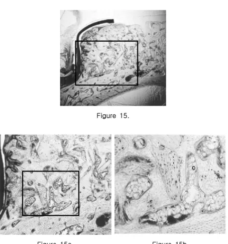
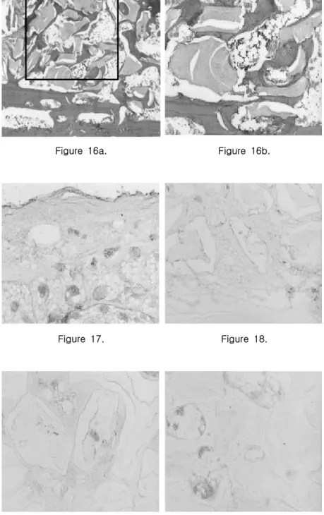
비탈회조직표본에서 막변연부위에서 차폐막과 하 방골의 긴밀한 접촉을 관찰할 수 있었고 4주군에 비 하여 골이식재 주위로 신생골의 형성이 뚜렷이 증가 된 것을 볼 수 있었다. 또한 골아세포가 뚜렷이 증가 되어 전반적으로 분포되어 있는 양상을 보였다 (Figure.15, 15a, 15b). 탈회조직표본에서도 신생 골의 형성이 4주군에 비하여 현저히 증가된 양상을 볼 수 있었으며 골수의 형성이 관찰되었다. 전반적 으로 분포하고 있는 신생골이 서로 융합하여 개조되 는 양상을 보였다(Figure.16, 16a, 16b).
5. 면역세포화학적 분석
4군 모두에서 특이적 염색반응이 일어나지 않았 다(Figure.17-20).
IV. 총괄 및 고찰
Schenk등(1994)56)은 비글견을 이용한 실험을 통해 골재생 및 형성과정을 시기에 따라 일차해면골 이 형성되는 첫 번째 단계, 일차해면골이 해면골과 치밀골로 변환되는 두 번째 단계, 그리고 형성된 골 이 개조되는 세 번째 단계의 세 단계로 분류하였는 데 첫 번째 단계는 약 8주경에 이루어지지만 두 번 째 단계는 16주에도 완전히 이루어지지 않는다고 하 였다. 이를 근거로 이번 실험에서는 골재생과 형성
과정을 8주까지 비교, 관찰하였다.
골유도재생술을 시행한 부위는 웅성백묘의 두개관 을 이용하였는데 두개관은 형태학적으로나 발생학적 으로 막성골이라는 점에서 안면의 막성골들과 유사성 을 가지며 해부학적으로 두개관은 중간에 해면골이 개재되어 있는 두 개의 피질골판으로 이루어져 있다 는 점에서 하악골과 유사하고 생리학적으로 두개관내 의 피질골은 퇴축된 하악골과 닮았다고 할 수 있다.57) 이러한 이유로 두개관을 이 실험에서 수술부위로 사 용하였다. 이번 실험에서 1-2 mm 깊이로 피질골을 제거한 것은 골수로부터 유래된 혈액공급 및 골형성 인자의 작용을 촉진하기 위하여58) 시행하였다. 또한 주변이 골로 둘러싸인 와동형의 골결손부는 주위 골 로부터 활발한 골재생이 골이식재의 골형성 효과를 구분하기 어렵게 하기 때문에59) 이번 실험에서는 기 존 골의 외부로 골형성을 유도하는 골결손부를 만들 어 다른 골재생에 관여하는 요소들을 배제한 상태에 서 골재생에 미치는 효과를 살펴보려고 하였다.
이번 실험에서 실험기간동안 티타늄강화차폐막에 의하여 형성되는 재생공간의 유지는 다른 연구결과
21-23,60,61)와 동일하게 전 기간동안 잘 이루어졌으며
염증반응이 없었던 것으로 볼 때 조직친화성도 좋은 것으로 나타났다.
차폐막 하방의 골충전재로 사용한 탈단백우골의 효과에 대해서는 상반된 연구들이 보고되고 있는데 Hämmerle등(1997)15)은 임플란트의 골결손부위에 탈단백우골을 비흡수성차폐막과 함께 사용한 결과 탈단백우골과 신생골의 접촉도는 80% 이상으로 탈 단백우골의 우수한 골전도성을 보고한 반면, Carma- gnola등(2002)42) (2003)43)은 개의 하악 과 사람의 발치창에 탈단백우골을 이식한 각각의 실 험에서 실험군의 신생골 형성이 골이식재를 사용하 지 않은 대조군에 비하여 현저히 적으며 중심부의 이식재는 결합조직에 둘러싸여 있음을 보고하였다.
Stavropoulos 등(2003)44)도 쥐의 하악지에 반구 형태의 테프론막과 탈단백우골을 함께 위치시키고 1 년 후 희생시켜 조직검사시 차폐막만 위치시킨 경우 에는 88%의 골형성을 보인 반면 탈단백우골을 함께
적용시킨 경우에는 23%의 골형성을 보였다고 하였 다. 또한 결합조직이 탈단백우골을 둘러싸고 있고 이식재의 흡수소견은 없으며 결합조직이 골로 전환 되는 소견도 관찰되지 않으며 이식재가 오히려 골형 성을 방해한다고 주장하였다. 인 등(2004)62)의 연 구에서도 탈단백우골은 골재생을 촉진시키지 않으며 오히려 골아세포로 분화될 세포의 증식을 방해한다 고 하였으며, 이번 실험에서도 탈단백우골은 충전재 의 역할을 할 뿐 골형성을 증가시키지 못하는 것으 로 나타났다.
차폐막의 고정은 골재생을 유도하는 중요한 요인 으로서 차폐막의 미세동요를 방지하기 위하여 고정용 핀을 사용하기도 한다.21)고정용 핀의 사용시 기저골 의 표면을 따라 형성될 수 있는 연조직의 침투를 방 지할 수 있으나 이번 실험에서는 골막봉합으로 차폐 막을 고정하여 차폐막과 기저골과의 적합이 충분하지 않았던 것으로 사료되어 차폐막의 동요로 인한 혈병 의 파괴와 주위 연조직 침투가 골형성에 영향을 미쳤 을 것으로 생각된다. Fugazzotto(2003)63)는 사람 에서 13개월이내에 탈단백우골의 대부분이 흡수되며 흡수를 방해하는 요인으로는 차폐막의 불완전한 고 정으로 혈병의 파괴, 연조직 침투 및 형성을 허용하 여 이식재의 흡수를 방해하는 경우와 이식재를 너무 치밀하게 채워 이식재의 소주구조가 부서져 이식재 의 흡수가 변연에서만 일어나는 경우를 제시하였다.
이번 실험에서의 차폐막의 불완전한 고정이 이식재 의 흡수를 방해했을 가능성도 배제할 수 없다.
골이식재를 대체할 수 있는 물질들 중 성장인자, 특 히 골형태형성단백질-2가 골유도를 촉진하는 것으로 보고되고 있으나,46,47) 골형태형성단백질-2는 원하지 않는 방향으로의 이소골형성을 야기할 수 있고, 흡수 가 빨리 일어나며, 체내에서 20-30분 후에 불활성화 되고, 또한 임상에서 사용하기에는 고비용이라는 단점 을 가지고 있다.46,64-66) 무기인산염에 대한 연구 중 Leyhausen 등(1998)54)은 많은 양의 무기인산염 (Inorganic polyphosphate)이 human mandi- bular-derived osteoblast-like cell에서 관찰되며 골광물화의 조절자로 작용하는 것으로 추측된다고 하
였다. 또한 Kawazoe 등(2004)67)은 쥐를 이용한 세 포실험을 통해 무기인산염이 골아세포 분화의 표지 (Marker)인 osteopontin과 osteocalcin의 발현을 증가시키고 alkaline phosphatase activity도 증가 시켰다고 하였으며, 무기인산염이 polyphosphatase 의 활성을 증가시킴으로써 세포의 골화를 촉진시킨다 고 보고하였다.
이 등(1999)68)은 토끼의 대퇴골에 교원질과 탈염 골분(demineralized human bone powder)을 매개 체로 2% 무기인산염의 효과에 관한 실험을 통해, 무 기인산염이 매개체의 골전도능을 향상시키거나 손상된 골조직의 골유도능을 증가시킴으로써 골재생을 촉진한 다고 보고하였다. 이번 실험에서는 무기인산염의 농도 에 따른 골재생 효과의 차이를 알아보고자 1%와 2%
로 농도를 달리하여 사용하였으며 1%의 무기인산염을 사용한 군에서는 별다른 골재생 효과를 관찰할 수 없 었으나 2%의 무기인산염 사용군에서는 뚜렷한 골재생 이 나타났다. 이는 무기인산염이 골세포나 골전구세포 들의 골결손부로의 이주를 유도하여 나타나는 것으로 생각할 수 있으며 이 등(1999)68)의 연구결과와 일치 한다. 골형성에 필요한 세포들의 골결손부로의 이주는 골유도에 있어서 중요한 과정이며69)무기인산염의 이 러한 기능을 고려할 때 무기인산염은 임상에 적용 가 능한 뛰어난 골유도물질 중의 하나라고 할 수 있다.
미분화세포의 기원과 조직 내에 존재하는 물질을 확인하기 위하여 PCNA(proliferating cell nu- clear antigen)를 이용한 면역세포화학적분석을 시 행하였으나 4군 모두에서 특이적 염색반응이 일어나 지 않았다.
무기인산염은 골재생을 증가시키는 것으로 보이지 만 그 정확한 기전에 대해서는 추가적인 연구가 필 요하며, 또한 차후에는 골유도재생술시 사용되는 티 타늄강화차폐막을 고정핀으로 고정하여 차폐막의 동 요로 인하여 야기될 수 있는 요인들을 제거해야 할 것으로 생각한다. 이러한 상태에서 무기인산염의 농 도를 증가시키고 다양한 매개체를 이용하여 골재생 에 미치는 효과를 관찰하는 연구가 계속적으로 이루 어져야 할 것으로 사료된다.
V. 결론
이번 연구는 무기인산염이 골재생 과정에 미치는 영향을 평가하기 위하여 생후 6주된 평균무게 2kg 의 웅성백묘 두개관에 일정한 크기로 피질골을 제거 한 후, 그 상방에 1%, 2%의 무기인산염에 침적한 탈단백우골을 채운 티타늄강화차폐막을 이용하여 골 유도재생술을 시행하였다. 티타늄강화차폐막만 사용 한 군을 대조군 I, 탈단백우골와 티타늄강화차폐막 을 사용한 군은 대조군 II, 1% 무기인산염에 침적한 탈단백우골과 티타늄강화차폐막을 사용한 군을 실험 군 III, 그리고 2%의 무기인산염에 침적한 탈단백우 골과 티타늄강화차폐막을 사용한 군을 실험군 IV로 분류하였다. 술 후 2, 4, 8주에 각각 희생시켜 차폐 막을 포함한 조직절편을 채취하여 통법에 따라 비탈 회표본과 탈회표본을 제작하고 조직학적 및 면역세 포화학적으로 비교, 관찰하여 다음과 같은 결론을 얻었다.
1. 티타늄강화차폐막은 공간유지능력과 세포차단성 이 우수하였으나 창상고정능력이 좋지 않았다.
2. 탈단백우골은 골재생을 촉진시키지 않았으나 공간충전재의 역할은 있었다.
3. 8주내에 탈단백우골은 완전히 흡수되지 않았다.
4. 1%의 무기인산염은 골형성을 증가시키지 않 았으나 2%의 무기인산염은 골재생을 증가시 켰다.
이상의 결론을 토대로 2%의 무기인산염은 골재 생에 효과가 있는 것으로 생각된다.
VI. 참고 문헌
1. Dahlin C, Linde A, Gottlow J, Nyman S.
Healing of bone defects by guided tissue regeneration. Plastic Reconst Sur 1988;
81:672-677.
2. Dahlin C, Alberius P, Lindhe A. Osteo-
promotion for cranioplasty: An ex- perimental study in rats using a mem- brane technique. J Neurosurg 1991;74 :487-497.
3. Dahlin C, Andersson L, Lindhe A. Bone augmentation at fenestrated implants by an osteopromotive membrane technique:
A controlled clinical study. Clin Oral Impl Res 1991;2:159-165.
4. Dahlin C, Sennerby L, Leckholm U, Lindhe A, Nyman S. Generation of new bone around titanium implants using a membrane technique: An experimental study in rabbits. Int J Oral Maxillofac Implants 1989;4:19-25.
5. Dahlin C, Gottlow J, Lindhe A, Nyman S.
Healing of maxillary and mandibular bone defects using a membrane technique: An experimental study in monkeys. Scand J Plast Reconstr Surg 1990;24: 13-19.
6. Becker W, Dahlin C, Becker BE. The use of e-PTFE barrier membranes for bone promotion around titanium im- plants placed into extraction socket: A prospective multicenter study. Int J Oral Maxillofac Implants 1994;9:31-40.
7. Siebert J, Nyman S. Localized ridge aug- mentation in dogs: A pilot study using membranes and hydroxyapatite. J Perio- dontol 1990;61:157-165.
8. Becker W, Becker B, Handlesman M, Celletti R, Ochsenbein C, Hardwick R, Langer B. Bone formation at dehisced dental implant sites treated with im- plant augmentation material: A pilot study in dogs. Int J Periodont Rest Dent 1990; 10:93-101.
9. Werrer K, Gotfredsen K, HjrtingHan-
sen E, Karring T. Guided tissue re- generation of dental implants placed in- to extraction sockets: An experimental study in monkeys. Clin Oral Impl Res 1991;2:166-171.
10. Jovanovic SA, Spiekermann H, Richter EJ. Bone regeneration around titanium implants in dehisced defect sites: A clinical study. Int J Oral Maxillofac Implants 1992;7:233-245.
11. Becker W, Becker B, Handelsman M, Ochsenbein C, Albrektsson T. Guided tissue regeneration for implant placed into extraction sockets: A study in dogs.
J Periodontol 1991;62: 703-709.
12. Bragger U, Hammerle CHF, Lang NP.
Immediate transmucosal implants using the principle of guided tissue regeneration II: A cross-sectional study comparing the clinical outcome 1 year after immediate to standard implant placement. Clin Oral Impl Res 1997;7: 268-276.
13. Buser D, Bragger U, Land NP, Nyman S. Regeneration and enlargement of jaw bone using guided tissue regeneration.
Clin Oral Impl Res 1990;1:22-32.
14. Dahlin C, Lekholm U, Becker W, Becker B, Higuchi K, Callens A, van Steen- berghe D. Treatment of fenestration and dehiscence bone defects around oral im- plants using the guided tissue regenera- tion technique: A prospective multi- center study. Int J Oral Maxillofac Im- plants 1995;10:312-318.
15. Hämmerle CHF, Olah AJ, Schmid J, Flückiger L, Gogolewski S, Winkler JR, Lang NP. The biological effect of de- proteinized bovine bone on bone neo-
formation on the rabbit skull. Clin Oral Impl Res 1997;8: 198-207.
16. Lang NP, Hämmerle CHF, Bragger U, Lehmann B, Nyman SR. Guided tissue regeneration in jaw bone defects prior to implant placement. Clin Oral Impl Res 1994;5:92-97.
17. Donos N, Kostopoulos L, Karring T.
Augmentation of the rat jaw with auto- genic cortico-cancellous bone grafts and guided tissue regeneration. Clin Oral Impl Res 2002;13:192-202.
18. Donos N, Kostopoulos L, Karring T.
Augmentation of the mandible with GTR and onlay cortical bone grafting. An ex- perimental study in the rat. Clin Oral Impl Res 2002;13: 175-184.
19. Von Arx T, Cochran DL, Hermann JS, Schenk RK, Buser D. Lateral ridge aug- mentation using different bone fillers and barrier membrane application. A histologic and histomorphometric pilot study in the mandible. Clin Oral Impl Res 2001;12:260-269.
20. Nevins M, Mellonig JT. Enhancement of the damaged edentulous ridge to receive dental implants: a combination of allog- raft and the GORE- TEX®membrane. Int J Periodont Rest Dent 1992;12: 97-111.
21. Simon M, Trisi P, Piattellli A. Vertical ridge augmentation using a membrane technique associated with osseointe- grated implants. Int J Periodont Rest Dent 1994;14:496-511.
22. Jovanovic SA, Schenk RK, Orsini M, Kenney Eb. Supracrestal bone formation around dental implants : an experimen- tal dog study. Int J Oral Maxillofac
Implants 1995;10:23-31.
23. Jovanovic SA, Nevins M. Bone formation utilizing titanium-reinforced barrier membranes. Int J Periodont Rest Dent 1995;15: 57-69.
24. Leckholm U, Becker W, Dahlin C, Bec- ker B, Donath K, Morrison E. The role of early versus late removal of GRAMⓇ membrane on bone formation at oral im- plants placed into immediate extraction sockets. An experimental study in dogs.
Clin Oral Impl Res 1993;4:121-129.
25. Donos N, Kostopoulos L, Karring T.
Alveolar ridge augmentation by combin- ing autogenous mandibular bone grafts and nonresorbable membranes. An ex- perimental study in the rat. Clin Oral Impl Res 2002;13: 185-191.
26. Malchiodi L, Scarano A, Quaranta M, Piattelli A. Rigid fixation by means of titanium mesh in edentulous ridge ex- pansion for horizontal ridge augmenta- tion in the maxilla. Int J Oral Maxil- lofac Implants 1998;13:701-705.
27. Slott C, Lundgren D. Augmentation of calvarial tissue using non-permeable sil- icone domes and bovine bone mineral.
An experimental study in the rat. Clin Oral Impl Res 1999;10:468-476.
28. Piattelli A, Scarano M, Corigliano Pia- ttelli M. Comparison of bone regenera- tion with the use of mineralized and dem- ineralized freezedried bone allografts: a histologic and histochemical study in man. Biomaterials 1996;17: 1127-1131.
29. Nevins M, Mellonig JT, Clem DSC III, Reiser GM, Buser DA. Implants in re- generated bone: long-term survival. Int
J Periodont Rest Dent 1998;18: 35-45.
30. Haas R, Haiolvogl D, Dőrtbudal O, Mailath G. Freeze-dried bone for maxil- lary sinus augmentation in sheep. Part II: Biomechanical findings. Clin Oral Impl Res 2002;13:581-586.
31. Feuille F, Knapp CI, Brunsvold MA, Mellonig JT. Clinical and histologic evaluation of bone- replacement grafts in the treatment of localized alveolar ridge defects. Part I : Mineralized free- ze-dried bone allograft. Int J Periodont Rest Dent 2003; 23:29-35.
32. 최성재, 권영혁, 박준봉. 탈회동결건조골과 합성 골이식재가 치조골 결손부 재생과정에 미치는 영향. 대한치주과학회지 1994;24:671-684.
33. Gross J. Bone grafting material for den- tal application: A practical guide. Com- pendium 1997; 18:1013-1036.
34. Slotte C, Lundgren D. Augmentation of calvarial tissue using non-permeable sil- icone domes and bovine bone mineral.
An experimental study in the rat. Clin Oral Impl Res 1999;10: 468-476.
35. Weibrich G, Trettin R, Gnoth SH, et al.
Analysis of the size of the specific sur- face area of bone regeneration materials by gas adsorption. Mund-Kiefer Gesi- chts Chir 2000;1-5.
36. Wetzel AC, Stich H, Caffesse RG. Bone apposition onto oral implants in the si- nus area filled with different grafting materials. Clin Oral Impl Res 1995;
6:155-163.
37. Piattelli M, Favero GA, Scarano A, Orsini G, Piattelli A. Bone reactions to anorganic bovine bone(Bio-Oss®) used in sinus augmentation procedure: A histo-
logic long term of 20 cases in humans.
Int J Oral Maxillofac Implants 1999;14:
835-840.
38. Klinge B, Alberius P, Isaksson S, Jon- sson J. Osseous response to implanted natural bone mineral and synthetic hy- droxyapatite ceramic in the repair of ex- perimental skull bone defects. J Oral Maxillofac Sur 1992;50:241-249.
39. Hurzeler MB, Quinones CR, Kirsh A, Gloker C, Schupbach P, Strub JR, Ca- ffesse R. Maxillary sinus augmentation us- ing different grafting materials and dental implants in monkeys. Part I. Evaluation of anorganic bovine-derived bone matrix.
Clin Oral Impl Res 1997;8 :476-486.
40. Berglundh T, Lindhe J. Healing around implants placed in bone defects treated with Bio-Oss. An experimental study in the dog. Clin Oral Impl Res 1997;8:
117-124.
41. Araujo M, Carmagnola D, Berglundh T, Thilander B, Lindhe J. Orthodontic movement in bone defects augmented with Bio-Oss. An experimental study in dogs. J Clin Periodontol 2001;28:73-80.
42. Carmagnola D, Berglundh T, Lindhe J.
The effect of a fibrin glue on the in- tegration of Bio-Oss® with bone tissue.
An experimental study in labrador dogs.
J Clin Periodontol 2002; 29:377-383.
43. Carmagnola D, Adriaens P, Berglundh T.
Healing of human extraction sockets fil- led with Bio-Oss. Clin Oral Impl Res 2003; 14:137-143.
44. Stavropoulos A, Kostopoulos L, Nyen- gaard, Karring T. Deproteinized bovine bone(Bio-Oss®) and bioactive glass(Bio-
gran®) arrest bone formation when used as an adjunct to guided tissue regenera- tion(GTR). An experimental study in the rat. J Clin Periodontol 2003;30:
636-643.
45. Slotte C, Lundgren D. Augmentation of calvarial tissue using non-permeable sil- icone domes and bovine bone mineral.
An experimental study in the rat. Clin Oral Impl Res 1999;10: 468-476.
46. Linkhart TA, Mohan S, Baylink DJ.
Growth factors for bone growth and re- pair: IGF, TGF-β and BMP. Bone 1996;
19(Suppl.): 1-12.
47. Bostrom MPG, Asnis P. Transforming growth factor beta in fracture repair.
Clin Orthop Rel Res 1998;355(Suppl.) :124-131.
48. Lanigan RS. Final report on the safety assessment of sodium metaphosphate, sodium trimetaphosphate, and sodium hexametaphosphate. Int J Toxicol 2001;
20(Suppl.3):75-89.
49. The United States Department of Agri- culture: Meat and poultry products:
phosphates and sodium hydroxide. Fed Regist 1982;47: 10779.
50. Kornberg A. Inorganic polyphosphate:
toward making a forgotten polymer un- forgettable. J Bacteriol 1995;177:491- 496.
51. Gabel NW, Thomas V. Evidence for the occurrence and distribution of inorganic polyphosphate in vertebrae tissues. J Neurochem 1971;18:1229- 1242.
52. Kumble KD, Kornberg A. Inorganic poly- phosphate in mammalian cells and ti- ssues. J Biol Chem 1995;270:5818-5822.
53. Fleisch H. Mechanisms of action of the bisphosphonates. Medicina(B Aires)1997
;57(Suppl.):65-75.
54. Leyhausen G, Lorenz B, Zhu H, Geur- tsen W, Bohnensack R, Müller WE, Schröder HC. Inorganic polyphosphate in human osteoblast-like cells. J Bone Miner Res 1998;13:803-812.
55. Shiba T, Nishimura D, Kawazoe Y, Onodera Y, Tsutsumi K, Nakamura R, Ohshiro M. Modulation of mitogenic ac- tivity of fibroblast growth factors by in- organic polyphosphate. J Bio Chem 2003;278:26788-26792.
56. Schenk RK, Hardwick WR, Dahlin C.
Healing pattern of bone regeneration in membrane-protected defects. A histo- logic study in the canine mandible. Int J Oral Maxillofac Implants 1994;9:13-29.
57. 이근혁, 권영혁, 박준봉, 허익. 골유도재생술시 그물형 티타늄막의 천공수가 골형성에 미치는 영향. 대한치주과학회지 2004;34:411-424.
58. Majzoub Z, Berengo M, Giardino R, Cor- dioli G. Role of intramarrow penetration in osseous repair: A pilot study in the rabbit calvaria. J Periodontol 1999;70:
1501-1510.
59. Lundgren D, Lundgren AK, Sennerby L, Nyman S. Augmentation of intramem- branous bone beyond the skeletal enve- lope using an occlusive titanium barrier.
Clin Oral Impl Res 1995;6:67-72.
60. Tinti C, Parma-Benfenati S, Pollizzi G.
Vertical ridge augmentation: what is the limit? Int J Periodont Rest Dent 1996;
16: 220-229.
61. Tinti C, Vincenzi GP. Expanded poly- tetrafluoroethylene titanium-reinforced membrane for regeneration of mucogin- gival recession defects: A 12-cases report. J Periodontol 1994;65:1088-1094.
62. 인영미, 권영혁, 박준봉, 허익. 골유도재생술시 탈단백우골이 세포증식에 미치는 영향. 대한치 주과학회지 2004;34:683-698.
63. Fugazzotto PA. GBR using bovine bone matrix and resorbable and nonresor- bable membrane. Part 1: Histologic re- sults. Int J Periodont Rest Dent 2003;
23:361-369.
64. Soldheim E. Growth factors in bone. Intl Orthop 1998;22: 410-416.
65. Schmitt JM, Hwang K, Winn SR, Hollinger JO. Bone morphogenetic proteins: an up- date on basic biology and clinical rel- evance. J Orthop Res 1999; 17:269-278 66. Illi OE, Feldmann CP. Stimulation of
fracture healing by local application of humoral factors integrated in bio- degradable implants. Eur J Pediatr Surg 1998;8:251-255.
67. Kawazoe Y, Shiba T, Nakamura R, Mizuno A, Tsutsumi K, Uematsu T, Yamaoka M, Shindoh M, Kohgo T. Induction of calcifi- cation in MC3T3-E1 cells by inorganic polyphosphate. J Dent Res 2004;83:
613-618.
68. 이진용, 류동목, 신제원. 무기인산염의 골재생 촉진효과에 관한 연구. 대한구강해부학회지 1999;23:219-228.
69. Reddi AH. Initiation of fracture repair by bone morphogenetic proteins. Clin Orthop Rel Res 1998;355(Suppl.):66-72.
사진 부도 설명
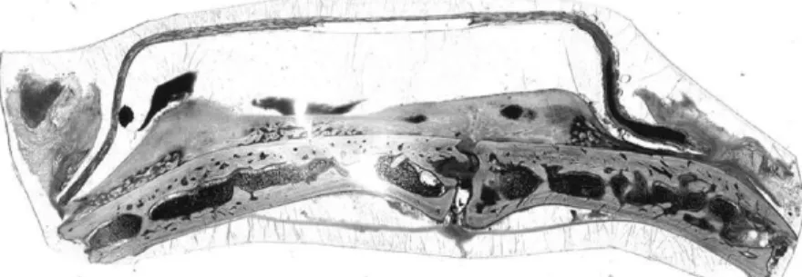
Figure. 1 - Figure. 4 : Control group I Figure. 1, 2(2 weeks after surgery)
Figure. 1 A little newly formed connective tissue existed under the TR-ePTFE menbrane.
(Original×40, Villanueva bone stain)
Figure. 2,a A little new bone formation was detected near the connective tissue in the pe- riphery(2: Original×40, a: Original×100, Hematoxylin and eosin stain).
Figure. 3(4 weeks after surgery)
Figure. 3,a A little new bone formation was detected on the remaining bone similar to the 2 weeks after srugery group, but the space under the membrane was filled with mostly soft tissue(3: Original×40, a: Original×100, Hematoxylin and eosin stain) Figure. 4(8 weeks after surgery)
Figure. 4,a Osteon was surrounded by new bone formation in the nondecalcified specimen(4:
Original×20, a: Original×100, Villanueva bone stain)
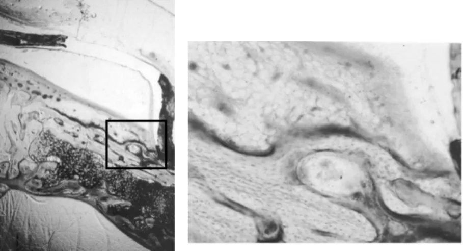
Figure. 5 - Fig. 9 : Control group II Figure. 5(2 weeks after surgery)
Figure. 5,a: DBBM(Deproteinized bovine bone mineral) particles was surrounded by con- nective tissue, but new bone formation was not detected(5: Original×20, a:
Original×40, Hematoxylin and eosin stain) Figure. 6, 7(4 weeks after surgery)
Figure. 6: DBBM particles was surrounded by connective tissues(Original×40, Villanue- va bone stain)
Figure. 7, a A little new bone formation was detected in the bottom of the defect area(7:
Original×40, a: Original×100, Hematoxylin and eosin stain) Figure. 8, 9(8 weeks after surgery)
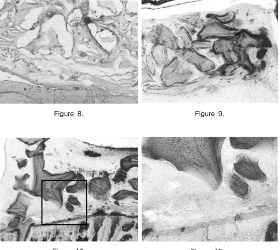
Figure. 8, 9 Distinct bone formation was not detected in the decalcified and nondecalcified specimen. DBBM particles was remained instead of being resorbed(8: Original×
40, Villanueva bone stain)(9: Original×100, Hema- toxylin and eosin stain)
Figure. 10 - Figure. 12 : Experimental group Ⅲ Figure. 10(2 weeks after surgery)
Figure. 10,a DBBM particles was seen and new bone formation was not detected (10: Original×40, a: Original×100, Villanueva bone stain)
Figure. 11(4 weeks after surgery)
Figure. 11,a Most of space was filled with connective tissue and osteoblast like cells were infiltrated. A little new bone formation was seen(11: Original×20, a: Original
×100, Hematoxylin and eosin stain) Figure. 12(8 weeks after surgery)
Figure. 12,a DBBM particles was remained instead of being resorbed. New bone formation increased when compared to 4 weeks after surgery group(12: Original×40, a:
Original×100, Hematoxylin and eosin stain)
Figure. 13 - Figure. 16 : Experimental group IV Figure. 13(2 weeks after surgery)
Figure. 13,a,bDBBM particles was seen in the nondecalcified specimen and soft tissue im- pingement was detected in the margin of the bone defect. (13: Original×20, a:
Original×40, b: Original×100 Villanueva bone stain) Figure. 14(4 weeks after surgery)
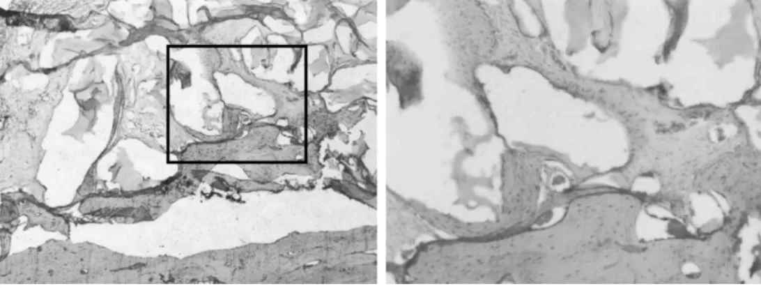
Figure. 14,a New bone was made from the bottom of the bone defect, but most of space was filled with DBBM particles(14: Original×20, a: Original×100 Villanueva bone stain)
Figure. 15, 16(8 weeks after surgery)
Figure. 15,a,b, New bone formation was clearly increased around the DBBM particles.
16,a,b Bone marrow was seen and remodeling of bone was also detected(15:
Original×20, a: Original×40, b: Original×100 Villanue-va bone stain) (16:
Original×20, a: Original×40, b: Original ×100 Hematoxylin and eosin stain) Figure. 17-20(Immunohistochemistric analysis)
All of groups were presented with non-specific staining(17-20: Original×100)
사진부도(Ⅰ)
Figure 1.
Figure 2. Figure 2a.
Figure 3. Figure 3a.
사진부도(Ⅱ)
Figure 4. Figure 4a.
Figure 5. Figure 5a.
Figure 6. Figure 7. Figure 7a.
사진부도(Ⅲ)
Figure 8. Figure 9.
Figure 10. Figure 10a.
Figure 11. Figure 11a.
사진부도(Ⅳ)
Figure 12. Figure 12a.
Figure 13. Figure 13a. Figure 13b.
Figure 14. Figure 14a.
사진부도(Ⅴ)
Figure 15.
Figure 15a. Figure 15b.
Figure 16.
사진부도(Ⅵ)
Figure 16a. Figure 16b.
Figure 17. Figure 18.
Figure 19. Figure 20.
-Abstract-
Effect of inorganic polyphosphate on guided bone regeneration 41)
Jong-Hyuk Chung, Young-Hyuk Kwon, Joon-Bong Park, Yeek Herr Department of Periodontology, Kyung Hee University, Seoul, Korea
This study was performed to evaluate the effect of inorganic polyphosphate on bone formation in the calvaria of rabbit in the procedure of guided bone regeneration with bovine cancellous bone graft and titanium reinforced expanded polytetrafluoroethylene(TR-ePTFE) membrane. The rabbits were divided into four groups. Control group I used only TR-ePTFE membrane, control group II used TR-ePTFE membrane and deproteinized bovine bone mineral soaked in saline, experimental group III and IV used TR-ePTFE membrane and deproteinized bovine bone mineral soaked in 1% or 2% inorganic polyphosphate respectively.
After decortication in the calvaria, GBR procedure was performed on 12 rabbits with titanium reinforced ePTFE membrane filled with deproteinized bovine bone mineral soaked in saline or inorganic polyphosphate. The animals were sacrificed at 2 weeks, 4 weeks, and 8 weeks after the surgery. Decalcified and non-decalcified specimens were processed for histologic and immunohistochemistric analysis.
1. Titanium reinforced ePTFE(TR-ePTFE) membrane showed good spacemaking and cell occlusiveness capability, but it showed poor wound stabilization.
2. The deproteinized bovine bone mineral did not promote bone regeneration, but it acted as a space filler.
3. There was no complete resorption of the deproteinized bovine bone mineral within 8 weeks.
4. 1% inorganic polyphosphate did not promote bone formation, but 2% inorganic poly- phosphate promoted bone formation.
Within the above results, 2% inorganic polyphosphate could be used effectively for bone regeneration.
Key words : Inorganic polyphosphate, GBR(guided bone regeneration), TR-ePTFE