Streptococcus mutans biofilm model에서 오배자( Galla Chinensis) 추출물과 칼슘의 법랑질 재광화효과와 항균효과에 관한 연구
김은정, 진보형
서울대학교 치의학대학원 예방치학교실
Galla chinensis extracts and calcium induce remineralization and antibacterial effects of enamel in a Streptococcus mutans biofilm model
Eun-Jeong Kim, Bo-Hyoung Jin
Department of Preventive and Social Dentistry, School of Dentistry, Seoul National University, Seoul, Korea
Copyright © 2018 by Journal of Korean Academy of Oral Health
This is an open-access article distributed under the terms of the Creative Commons Attribution Non-Commercial License (http://creativecommons.org/licenses/by-nc/4.0/), which permits unrestricted non-commercial use, distribution, and reproduction in any medium, provided the original work is properly cited.
Objectives: The purpose of this study was to investigate the combined effects of Galla chinensis ex- tract (GCE) and calcium (CA) on enamel remineralization. The antibacterial effect of G. chinensis on Streptococcus mutans biofilm was also evaluated by examining the bacterial growth, acidogenesis, and morphology of the biofilm in vitro.
Methods: S. mutans biofilm was formed on bovine enamel specimens over a 72-h period and treated for 10 min with 1.0 mol CA, 4,000 ppm aqueous solution of GCE, or a combination of the two (GCE+CA).
The enamel specimens were analyzed for enamel surface microhardness after remineralization. We tested the anti-cariogenic effects of GCE based on the inhibition of acid production, antibacterial activity, and morphological changes in S. mutans. The differences between the groups and antibacterial effects were analyzed using one-way analysis of variance.
Results: GCE+CA group showed the highest efficacy in enhancing remineralization. The GCE group showed the highest antibacterial activity against S. mutans biofilm. Although the GCE+CA group showed significant antibacterial activity, it was less than that of the GCE group (P<0.05). Both GCE and GCE+CA groups maintained a pH of approximately 7.0 for 1 h whereas the pH of the control group decreased rapidly from pH 7.3 to pH 6.1. SEM imaging revealed that S. mutans treated with GCE and GCE+CA showed irregular cell wall structure and showed fewer cells in the chain than the typical long chains observed in the control group.
Conclusions: This study found that natural G. chinensis significantly enhances enamel remineralization, and exerts synergistic effects with calcium. It also exerts strong bactericidal activity and inhibits acid pro- duction and changes in the microstructure of S. mutans biofilm.
Key Words: Anti-bacterial agents, Combined effect, Galla chinensis, Remineralization Received: July 11, 2018
Revised: September 12, 2018 Accepted: September 12, 2018
Corresponding Author: Bo-Hyoung Jin Department of Preventive and Social Dentistry, School of Dentistry, Seoul National University, 28 Yeongun-dong, Jongro-gu, Seoul 03080, Korea Tel: +82-2-740-8783 Fax: +82-2-766-8781 E-mail: [email protected]
JKAOH is available at http://www.jkaoh.org pISSN 1225-388X / eISSN 2093-7784
Introduction
Enamel is highly mineralized and is the strongest biological hard tissue in the human body1). In contrast to other tissues, dental enamel cannot heal itself and must be re-hardening by a physiochemical process involving inorganic constituents from saliva or solutions2). Accordingly, high levels of mineral supplements, such as calcium, fluoride, and phosphates, have preventive effects on enamel mineral loss. However, some of these substances may have side effects in long-term use. For these reasons, there is growing interest in finding new com- pounds for long-term use3). Among them are several natural extracts that show the ability to have a better effect on bal- ance tooth de-/remineralization of dental enamel4,5).
Galla Chinensis (G. Chinensis), one of the traditional natural, non-toxic Chinese herb, has been used for the past 2,000 years. Previous studies have indicated that G. Chinen- sis had an ability to inhibiting cariogenic bacteria6,7), enamel demineralization, and enhancing remineralization8,9). It is necessary to evaluate the effect of their chemical compounds on promoting remineralization of dental enamel. A previous study reported those co-operative effects of fluoride and the chemical compounds of G. Chinensis on enhancing reminer- alization of dental enamel10), however, experiments have not been performed to investigate the co-operative effects of cal- cium and the G. Chinensis on enhancing the remineralization underneath a biofilm model. Moreover, some experiment also performed the potential rehardening effect of G. Chinensis under pH-cyclic conditions, but since this does not reflect the complex environment in the mouth, we performed this study to assess the effect of G. Chinensis on enamel by reproduc- ing the oral ecological environment as much as possible us- ing biofilm model. So far, no biofilm model has persuasively addressed the effectiveness of caries-preventive agents such as traditional herb on remineralization of dental hard tissue.
Thus, it is of interest to study remineralization underneath a biofilm.
Therefore, this study tested the hypothesis that the com- bination of calcium and G. Chinensis would have a synergistic effect on the remineralization underneath a biofilm model.
The purpose of this study was to investigate the effects of G.
Chinensis with calcium on enhancing remineralization, and also the antibacterial effect of G. Chinensis underneath a S.
mutans biofilm was evaluated by examining the bactericidal activity, acidogenesis and morphology in vitro.
Materials and Methods
1. Preparation of enamel specimens
Sound bovine incisors without cracks, infection or any le- sions under Quantitative Light-Induced Fluorescence (QLF, QLF Pro®, Inspektor Research System BV, Amsterdam, Neth- erlands) were selected in this study. Cylindrical cores 5 mm in diameter were punched out at the top of the bovine enamel surface. Samples were placed in 1.2×1.0×0.8 cm molds and mounted in acrylic resin. The specimens were ground flat and polished using wetted silicon carbide paper (600-2,000 grid).
Specimens were rinsed thoroughly with distilled water and stored in a 100% humidified atmosphere before use. A total of 84 specimens were used in the experiment. Only those specimens which enamel surface hardness raged from 300- 330 VHN were selected. The selected specimens were treated with a pH 5.0 solution containing 0.2% Carbopol (#980, No- veon Inc, Cleveland, USA) with 0.1 M lactic acid containing 50% calcium hydroxide phosphate for 72 hours to form initial artificial caries enamel. The VHN of demineralized specimens was measured and 84 specimens having the surface hardness of the initial dental enamel with an average VHN of 35-55 were selected. For each group, GCE, GCE+CA, and CA groups were assigned to 24 and control to 12.
2. G. Chinensis extract (GCE)
The effective components of G. Chinensis (produced in the Gyeongbuk province of the Republic of Korea) were ex- tracted as previously reported11). Briefly, it (1 kg) was dried in an oven at 60°C for 72 hours, finely powdered and added to 600 ml of distilled water. The mixture was stirred for 10 h at 60°C and then filtered. The extract was re-extracted with dis- tilled water under the same conditions. Then, the extract was dissolved in 500 ml of ethanol (100%) for 48 h at 60°C and an agitator speed of 150 rpm. After filtration and evaporation of the ethanol, the remaining extract was lyophilized to provide a powder.
3. Bacteria strain, media, growth conditions
Streptococcus mutans ATCC 25175 was provided from Korean collection for Oral Microorganisms in Seoul National University and cultivated with a tryptic soy broth (TSB) at 37 C and 5% CO2. The S. mutans genome sequence was determined using a shotgun high-throughput sequencing approach as described. The detailed methods are published as supporting information on the PNAS web site (www.pnas.org)11). Biofilms of S. mutans were formed on bovine specimens in a 50 ml
tube. Each specimen was transferred daily to fresh medium over a 3-day period12). The S. mutans biofilms on each speci- men contained approximately 2×10⁷ colony forming units per milliliter before experiment start.
4. Remineralization process
After exposing the biofilm to each solution (1.0 M calcium, a 4,000 ppm aqueous solution of GCE and a 4,000 ppm aque- ous solution of GCE containing 1.0 M calcium) for 10 min, and then they were placed in a 50 ml tube containing a sterile sa- line solution. The specimens in the tube were ultra-sonicated at 50 W (Branson Sonic, USA) using 3×10 sec pulses with 2×
5 sec intervals before measuring.
5. Measurement of bacterial viability
Using sterile bovine specimens that did not process any- thing, after exposing the biofilms to the solutions (1.0 M cal- cium, a 4,000 ppm GCE and a 4,000 ppm aqueous solution of GCE containing 1.0 M calcium) for 1, 5, 10 min and 1 hour, they were placed in 50 ml tube containing the sterile saline solution. The bovine specimens in the 50 ml tube were ultra- sonicated using 3×10 sec pulses with 2×5 sec intervals12,13). The suspension was diluted serially from 10―1 to 10―6, and plated on tryptone soy agar. The plates were incubated in 5%
CO2 at 37°C for 48 h, and the CFU were determined by count- ing the number of colonies.
6. Measurement of acid production
The level of acid production from the S. mutans biofilms treated with the compounds was determined by measuring the pH14). The pH was measured using a pH electrode (Orion ROSSTM, 8102BNUWP, Beverly, MA, USA) connected to a pH meter (Orion StarTM, Beverly, MA, USA). After a 5, 10 min, and 1-hour treatment with the test compounds, the pH of the media was measured each time point. These assays were re- peated at least three times.
7. SEM analysis
Scanning electron microscopy (SEM) S-4700 (Hitachi, Japan) was used to examine the changes in the S. mutans morphology. The bovine enamel specimens were fixed in 4%
paraformaldehyde in 0.1 M PBS for 1 h at room temperature.
The fixed samples were then washed 2 times with PBS and distilled water, and sputter-coated with platinum and ob- served by SEM.
8. Assessment of remineralization effect
The surface microhardness of enamel specimens was as- sessed using a Vickers microhardness tester (Shimadzu, HMV- 2, Kyoto, Japan) at the beginning of the experiment, and after being immersed in a mineral and natural supplement. Inden- tations were measured for 10 s using diamonds at 9.807 N with a magnification of 40 X. The average microhardness was calculated.
9. Statistical analysis
The differences between the groups and antibacterial effects were analyzed using one-way analysis of variance (ANOVA) and a Tukey’s post hoc honestly significant differ- ences (HSD) test using the studentized range. The level of significance was P<0.05. The SPSS (Statistical Packages for Social Science, Ver. 19.0, Chicago, IL, USA) statistical program was used for all statistical analyzes.
Results
1. Enamel microhardness changes after the experimental procedure
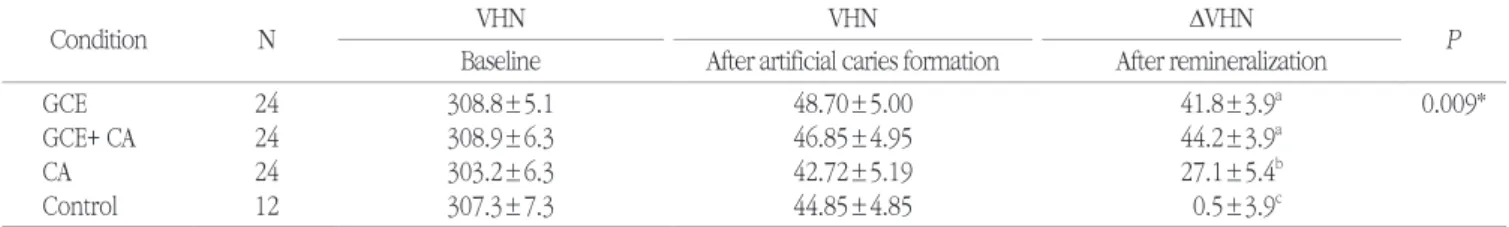
Table 1 shows the mean enamel surface hardness after exposure to the remineralization solutions (GCE; G. Chinensis extract, GCE+CA; G. Chinensis extract + calcium, and CA;
calcium). The GCE+CA groups showed the most enhanced remineralization; 44.2 DVHN; the lowest remineralization ef-
Morphological change (SEM) Bacteria viability Acidogencity
Cariogenic properties Analysis Galla Chinensis extract
S.mutansbiofilm formation
4,000 ppm GCE Sample preparation
Biofilm
0min 7.5 7.3 7.1 6.9 6.7 6.5 6.3 6.1 5.9 5.7
pH
Exposure time
5.5 1min5min10min1hour
GCE GCE+CA Control CA
Remineralization effect Analysis Calcium 1.0
mmol
Calcium 1.0 mmol 4,000 ppm GCE+
Remineralization process underneath aS.mutansbioflim
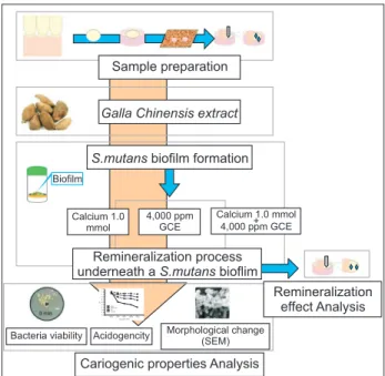
Fig. 1. Flowchart of the in vitro experimental study design.
fect was found in the CA group; 27.1 DVHN. As a result of one-way ANOVA analysis, the CA group were significantly different from GCE group and GCE+CA group (P<0.05).
2. Antibacterial activity of GCE for S. mutans biofilm After the formation of S. mutans biofilm on bovine enamel species, the biofilm was treated with three different solutions.
The GCE and GCE+CA groups showed significantly lower numbers of surviving S. mutans colony forming units (CFU) than those of the control groups (P<0.05). The GCE showed the highest level of antibacterial activity for S. mutans biofilm, and the GCE+CA also significant antibacterial activity but less than GCE. The GCE+CA exhibited similar bactericidal activ- ity to GCE. The GCE and GCE+CA groups for 5 min showed 91.0% and 87.5% fewer CFU, respectively than the control group. The GCE group for 10 min showed greater bacteri- cidal activity (94.6%) than that of the GCE+CA and CA group exposed at the same time. However, the CA group (39.3%) showed antibacterial activity but not as much as GCE or GCE+CA (Table 2).
3. Inhibition of acid production
The pH of culture medium was recorded during 1 hour after each solution treatment to determine the effect of GCE on acid production. The pH patterns of the GCE were signifi- cantly different from control group after 1 min (P<0.05). Both GCE and GCE+CA groups maintained a pH of approximately 7.0 for 1 hour whereas the pH of the control group decreased rapidly from pH 7.3 to pH 6.1 after 1 hour (Fig. 2).
4. Morphological changes in S. mutans biofilms The mechanism responsible for the antimicrobial activ- ity of GCE was investigated by observing the morphological changes of S. mutans by SEM after treating the biofilm with the treatment solution for 10 min. SEM showed less mor- phological and intracellular content in the GCE and GCE+CA groups for 10 min compared to the control group. Also, GCE and GCE+CA groups showed irregular cell wall structure and showed fewer cells in the chain than the typical long chains observed in the control group (Fig. 3).
Table 1. Comparison of the surface microhardness of different groups after remineralization
Condition N VHN VHN DVHN
Baseline After artificial caries formation After remineralization P
GCE 24 308.8±5.1 48.70±5.00 41.8±3.9a 0.009*
GCE+ CA 24 308.9±6.3 46.85±4.95 44.2±3.9a
CA 24 303.2±6.3 42.72±5.19 27.1±5.4b
Control 12 307.3±7.3 44.85±4.85 0.5±3.9c
Values are mean±SD.
DVHN = After remineralization VHN - Before treatment VHN (baseline).
CA, immersed in 1.0 mol CaCl2 for 10 min; GCE, immersed in 4,000 ppm GCE for 10 min; CA+GCE, immersed in 1.0 mol CaCl2 and 4,000 ppm GCE for 10 min; Control, no treatment.
*Statistically significant by repeated measured ANOVA at the a=0.05 level.
a-cThe different lower case letters indicate statistically significant differences between same groups by Tukey’s HSD post-hoc test at P<0.05.
Table 2. Antibacterial effects of the GCE against S. mutans biofilms Condition
Exposure time CFU (×108)
0 min 5 min 10 min
GCE 48.8±10.6 5.2±1.4a 3.1±0.9a
GCE+CA 49.5±6.7 7.8±0.2a 6.5±2.2a
CA 51.2±8.8 34.1±7.8b 30.1±8.4b
Control 50.5±9.7 56.5±9.7c 60.5±10.8c
The data shown are the Mean±SD.
The different superscripts in the same column indicate statistically sig- nificant difference from each group (P<0.05).
0 min 7.5
7.3 7.1 6.9 6.7 6.5 6.3 6.1 5.9 5.7
pH
Exposure time 5.5
1 min 5 min 10 min 1 hour GCE
GCE+CA
Control CA
Fig. 2. Acidogenicity of S. mutans biofilms was determined by mea- suring the pH of media.
Discussion
Dental plaque is a representative example of a biofilm, which plays an essential role in the pathogenesis of dental caries. A biofilm is a multicellular aggregate of microorgan- ism attached and accumulates on the surface. One approach to controlling oral biofilm is to remove or reduce the biofilm mass or its acidogenicity by using an antimicrobial agent.
Synthetic compounds such as chlorhexidine have been used as an antimicrobial agent to inhibit the growth of bacteria and reduce the adhesion of biofilm to prevent dental car- ies. However, excessive use leads to side effects of synthetic compounds including alteration of the oral cavity, the bacte- rial tolerance, taste disorders, dry mouth, and tooth discolor- ation15,16). The search for natural antiplaque agents with safe efficacy and potent activity has focused on reducing the use of synthetic antimicrobials in daily oral care products17). G.
Chinensis has been widely studied as a Chinese herbal medi- cine to reconstruct tooth enamel following enamel mineral loss and discussed as an effective preventative caries agent due to its unique potential remineralization effects18,19) and antibacterial effects6,7). Also, previous studies on the safety of G. Chinensis showed that G. Chinensis did not cause toxicity to the cells20). However, some studies on antibacterial activity on biofilm model have been conducted, none of the studies investigate the effect of chemical compounds of G. Chinensis on the remineralization and antibacterial effects underneath a biofilm model. Thus, the present study investigated the effect of G. Chinensis and the combined effect of G. Chinensis with calcium on the remineralization and antibacterial effects of enamel underneath a S. mutans biofilm in vitro.
Our data showed that the GCE exhibits an apparent rem- ineralization effect on bovine enamel. In the present study, surface hardness change was assessed with microhardness measurement. Since the enamel surface is not uniform to remineralization, we tried to measure it at the same spot, and it was repeatedly measured three times when measure- ment after experiment process, for reduce errors during the measurement process. The measurement of surface enamel hardness using microhardness determination is judged as a suitable tool to investigate the surface softening of enamel21). The main finding in this study was that enamel remineralized with GCE and GCE+CA showed more deposited than those of CA group. The remineralization effect of GCE was previously observed by Chu et al.22), Huang et al.23). It showed a thick layer was formed on the surface of the enamel in the GCE group. The reason for this is not apparent, but maybe these results indicate that GCE has more ion channels to the lesion body, so makes minerals to deposit more. In addition, when GCE was combined with calcium, a higher remineralization effect was seen compared to the calcium group (Table 2). It indicated that the combined use of calcium and GCE has a synergistic effect in improving remineralization on enamel underneath biofilm model. The results of this study cor- responded well with those of an earlier study that reported that a chemical compound in GCE might act as a calcium ion carrier, supplying the caries lesion with calcium ions from the remineralization solution24). Cheng et al.8) also proposed that some component of GCE might combine with the enamel crystals of a surface layer and inhibit the demineralization of enamel. This may be mainly due to their different mechanisms of action for remineralization. Based on all these results, it can be shown that GCE can directly affect remineralization of the enamel surface and can affect the calcium deposition on the demineralized enamel surface in the combined group during the remineralization process.
There were significantly fewer CFUs of S. mutans in the group exposed to GCE and GCE+CA than in the group ex- posed to CA and control group (P<0.05). In addition, the bio- film exposed to GCE and GCE+CA maintained a constant pH around 7. This result shows that GCE can stop the additional acid production of S. mutans. This is similar to the results of previous studies on the antibacterial effect of CHX, the most effective antibacterial agent25). From the above results, GCE has potential as an antimicrobial agent against S. mutans in- stead of CHX, which has many side effects when used for a long time. According to the SEM images in this study, GCE might have destroyed S. mutans chain. S. mutans exposed Fig. 3. SEM images of the S. mutans biofilm after 10 min treatment. (A)
GCE, (B) GCE+CA, (C) CA, and (D) Control.
B
C D
A
to GCE also showed morphology change compared with the control group. The antimicrobial activity of G. Chinensis on common oral bacteria has been confirmed, as its main component, gallotannins, was found to be bactericidal for S.
mutans strains26). GCE may additionally function by adjusting biofilm structure, composition, and glucosyltransferase activ- ity besides directly inhibiting both bacteria growth and lactic acid formation. Also, GCE has been proven to limit acidic ac- cumulation from carbohydrate metabolism and reduce the proportion of cariogenic bacteria in the biofilm and inhibit demineralization. These results are similar to those of previous studies of natural materials. A grapefruit seed extract is a nat- urally antibacterial material. It can weaken the function of the physiologically active enzyme in microorganism cells and also destroy the cell wall function27). And also, Kim et al.25) studied about Curcuma Xanthorrhiza extract, it has strong bacteri- cidal acidity, inhibitory effects on acidogenesis, and alters the microstructure of S. mutans biofilm.
There were several limitations to this study. This study performed using bovine enamel instead of human enamel specimens. However, bovine enamel specimens instead of hu- man enamel specimens were primarily chosen due to several reasons. Human enamel is often difficult to obtain in sufficient quantity and with adequate quality, due to extensive caries lesions or other defects. And also, it can cause large variations in the outcome measures due to the source and age of the collected human teeth28). Bovine teeth have been used instead of human teeth, as in other investigations8,29). Camargo et al.27) revealed no significant difference between bovine and human teeth in the pH measurement. That is why we used bovine teeth in this study. Since this study only analyzed the antimi- crobial effect of S. mutans, further studies will be needed to investigate the efficacy of G. Chinensis on other cariogenic bacteria other than S. mutans. Further, since this study is an in vitro study, future studies using clinical trials will be needed to determine clinical relevance.
Conclusion
This study found that natural G. Chinensis has a signifi- cant effect on enhancing the remineralization of enamel le- sion, and it had combined synergic effects with calcium in improving remineralization. And G. Chinensis also has potent bactericidal activity, inhibits acid production, and changes the microstructure of S. mutans biofilm. These results suggest that G. Chinensis may act as a preventive dental caries agent.
References
1. Cheng L, Li JY, Huang S, Zhou XD. Effect of Galla Chinensis on enhancing remineralization of enamel crystals. Biomed Mater 2009;4:1-6.
2. Zero DT. Dental caries process. Dent Clin North Am 1999;43:635- 664.
3. Phan TN, Marquis RE. Triclosan inhibition of membrane enzymes and glycolysis of Streptococcus mutans in suspensions and biofilms.
Can J Microbiol 2006;52:977-983.
4. Jeon J, Osalen P, Falsetta M, Koo H. Natural products in caries research: current (limited) knowledge, challenges and future per- spective. Caries Res 2011;45:243-263.
5. Palombo EA. Traditional medicinal plant extracts and natural prod- ucts with activity against oral bacteria: potential application in the prevention and treatment of oral disease. Evid Based Complement Alternat Med 2011;1-15.
6. Huang Z, Zhou X, Li J, Liu T, Li H, Zhu B. The effects of traditional Chineses medicines on the adherence of Streptococcus mutans to salivary acquired pellicle in vitro. Sichuan Da Xue Xue Bao Yi Xue Ban 2003;34:135-137.
7. Xie Q, Li JY, Zuo YL, Zhou XD. The effect of Galla Chinensis on the growth of cariogenic bacteria in vitro. Hua Xi Kou Qiang Yi Xue Za Zhi 2005;23:82-84.
8. Cheng L, ten Cate JM. Effect of Galla Chinensis on the in vitro rem- ineralization of advanced enamel lesions. Int J Oral Sci 2010;2:15- 20.
9. Liu Z, Liu T, Li J, Zhou X, Zhang J. The effect of Galla Chinensis on the demineralization of enamel. Sichuan Da Xue Xue Bao Yi Xue Ban 2003;34:507-509.
10. Lei C, Jiyao L, Yuquing H, Xuedong Z. Effect of compounds of Galla Chinensis and their combined effects with fluride on remineraliza- tion of initial enamel lesion in vitro. J Dent 2008;36:369-373.
11. Chu JP, Li JY, Hao YQ, Zhou XD. Effect of compounds of Galla Chi- nensis on remineralisation of initial enamel carious lesion in vitro. J Dent 2007;35:383-387.
12. Koo H, Hayacibara MF, Schobel BD, Cury JA, Rosalen PL, Park YK, et al. Inhibition of Streptococcus mutans biofilm accumulation and polysaccharide production by apigenin and tt-farnesol. J Antimi- crob Chemother 2003;52:782-789.
13. Koo H1, Pearson SK, Scott-Anne K, Abranches J, Cury JA, Rosalen PL, et al. Effects of apigenin and tt-farnesol on glucosyltransferase activity, biofilm viability and caries development in rats. Oral Mi- crobiol Immunol. 2002;17:337-343.
14. Koo H, Seils J, Abranches J, Burne RA, Bowen WH, Quivey RG Jr.
Influence of apigenin on gtf gene expression in Streptococcus mu- tans UA159. Antimicrob Agents Chemother 2006;50:542-546.
15. Flötra L, Gjermo PER, Rölla G, Waerhaug J. Side effects of chlorhex- idine mouth washes. Scand J Dent Res 1971;79:119-125.
16. Flötra L. Different modes of chlorhexidine application and related local side effects. J Periodontal Res 1973;8:41-44.
17. Xie Q, Li J, Zhou X. Anticaries effect of compounds extracted from Galla Chinensis in a multispecies biofilm model. Oral Microbiol Im- munol 2008;23:459-465.
18. Grobler SR, van der Horst G. Biochemical analysis of various cool drinks with regard to enamel erosion, de- and remineralization. J Dent Assoc S Afr 1982;37:681-684.
19. Li Y, Tang R. Study of effects of Chinese nutgall on normal enamel and non-organic enamel demineralization. J Clin Stomatol 2006;22:344-346.
20. Lee YS, Han OK, Bae MJ, Kim KJ, Shin SW, Lee SK et al. Antimi- crobial and anticancer effects of Galla Rhois on pathogens isolated from oral and KB human oral epidermoid carcinoma cells. Korean J
Oriental Physiology & Pathology 2003;17:1427-1432.
21. Curzon MEJ, Hefferren JJ. Modern methods for assessing the cario- genic and erosive potential of foods. Br Dent J 2001;191:41-46.
22. Chu JP, Li JY, Hao YQ, Zhou XD. Effect of compounds of Galla Chi- nensis on remineralisation of initial enamel carious lesion in vitro. J Dent 2007;35:383-387.
23. Kang MS, Oh JS, Kang IC, Hong SJ, Choi CH. Inhibitory effect of methyl gallate and gallic acid on oral bacteria. J Microbiol 2008;46:744-750.
24. Tian F, Li B, Ji B, Yang J, Zhang G, Chen Y, et al. Antioxidant and antimicrobial activities of consecutive extracts from Galla Chinen- sis: The polarity affects the bioactivities. Food Chem 2009;113:173- 179.
25. Kim JE, Kim HE, Hwang JK, Lee HJ, Kwon HK, Kim BI. Antibacterial characteristics of Curcuma xanthorrhiza extract on Streptococcus
mutans biofilm. J Microbiol 2008;46:228-232.
26. Wu-Yuan CD, Chen CY, Wu RT. Gallotannins inhibit growth, water-insoluble glucan synthesis, and aggregation of mutans strep- tococci. J Dent Res 1998;67:51-55.
27. Camargo CH, Bernardineli N, Valera MC, de Carvalho CA, de Oliveira LD, Menezes MM, et al. Vehicle influence on calcium hy- droxide pastes diffusion in human and bovine teeth. Dent Trauma- tol 2006;22:302-306.
28. Yassen GH, Platt JA, Hara AT. Bovine teeth as substitute for hu- man teeth in dental research: a review of literature. J Oral Sci 2011;53:273-282.
29. Huang S, Gao S, Cheng L, Yu H. Combined effects of nano- hydroxyapatite and Galla Chinensis on remineralization of initial enamel lesion in vitro. J Dent 2010;38:811-819.