Primary torsion of the greater omentum with omental infarction is a rare condition. Infarction of the omentum causes localized acute abdominal pain and can therefore mimic other surgical emergencies. There is little infor- mation on this condition to be found in the radiological literature, because it is not usually diagnosed until surgi- cal intervention, being previously diagnosed as acute ap- pendicitis or acute cholecystitis (1, 2). There are only a few radiological reports of secondary torsion of the greater omentum caused by the hernial sac (1-3). In this report, we present the CT findings of a case of pri- mary torsion of the greater omentum with segmental in- farction, which should contribute to the correct preoper- ative diagnosis of future occurrences of this condition.
Case Report
A 43-year-old man presented with a one-day history of right lower quadrant pain, without any previous history of such problems. He underwent laparotomy in a local clinic due to suspected acute appendicitis. Upon laparo- tomy, multiple omental masses, omental necrosis and old bloody ascites in the peritoneal cavity were ob- served along with a normal appendix. After closure of the abdomen, the patient was referred to our surgical department for proper treatment of the omental mass.
On admission, the patient was afebrile with normal lab- oratory findings. Leukocytosis was not present.
In an attempt to identify the of the omental mass, we performed an abdominal CT scan. Contrast-enhanced CT revealed a counter clockwise whirling patterned greater omentum with an inner high-density vascular structure (Fig. 1A). Also, contrast-enhanced CT revealed an irregular shaped soft tissue infiltration of the greater
J Korean Radiol Soc 2004;50:437-440
─ 437 ─
CT Findings of Primary Torsion of the Greater Omentum with Segmental Infarction: Case Report1
Yong Sun Jeon, M.D., Soon Gu Cho, M.D., Won Hong Kim, M.D., Mi Young Kim, M.D., Chang Hae Suh, M.D.
1Departments of Radiology, Inha University College of Medicine Received February 20, 2004 ; Accepted April 29, 2003
Address reprint requests to : Yong Sun Jeon, M.D., Department of Radiology, Inha University Hospital, 7-206, 3rd St., Shinheung-dong, Choong-gu, Inchon 400-711, Korea
Tel. 82-32-890-2769 Fax. 82-32-890-2743 E-mail: [email protected]
Herein, we report on a case of primary torsion of the greater omentum with segmen- tal infarction, which should provide useful information for the preoperative diagnosis of future such cases. Primary torsion of the greater omentum with omental infarction is a rare condition. There are only a few radiological reports of secondary torsion of the greater omentum caused by the hernial sac. During surgical exploration, infarction of the greater omentum was identified, due to the observation of omental torsion with- out any underlying cause. We describe a patient with characteristic computed tomog- raphy (CT) findings of primary omental torsion with segmental infarction, which cor- related with the operative and pathologic results.
Index words :Omentum Torsion Images
Computed tomography (CT)
omentum at the level of the mid and lower abdomen, without any intra-abdominal mass (Fig. 1B). There was no evidence of bowel ischemia or infarction on the CT findings.
The patient underwent exploration. During surgery, omental torsion and omental infarction were identified without any underlying cause being identified (Fig. 1C).
The transverse colon and mesentery were normal.
Partial omental resection and incidental appendectomy were performed.
During the histopathologic examination, the omental venous structure showed congestion and a blood clot.
The histological diagnosis was diffuse omental fat necro- sis, hemorrhage, fibrosis and acute inflammation (Fig.
1D).
Discussion
Omental torsion means a partial or total rotation of the omentum around its main axis. Primary omental torsion is unipolar, where one end of the omentum remains fixed while the other end is free, whereas secondary omental torsion is bipolar with the free end attached ei- ther to adhesions or to some other pathological condi- tion (4, 5). Primary torsion has several predisposing fac- tors, such as a bifid omentum or tongue like omental projections, and changes in the omental consistency (4, 5). Secondary torsion is more common and its etiology
Yong Sun Jeon, et al: CT Findings of Primary Torsion of the Greater Omentum with Segmental Infarction
─ 438 ─
A B
C D
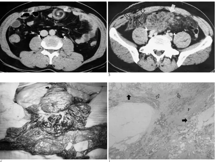
Fig. 1. A. Contrast-enhanced CT section of the mid-abdomen demonstrating a whirling patterned greater omentum with an inner high-density vascular structure (arrow).
B. Contrast-enhanced CT section of the lower abdomen showing irregular shaped increased density (arrow) in the greater omen- tum. There is focal free air (open arrows) in the right omentum and abdominal wall due to a previous operation.
C. On exploration, a primary omental torsion (arrow) and omental infarction (open arrow) were observed. Venous congestion and a blood clot (arrow) were observed in the central portion of the omentum.
D. Photomicrograph (hematoxylin & eosin staining, ×100) of the omentum shows fat necrosis (arrows), hemorrhage (open arrows) and fibrosis (F).
includes intraabdominal hernia, inflammation, cyst, tu- mor and previous laparotomy (4, 5). When torsion oc- curs, venous return is compromised, with the result that the distal portion of the omentum becomes congested and edematous with hemorrhagic extravasation of serosangeneous fluid into the peritoneal cavity, and aseptic peritonitis ensues (4). As the torsion proceeds, ar- terial occlusion leads to hemorrhagic infarction and fat necrosis, followed by an inflammatory reaction (4).
The symptoms of omental torsion are nonspecific and largely dependent on the degree and duration of torsion (4). Nausea, anorexia and vomiting are uncommon symptoms, occurring in less than half of the patients af- fected by this condition (4). Focal tenderness with vary- ing degrees of peritonism is found on examination (1).
This disease can cause acute, as occurred in our patient, with tenderness and guarding of the right lower quad- rant (4, 7). When the pain is on the right, acute appen- dicitis, cholecystitis or right renal colic may be consid- ered in the differential diagnosis, depending on the find- ings of the clinical examination. There may be fever or leukocytosis (4, 7).
Cross-sectional imaging can establish a diagnosis of omental infarction in the proper clinical settings. So CT and/or ultrasound (US) can be extremely helpful in es- tablishing the diagnosis. The US examination shows a hyperechoic, localized, usually paraumbilical mass (8, 9). CT demonstrates a heterogeneous fatty mass anterior to the colon; adherent to an inflamed parietal peri- toneum and containing strands of soft tissue attenuation (8, 9). The appearance of irregular shaped soft tissue le- sions with infiltration of the greater omentum at the lev- el of the mid and lower abdomen enables the radiologist to differentiate omental infarction from a mass of omen- tal origin.
In our case, omental torsion was observed, which be established preoperatively with cross-sectional imaging.
Our CT showed whirling fatty tissue in the greater omentum with an inner high density corresponding to a central vessel. On correlation with the gross section, the intravascular high density was identified as venous con- gestion and a blood clot. The lesion had a characteristic superficial paraumbilical location situated between the rectus abdominis muscle and the transverse colon, cor- responding to the greater omentum. A similar whirling pattern may also be seen in small bowel volvulus, but it is usually associated with small bowel obstruction and is
centrally located in the mesentery (6).
The management of patients with segmental infarc- tion of the omentum is controversial. To reduce the pos- sibility of subsequent adhesion formation, resection of the involved segment is recommended in the surgical lit- erature (4, 5). However, in some literatures it was re- ported that a right-sided segmental infarction of the greater omentum could be managed by conservative treatment with good results (8-10). The natural history is that of resolution of the inflammatory process with re- traction, fibrosis and either complete resolution or au- toamputation (1). Based on the information at hand, con- servative management with symptomatic relief and clin- ical and radiological follow up would seem to constitute a reasonable approach.
In conclusion, we report on a case of primary omental torsion with segmental infarction and characteristic CT findings. The diagnosis of this condition is hardly ever made purely on clinical grounds, as it mimics the symp- toms of appendicitis. It is possible to obtain a correct preoperative diagnosis by means of CT, in which case conservative treatment represents a viable alternative, thus avoiding surgical resection.
References
1. Stella DL, Schelleman TG. Segmental infarction of the omentum secondary to torsion: ultrasound and computed tomography diag- nosis. Australas Radiol 2000;44:212-215
2. Maeda T, Mori H, Cyujo M, Kikuchi N, Hori Y, Takaki H. CT and MR findings of torsion of greater omentum: a case report. Abdom Imaging 1997;22:45-46
3. Aoun N, Haddad-Zebouni S, Slaba S, Noun R, Ghossain M. Left- sided omental torsion: CT appearance. Eur Radiol 2001;11:96-98 4. Karayiannakis AJ, Polychronidis A, Chatzigianni E, Simopoulos C.
Primary torsion of the greater omentum: report of a case. Surg Today 2002;32:913-915
5. Leitner MJ, Jordan CG, Spinner MH, Reese EC. Torsion, infarction and hemorrhage of the omentum as a cause of acute abdominal distress. Ann Sur 1952;135:103-110
6. Jaramillo D, Raval B. CT diagnosis of primary small bowel volvu- lus. AJR Am J Roentgenol 1986;147:941-942
7. Schnur PL, McIlrath D, Carney JA, Whittaker LD. Segmental in- farction of the greater omentum. Mayo Clin Proc 1972;47:751-757 8. Puylaert JB. Right-sided segmental infarction of the omentum:
clinical, US and CT findings. Radiology 1992;185:169-172
9. Karak PK, Milmond SH, Neumann D, Yamase HT, Ramsby G.
Omental infarction: report of three cases and review of the litera- ture. Abdom Imaging 1998;23:96-98
10. Noordzij J, Puylaert J, Smitthuis R, Langezaal O. Right-sided seg- mental infarction of the omentum. Eur J Surg 1994;160:703-705 J Korean Radiol Soc 2004;50:437-440
─ 439 ─
Yong Sun Jeon, et al: CT Findings of Primary Torsion of the Greater Omentum with Segmental Infarction
─ 440 ─
대한영상의학회지 2004;50:437-440
부분 경색을 동반한 원발성 대망염전의 CT 소견: 증례 보고1
1인하대학교 의과대학 방사선과학교실
전용선・조순구・김원홍・김미영・서창해
저자들은 부분 경색을 동반한 원발성 대망 염전의 CT 소견 1예를 보고 하고자 한다. 부분 경색을 동반한 원발성 대망 염전은 매우 드문 질환으로, 대부분의 보고는 헤르니아에 의한 이차성 대망 경색 이다. 저자들의 증례는 수술 소견상, 다 른 원인 없이 발생한 원발성 대망 염전에 의해서 부분 경색이 발생된 것으로 나타났다. 이제 저자들은 부분 경색을 동반 한 원발성 대망 염전의 특징적인 CT 소견을 수술 소견과 조직 검사 결과와 연관하여 보고 하고자 한다.