752
통신저자:배 대 경
서울시 동대문구 회기동 1 경희대학교 의과대학 정형외과학교실 TEL: 02-958-8366ㆍFAX: 02-964-3865 E-mail: [email protected]
*본 논문의 요지는 2007년도 대한슬관절학회 춘계학술대회에서 발표되었음.
Address reprint requests to Dae Kyung Bae, M.D.
Department of Orthopedic Surgery, School of Medicine, Kyung Hee University, 1, Hoegi-dong, Dongdaemun-gu, Seoul 130-702, Korea
Tel: +82.2-958-8366, Fax: +82.2-964-3865 E-mail: [email protected]
퇴행성 슬관절염에서 시행한 미세천공술의 임상적 및 방사선적 중기 추시 결과
배대경ㆍ윤경호ㆍ송상준ㆍ노정호ㆍ김만호 경희대학교 의과대학 정형외과학교실
Midterm Clinical and Radiological Results after Microfracture in Osteoarthritic Knees
Dae Kyung Bae, M.D., Kyoung Ho Yoon, M.D., Sang Jun Song, M.D., Jung Ho Noh, M.D., and Man Ho Kim, M.D.
Department of Orthopedic Surgery, School of Medicine, Kyung Hee University, Seoul, Korea
Purpose: To evaluate midterm results after microfracture in osteoarthritic knees.
Materials and Methods: Between October 1997 and April 2006, 67 osteoarthritic knees, with minimum 4-year follow-up, underwent microfracture. Baumgaertner scores were evaluated to determine clinical results. Radiological results were assessed based on joint space widening and improvement of mecha- nical axis deviation. Joint space widening was calculated by comparing the preoperative joint space with the final follow-up joint space. Varus deformity was evaluated on orthoroentgenogram and recorded as a percentile of the point at which the mechanical axis intersected a line extending from the center of the knee to medial border of the medial tibial condyle. The figure was expressed as MA%.
Results: The average Baumgaertner score at final follow-up was 7.0. The average joint space changed from 2.74 mm to 4.22 mm on AP radiographs and from 1.91 mm to 3.85 mm on lateral radiographs.
Average MA% was 57.5% preoperatively and 45.8% at final follow-up. Clinical and radiological improve- ments were maintained in most cases followed for more than 4 years. Four patients had total knee arthroplasty after an average of 4.1 years.
Conclusion: We noted pain relief, joint space widening, and improvement of mechanical axis after microfracture for degenerative arthritis of the knee. Maintenance of clinical and radiological improvement was observed at midterm follow-up.
Key Words: Knee, Osteoarthritis, Arthroscopy, Microfracture
서 론
퇴행성 슬관절염에서 손상된 관절 연골을 치유하는 방 법은 여러가지가 있는데5,7,8,10,26)
, 그 중에서 미세천공술 은 술기가 쉽고 간단하며 여러 저자들에 의해 임상적 우 수성과 연골의 조직학적 재생이 확인되었다2,3,18,20,23,24)
. Bae 등3)은 미세천공술을 시행한 후, 재생된 연골의 조직
학적 분석을 통하여 섬유연골(fibrocartilage)과 초자양 연골(hyaline like cartilage)로 이루어진 혼합성 연골과 제2형 교원질의 존재를 보고하였다. 그러나, 이들 재생 된 연골 조직이 지속적인 체중부하에 생역학적 변화를 일 으키지 않고 얼마나 유지할 수 있는지에 대한 논란이 있 으며13), 미세천공술 후 이학적 검사 및 설문조사 등을 통
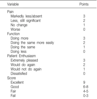
Table 1. Baumgaertner’s Nine-point Scale
Variable Points
Pain
Markedly less/absent 3
Less, still significant 2
No change 1
Worse 0
Function
Doing more 3
Doing the same more easily 2
Doing the same 1
Doing less 0
Patient Enthusiasm
Extremely pleased 3
Would do again 2
Would not do again 1
Dissatisfied 0
Score
Excellent 9
Good 6-8
Fair 4-5
Fail 0-3
Fig. 1. (A) Joint space on the AP radiograph was measured by the distance between the medial femoral condyle and the midpoint from medial tibial spine to medial border of the medial tibial condyle. (B) Joint space on the lateral radio- graph was measured by the nearest of distance between medial femoral condyle and 한 임상적 결과에 대한 보고는 있으나, 방사선학적 변화
에 대한 보고는 드물다. 이에 저자들은 미세천공술 후 중 기 추시시 임상적 및 방사선학적 결과를 분석하고, 술 전 내반 변형 정도에 따라 임상적, 방사선학적 결과를 비교 하고자 하였다.
대상 및 방법
1997년 10월부터 2006년 4월까지 퇴행성 슬관절염으
로 편측 슬관절에 관절경적 미세천공술을 시행한 236예 중 4년 이상 추시가 가능하였던 67예를 대상으로 하였다.
이 중 남자는 10예, 여자는 57예 이었다. 추시 기간은 평균 5년 9개월(4-9년)이었으며, 수술 당시 연령은 평균 59.4세(46-84세)였다. 전 예에서 방사선학적 평가를 시 행하였고, 4년 이상 매년 추시가 가능하였던 환자는 65 예이었다. 병력상 슬관절면 주위의 동통, 이학적 검사상 연골 결손 부위의 압통과 마찰음(crepitus)이 있고, 방사 선 사진상 관절 간격의 감소, 골 스캔상 국소 열소가 관찰 되는 환자로, 최소 3개월 이상 보존적 치료에도 불구하고 증상이 호전되지 않고 일상생활에 지장이 있으며, 체중 부하 단순 전후방 사진상 Kellgren-Lawrence 분류11)에 따라 1, 2도의 퇴행성 변화를 보인 환자를 대상으로 관절 경 검사를 시행하여 Outerbridge 분류17)에 따라 제3, 4 등급의 연골 결손이 관찰된 경우에 미세천공술을 시행하 였다. 관절의 불안정성, 심한 부정정렬, 5o 이상의 굴곡 구축, 박리성 골연골염, 염증성 관절염, 골괴사증, 협조 가 잘 되지 않아 술 후 재활치료가 어려울 것으로 예상되 는 경우는 대상에서 제외하였다. 수술 방법과 재활치료 는 2001년 저자들1)이 소개한 것과 동일하였다.
임상적 평가는 Baumgaertner5)의 슬관절 기능평가방 법을 이용하였다(Table 1). 동통, 기능, 환자의 만족도에 따라서 각 항목별로 0-3점까지 9점 만점으로 하였고 9 점을 우수(excellent), 6-8점을 양호(good), 4-5점은 보통(fair), 3점 이하는 불량(fail)으로 하였다.
Fig. 2. The extent of varus deformity shown on preoperative or- thoroentgenogram was evaluated by MA% [(b/a)×100]. It shows a medial deviation of the mechanical axis with joint space narrowing of medial compartment: (a) is the distance from the center of the knee to the medial border of the medial tibial condyle and (b) is the distance from the center of the knee to the point at which the mechanical axis intersects the knee joint line.
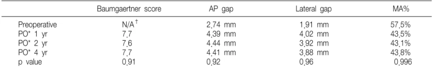
Table 2. Baumgaertner Score, AP Gap, Lateral Gap, and MA% in Patients with Follow-up for More Than 4 Years
Baumgaertner score AP gap Lateral gap MA%
Preoperative N/A† 2.74 mm 1.91 mm 57.5%
PO* 1 yr 7.7 4.39 mm 4.02 mm 43.5%
PO* 2 yr 7.6 4.44 mm 3.92 mm 43.1%
PO* 4 yr 7.7 4.41 mm 3.88 mm 43.8%
p value 0.91 0.92 0.96 0.996
*, Post-operative; †, not available.
방사선학적 평가는 술 전과 최종 추시시 관절 간격과 역학적 축을 계측하였다. 관절 간격은 체중부하 전후방 방사선 사진상 내측 경골극과 경골 내과 내측변 사이의 중앙점에서 대퇴골 내과까지의 길이와 체중부하 측면 방 사선 사진상 대퇴골 내과와 내측 경골 고평부 사이의 최 단 거리로 계측하였다(Fig. 1). 슬관절의 내반 변형 정도 는 체중부하 하지 전장 사진상 슬관절의 중심에서 역학적 축까지의 직선거리를 슬관절 중심에서 슬관절 내측까지 의 길이로 나누어 백분율로 환산한 값(MA%)으로 정의하 였다(Fig. 2). 체중부하 하지 전장 사진은 환자가 슬관절 을 최대한 신전하고, 체중부하를 하지에 균등하게 한 기 립 상태에서 방사선 투과선이 슬관절면에 수직이 되도록 촬영하였다. 고관절부터 족근관절까지 하지 전장이 포함
되도록 하였으며, 하지의 회전으로 인한 오차를 막기 위 해 환자의 슬개골 중심이 슬관절의 정중앙에 위치하도록 촬영하였다. 계측치는 한 사람의 연구자(정형외과 전문 의)가 두 차례 측정하여 그 평균치를 구하였다. 술 전 MA%를 25% 구간마다 나누어, 25% 이하(I군, 15예), 25-50% (II군, 18예), 50-75% (III군, 21예), 75% 초과 (IV군, 13예)로 나누어 각 군에서 매년 추시시 임상적, 방사선학적 평가를 시행하였다. 추시 기간에 따른 임상 적, 방사선학적 결과를 비교하였다(One Way ANOVA test). SPSS version 12.0을 이용하여 통계처리 하였으 며 유의성 판정은 p<0.05로 하였다.
결 과
1. 임상적 결과
매년 추시한 평균 Baumgaertner 점수는 술 후 1년째 7.7, 2년째 7.6, 4년째 7.7이었다(p=0.91). 최종 추시시 Baumgaertner에 의한 슬관절 점수는 평균 7.0이었고, 우수 6예, 양호 43예, 보통 14예, 불량 4예로 49예(73.1%) 에서 양호 이상의 결과를 보였다. 각 군에 따른 임상적 결 과는 I군 7.7, II군 7.6, III군 6.5, IV군 6.3이었다.
2. 방사선학적 결과
전후방 사진상 관절 간격은 술 전 평균 2.74 mm, 최종 추시시 평균 4.22 mm로 1.48 mm 증가하였고, 술 후 1년째 4.39 mm, 2년째 4.44 mm, 4년째 4.41 mm이었 다(p=0.92). 측면 사진상 관절 간격은 술 전 평균 1.91 mm, 최종 추시시 평균 3.85 mm로 1.94 mm 증가하였고, 술 후 1년째 4.02 mm, 2년째 3.92 mm, 4년째 3.88 mm 이었다(p=0.96). MA%는 술 전 평균 57.5%, 최종 추시시 평균 45.8%로 11.7% 감소하였고, 술 후 1년째 43.5%, 2년 째 43.1%, 4년째 43.8%이었다(p=0.996) (Table 2).
각 군에 따른 관절 간격의 증가는 전후방 사진상 I군
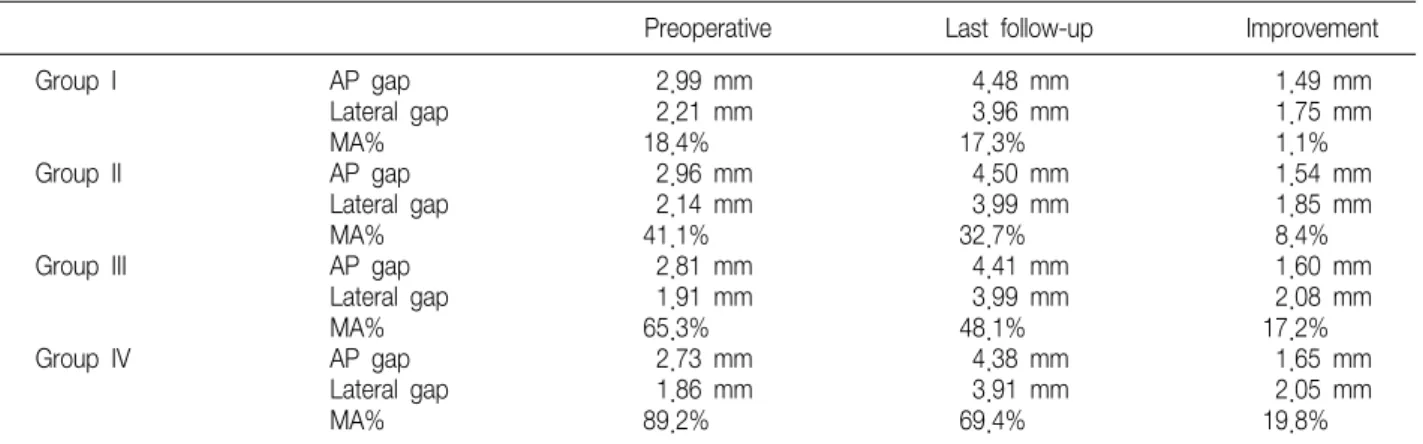
Table 3. AP Gap, Lateral Gap, and Mechanical Axis Percentage (MA%) according to Preoperative MA%
Preoperative Last follow-up Improvement
Group I AP gap 2.99 mm 4.48 mm 1.49 mm
Lateral gap 2.21 mm 3.96 mm 1.75 mm
MA% 18.4% 17.3% 1.1%
Group II AP gap 2.96 mm 4.50 mm 1.54 mm
Lateral gap 2.14 mm 3.99 mm 1.85 mm
MA% 41.1% 32.7% 8.4%
Group III AP gap 2.81 mm 4.41 mm 1.60 mm
Lateral gap 1.91 mm 3.99 mm 2.08 mm
MA% 65.3% 48.1% 17.2%
Group IV AP gap 2.73 mm 4.38 mm 1.65 mm
Lateral gap 1.86 mm 3.91 mm 2.05 mm
1.49 mm, II군 1.54 mm, III군 1.60 mm, IV군 1.65 mm이었고, 측면 사진상 I군 1.75 mm, II군 1.85 mm, III군 2.08 mm, IV군 2.05 mm이었다.
각 군에 따른 MA%의 변화는 I군 1.1%, II군 8.4%, III군 17.2%, IV군 19.8% 감소하였다(Table 3).
3. 관절경 검사 소견
대부분의 환자에서는 내측 구획에 연골의 병변이 있었 다. 5예는 내측과 외측 구획 모두에, 2예는 외측 구획에 만 연골 결손이 국한되어 있었다. 2예에서는 슬개골에 연 골 결손을 보였다. 연골 결손의 평균 크기는 대퇴골에서 5.5 cm2 (0.18-28 cm2), 경골에서 3.4 cm2 (0.6-20 cm2)이었다. 연골 결손의 크기(3 cm2 미만 군과 3 cm2 이상인 군)에 따른 최종 Baumgaertner 점수는 통계학 적으로 유의한 차이를 보이지 않았다(p=0.441). 16예에 서 전방십자인대의 퇴행성 변화를 보였으며 변연 절제술, 다듬기(trimming) 등의 최소한의 처치만을 시행하였고 재건술 또는 봉합술을 시행한 예는 없었다. 53예(79.1%) 에서 반월상 연골의 퇴행성 변화를 보였으며, 21예 (31.3%)에서 반월상 연골의 파열을 보였다. 내측 반월상 연골의 파열 14예, 양측 반월상 연골의 파열 6예, 외측 반월상 연골의 파열 1예 있었다. 반월상 연골 파열에 대 해서는 부분 절제술 및 아전 절제술을 시행하였고 봉합술 을 시행한 예는 없었다. 반월상 연골 파열에 따른 최종 Baumgaertner 점수는, 파열이 있는 21예에서 6.9, 파 열이 없는 46예에서 7.0으로 유의한 차이가 없었다(p=
0.857).
4. 장기 추시 결과와 슬관절 전치환술로의 전환 8년 이상 추시한 8예 중 7예에서는 관절 간격이 유지 되었으며, 1예에서는 술 후 5년째까지 유지되었지만, 그 후 감소하였다. Baumgaertner 점수는 우수 3예, 양호 4예, 보통 1예이었다.
슬관절 전치환술로의 전환은 67예 중 4예(6.0%)에서 술 후 평균 49.5개월(41-56개월)에 시행하였다.
고 찰
관절 연골은 무혈성의 특성을 가지고 있어서 이의 치유 과정은 일반 조직의 치유과정과 달라 많은 연구와 논란의 대상이 되어 왔다. Urist25)에 의해 관절내의 손상이 초자 양 연골(hyaline like cartilage)에 의해 치유될 수 있다 고 알려진 후, Bassett4)는 간엽 세포(pluripotential mesenchymal cell)가 주위 환경의 변화에 따라 다르게 분화될 수 있다고 발표하였으며 산소 분압 및 물리학적 압력이 인자가 될 수 있다고 주장하였다. 관절 연골이 치 유되기 위해서는 기원 세포와 기질의 존재, 응력의 소실, 연골하 골의 보존, 적절한 기계적 자극이 필요하다고 알 려져 있다19).
미세천공술은 연골하 골에 미세한 손상을 주어 골수내 간엽세포(mesenchymal stem cell)의 분화에 의해 마모 된 관절 연골을 재생시킨다는 이론에 근거하고 있는데21-24), 대부분의 연구에서 미세천공술 후 70-90%에서 슬관절 의 기능이 호전되었다고 보고하였다12,14,16,22,24)
. Stead- man 등21)은 미세천공술 후 평균 11년 추시시 동통 및 기능이 80%에서 호전되었다고 보고하였고, Miller 등15) 은 평균 2.6년 추시시 Lysholm 점수가 술 전 평균 53.8
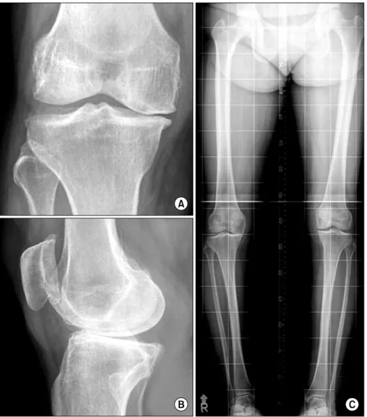
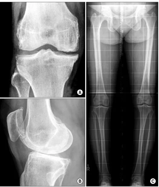
Fig. 3. Preoperative radio- graphs of fifty five-year-old wo- man with symptomatic degene- rative osteoarthritis on right knee. (A) AP gap was 2.21 mm on anterior-posterior view of the knee. (B) Lateral gap was 1.19 mm on lateral view of the knee. (C) MA% was 99.8% on orthoroentgenogram.
에서 최종 추시시 83.1로 호전되었고 환자 만족도는 10 점 만점에 8.3점이었다고 보고하였다. 많은 연구에서 미 세천공술 후 2년 이내에 의미있는 증상 호전을 보였 고6,9,12,21,24)
, 저자들의 경우 대부분의 환자에서 술 후 1 년 이내에 임상적 호전을 보였으며 연골조직의 재생을 간 접적으로 알 수 있는 체중부하 전후방 방사선 사진 및 측 면 사진을 통한 관절 간격의 증가와 체중부하 하지 전장 사진을 통한 내반 변형의 호전을 관찰하였다(Fig. 3, 4).
평균 5년 9개월의 추시시 Baumgaertner5)의 슬관절 기 능평가상 73.1%에서 양호 이상의 만족할 만한 결과를 보 였으며 최소 4년간 추시한 결과 기능이 감소하지 않았다.
Miller 등15)은 미세천공술을 시행한 환자 중 5.9%에서 3년 이내에 재수술 또는 인공관절 치환술을 시행하였다.
Marder 등14)은 10%의 환자에서 골연골 이식, 자가 연골 세포 이식 등의 이차 수술을 시행하였다. 저자들의 경우 는 6.0%에서 술 후 평균 49.5개월에 슬관절 전치환술을 시행하였다.
저자들의 경우 미세 천공술 후 관절 연골 두께의 증가 와 내반 변형의 호전을 관찰할 수 있었다(Table 3). 1군 에서는 연골 결손의 크기가 작고 국소적이기 때문에 술 전 내반 변형이 심하지 않고(MA%, 18.4%) 연골의 재생 이 이루어지더라도 내반 변형의 변화가 작았다(MA%, 17.3%). 4군에서는 연골 결손의 크기가 크고 광범위하며 술 전 상대적으로 심한 내반 변형을 동반하여(MA%, 89.2%) 연골의 재생 후 연골 두께의 증가와 내반 변형의 호전(MA%, 69.4%)이 많았다.
Fig. 4. Radiographs 4 years after microfracture of fifty five- year-old woman with sympto- matic degenerative osteoar- thritis on right knee. (A) AP gap was 4.40 mm on anterior- posterior view of the knee. (B) Lateral gap was 2.48 mm on lateral view of the knee. (C) MA% was 52.1% on orthoro- entgenogram.
배 등1)의 연구에서 동반 손상, 연골 결손의 크기에 따른 임상 결과는 차이가 없었으며, 본 연구에서도 관절경 소 견상 연골 결손의 크기 및 반월상 연골 파열 유무와 Baumgaertner5)의 슬관절 점수는 유의한 상관관계를 보 이지 않았다.
결 론
퇴행성 슬관절염 환자에서 관절경을 이용한 미세천공 술의 임상적 결과는 73.1%에서 양호 이상의 결과를 얻었 으며 대부분의 환자에 있어서 관절 간격의 증가 및 내반 변형 정도가 호전되었다. 수술 후의 임상적, 방사선학적 결과는 중기 추시에서도 대부분 변화없이 지속되고 있었 다. 술 전 내반 변형 정도에 따른 최종 추시시의 임상적,
방사선학적 결과로, 술 전 내반 변형 정도가 클수록 최종 추시시 내반 변형의 교정과 관절 간격이 많이 관찰되었으 며, 술 전 내반 변형 정도가 작을수록 임상적 결과가 더 많이 호전되었다.
참고문헌
1. Bae DK, Ko BW, Kim SK: Results of microfracture surgery in osteoarthritic knee. J Korean Orthop Assoc, 36: 555-560, 2001.
2. Bae DK, Yim CM, Kim JM, Park YK: Microfracture surgery for cartilage regeneration in degenerative arthritis of the knee. J Korean Orthop Assoc, 35: 231-238, 2000.
3. Bae DK, Yoon KH, Song SJ: Cartilage healing after
microfracture in osteoarthritic knees. Arthroscopy, 22: 367- 374, 2006.
4. Bassett CA: Bibliography of bone formation. Transplant Bull, 29: 104-109, 1962.
5. Baumgaertner MR, Cannon WD Jr, Vittori JM, Schmidt ES, Maurer RC: Arthroscopic debridement of the arthritic knee. Clin Orthop Relat Res, 253: 197-202, 1990.
6. Blevins FT, Steadman JR, Rodrigo JJ, Silliman J:
Treatment of articular cartilage defects in athletes: an analysis of functional outcome and lesion appearance. Orthopedics, 21:
761-767, 1998.
7. Dandy DJ: Arthroscopic debridement of the knee for osteo- arthritis. J Bone Joint Surg Br, 73: 877-878, 1991.
8. Friedman MJ, Berasi CC, Fox JM, Del Piazzo W, Snyder SJ, Ferkel RD: Preliminary results with abrasion arthro- plastyin the osteoarthritic knee. Clin Orthop Relat Res, 182:
200-205, 1984.
9. Gobbi A, Nunag P, Malinowski K: Treatment of full thickness chondral lesions of the knee with microfracture in a group of athletes. Knee Surg Sports Traumatol Arthrosc, 13:
213-221, 2005.
10. Johnson LL: Arthroscopic abrasion arthroplasty historical and pathologic perspective: present status. Arthroscopy, 2: 54-69, 1986.
11. Kellgren JH, Lawrence JS: Radiological assessment of osteo-arthrosis. Ann Rheum Dis, 16: 494-502, 1957.
12. Knutsen G, Engebretsen L, Ludvigsen TC, et al: Auto- logous chondrocyte implantation compared with microfracture in the knee. A randomized trial. J Bone Joint Surg Am, 86:
455-464, 2004.
13. Kuo AC, Rodrigo JJ, Reddi AH, Curtiss S, Grotkopp E, Chiu M: Microfracture and bone morphogenetic protein 7 (BMP-7) synergistically stimulate articular cartilage repair.
Osteoarthritis Cartilage, 14: 1126-1135, 2006.
14. Marder RA, Hopkins G Jr, Timmerman LA: Arthroscopic microfracture of chondral defects of the knee: a comparison of two postoperative treatments. Arthroscopy, 21: 152-158, 2005.
15. Miller BS, Steadman JR, Briggs KK, Rodrigo JJ, Rodkey
WG: Patient satisfaction and outcome after microfracture of the degenerative knee. J Knee Surg, 17: 13-17, 2004.
16. Mithoefer K, Williams RJ 3rd, Warren RF, et al: The microfracture technique for the treatment of articular cartilage lesions in the knee. A prospective cohort study. J Bone Joint Surg Am, 87: 1911-1920, 2005.
17. Ogilvie-Harris DJ, Fitsialos DP: Arthroscopic management of the degenerative knee. Arthroscopy, 7: 151-157, 1991.
18. Passler HH: Microfracture for treatment of cartilage defects.
Zentralbl Chir, 125: 500-504, 2000.
19. Qui YS, Shahgaldi BR, Revel WJ, Heatley FW: Obser- vation of subchondral plate advancement during osteochondral repair: a histomorphometric and mechanical study in the rabbit femoral condyle. Osteoarthritis Cartilage, 11: 810-820, 2003.
20. Rodrigo J, Steadman JR, Syftestad G, Benton H, Silli- man J: Effects of human knee synovial fluid on chondrogenesis in vitro. Am J Knee Surg, 8: 124-129, 1995.
21. Steadman JR, Briggs KK, Rodrigo JJ, Kocher MS, Gill TJ, Rodkey WG: Outcomes of microfracture for traumatic chondral defects of the knee: average 11-year follow-up. Arth- roscopy, 19: 477-484, 2003.
22. Steadman JR, Miller BS, Karas SG, Schlegel TF, Briggs KK, Hawkins RJ: The microfracture technique in the treatment of full-thickness chondral lesions of the knee in National Football League players. J Knee Surg, 16: 83-86, 2003.
23. Steadman JR, Rodkey WG, Briggs KK, Rodrigo JJ: The microfracture technic in the management of complete cartilage defects in the knee joint. Orthopade, 28: 26-32, 1999.
24. Sterett WI, Steadman JR: Chondral resurfacing and high tibial osteotomy in varus knee. Am J Sports Med, 32: 1243- 1249, 2004.
25. Urist MR: The repair of articular surfaces following arthroplasty of the hip. Clin Orthop, 12: 209-229, 1958.
26. Woo SL, Kwan MK, Lee TQ, Field FP, Kleiner JB, Coutts RD: Perichondral autograft for articular cartilage.
Shear modulus of neocartilage studied in rabbits. Acta Orthop Scand, 58: 510-515, 1987.
= 국문초록=
목적: 퇴행성 슬관절염 환자에서 미세천공술 후 중기 추시 결과를 보고하고자 한다.
대상및방법: 1997년 10월부터 2006년 4월까지 관절경적 미세천공술을 시행받은 환자 중 4년 이상 추시가 가능 하였던 67예를 대상으로 하였다. 임상적 평가는 Baumgaertner의 슬관절 기능평가방법을 이용하였다. 방사선학 적 평가는 술 전과 최종 추시시 슬관절 방사선사진에서 관절 간격의 변화와 역학적 축의 내측 전위 정도를 계측하였다.
결과: Baumgaertner 점수는 최종 추시시 7.0이었고, 73.1%에서 양호 이상이었다. 전후면 및 측면 방사선사진상 관절 간격은 술 전 2.74 mm, 1.91 mm, 최종 추시시 4.22 mm, 3.85 mm이었다. 역학적 축의 내측 전위 정도는 술 전 평균 57.5%, 최종 추시시 45.8%이었다. 매년 추시시 임상적 및 방사선학적 결과가 대부분에서 변화 없이 유지되었으나, 4예에서 술 후 평균 4.1년에 슬관절 전치환술을 시행하였다.
결론: 퇴행성 슬관절염 환자에서 미세천공술 후 동통의 감소, 관절 간격의 증가, 하지 정열의 호전을 관찰할 수 있었다. 중기 추시시 임상적, 방사선학적 결과는 지속적인 체중부하에도 변화를 일으키지 않고 유지되었다.
색인 단어: 슬관절, 퇴행성 관절염, 미세천공술