Introduction
Cephalometry was defined as the measurement of the head, and its up-to-date definition is refined as a scientific study of the measurement of the head with relation to specific reference points to assess facial growth and devel- opment.1,2 In particular, in order to evaluate facial asym- metry, the definition of the midline in two-dimension (2D) and the midsagittal plane (MSP) in three-dimension (3D) have been generally used to evaluate facial asymmetry in
cephalometric analysis.3
With the recent advancement of computed tomography (CT), pre-surgical 3D analysis has been frequently used in surgery and orthodontics. The references with various planes, angles, and points have been proposed to develop 3D cephalometric analyses on CT with other studies.4,5 Recently a reference point defining the coordinate-system using 3D CT image was proposed.6Olszewski et al showed the actual necessity to establish the appropriate reference on CT image like the conventional cephalometric radio- graph.7Even though the volumetric method was introduced, the practical use for cephalometric analysis has been lim- ited. The quantification of 3D CT volume modeling was presented without a specific reference.8This method cannot
Determination of midsagittal plane for evaluation of facial asymmetry using three-dimensional computed tomography
Tae-Young Kim, Jee-Seon Baik*, Joo-Young Park**, Hwa-Sung Chae***, Kyung-Hoe Huh, Soon-Chul Choi
Department of Oral and Maxillofacial Radiology and Dental Research Institute, School of Dentistry, Seoul National University, Seoul, Korea
*Department of Oral and Maxillofacial Surgery, Ilsan Paik Hospital, Inje University, Goyang, Korea
**Department of Oral and Maxillofacial Surgery, Seoul National University Dental Hospital, Seoul, Korea
***Section of Orthodontics, Samsung Medical Center, Seoul, Korea ABSTRACT
Purpose : The aim of the present study was to investigate the disagreement of cephalometric analysis depending on the reference determination of midsagittal plane on three-dimensional computed tomography.
Materials and Methods : A total of 102 young women with class III dentofacial deformity were evaluated using three-dimensional computed tomography. The cranial and facial midsagittal planes were defined and the amounts of jaw deviation were calculated. The amounts of jaw deviation were compared with paired t-test (2-tailed) and Bland- Altman plot was drawn.
Results : The landmark tracing were reproducible (r›.978). The jaws relative to the cranial midsagittal plane were 10-17 times more significantly deviated than to the facial midsagittal plane (P⁄.001). Bland-Altman plot demonstrated that the differences between the amounts of jaw deviation from two midsagittal planes were not normally distributed versus the average of the amounts of jaw deviation from two midsagittal planes.
Conclusion : The cephalometric analyses of facial asymmetry were significantly inconsistent depending on the reference determination of midsagittal plane. The reference for midsagittal plane should be carefully determined in three-dimensional cephalometric analysis of facial asymmetry of patients with class III dentofacial deformity.
(Imaging Sci Dent 2011; 41 : 79-84)
KEY WORDS : Cephalometry; Facial Asymmetry; Tomography, X-Ray Computed; Imaging Three-Dimensional
Received March 31, 2011; Revised April 25, 2011; Accepted May 2, 2011 Correspondence to : Prof. Kyung-Hoe Huh
Department of Oral and Maxillofacial Radiology, School of Dentistry, Seoul National University, 275-1 Yeongeon-dong, Jongno-gu, Seoul 110-768, Korea
Tel) 82-2-2072-3498, Fax) 82-2-744-3919, E-mail) [email protected]
Copyright ⓒ 2011 by Korean Academy of Oral and Maxillofacial Radiology
This is an Open Access article distributed under the terms of the Creative Commons Attribution Non-Commercial License (http://creativecommons.org/licenses/by-nc/3.0) which permits unrestricted non-commercial use, distribution, and reproduction in any medium, provided the original work is properly cited.
Imaging Science in Dentistry∙pISSN 2233-7822 eISSN 2233-7830
be actually applied to the 3D analysis of facial asymmetry without defining the MSP.
It was suggested that the evaluation of facial asymmetry may result differently according to the different references on 2D cephalometry.3 Therefore, it is presumed that the carefully selected references in 3D cephalometry may be as important as in 2D cephalometry. The MSP can vary according to the reference determination and therefore the appropriate determination of reference for MSP is indis- pensible in 3D cephalometry. Until now, none of the study has so far shown the inconsistency of cephalometric analy- sis according to the determination of reference on 3D CT.
The purpose of the present study was to investigate the disagreement of cephalometric analysis depending on the reference determination of midsagittal plane for evaluation of facial asymmetry of patients with class III dentofacial deformity using 3D CT. The amounts of jaw deviation relative to two differently determined MSPs were compared to suggest the importance of reference determination on 3D cephalometric analysis.
Materials and Methods
Patient selection
After the approval by the institutional review board in Seoul National University Dental Hospital, the subjects, who undertaken CT examination, were reviewed retrospec- tively from February of 2005 to May of 2010. The inclu- sion criteria were as follows: (1) the age ranged from 20
to 29 years (mean 21.551±2.644 years) (2) female adults (3) ANB angle less than 0�(mean -3.073±2.445�) on the lateral cephalometric radiograph (4) no disease affect- ing craniofacial growth. A total of 102 subjects from 920 patients matched with those criteria. In order to avoid any skewing effects such as sex, age, and skeletal classification, only young women with class III dentofacial deformity were included.
Image acquisition and analysis
Multi-detector spiral CT (Somatom Sensation 10, Sie- mens, Erlangen, Germany) was taken with the following specifications: 512×512 matrix, 0.75 mm slice thickness, 120 kVp, and 80 mA. The subjects were asked to close their mouth at maximum intercuspation. The acquired CT raw data were imported to 3D image software (InVivoDen- tal, version 5.0, Anatomage Inc., San Jose, California) for the subsequent image analysis.
The subjects’ Frankfurt (FH) plane was adjusted to the horizontal plane with the landmarks of bilateral porions and left orbitale in 3D image software. The landmarks were identified based on every cross-sectional view and 3D model by one examiner to acquire the coordinates (x, y, z) of the landmarks.9The distance between two points was calculated with the distance formula in 3D Cartesian coordinate system.
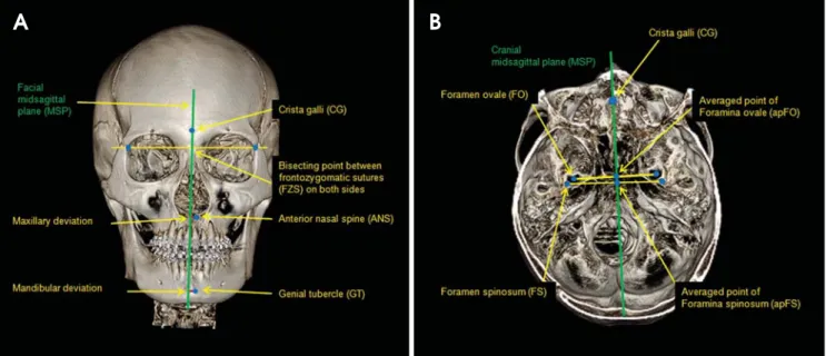
Table 1 and Fig. 1 show the definitions of the landmarks used. Crista galli (CG) was used as the common reference point of the face and cranium for MSP (Fig. 1). The facial
A B
Fig. 1.Landmarks and jaw deviation. A. The jaw deviation is determined as a perpendicular distance between midpoint (ANS and GT) and midsagittal plane (MSP), B. The averaged points of foramina ovale (FO) and foramina spinosum (FS) on both sides are depicted.
MSP was defined as the sagittal plane which crossed CG and also vertically bisected a line formed by FZSs on both sides. The cranial MSP was defined as the sagittal plane crossing CG, the averaged point (apFO) of foramina ovale and the averaged point (apFS) of foramina spinosum on both sides (Table 1 and Fig. 1).
The jaw deviation was determined as the perpendicular distance from MSP to the midpoint (Table 2). The jaw deviation was measured respectively (Fig. 1A). Anterior
nasal spine (ANS) and genial tubercle (GT) were used to indicate the midpoints of maxilla and mandible. The detail- ed definitions and abbreviations are presented in Table 2.
Statistical analysis
Landmark tracing was performed again two weeks apart by one examiner for the test of reproducibility according to intraclass correlation (ICC) coefficients.10 The coordi- nate of each axis in the landmark was used as a variable
Table 2.Definitions of the amounts of jaw deviation
Abbreviation Definition
DC (mm) The absolute value of the perpendicular distance from the midpoint to the cranial midsagittal plane (MSP) DF (mm) The absolute value of the perpendicular distance from the midpoint to the facial midsagittal plane (MSP) DCmx(mm) DC in the maxilla using anterior nasal spine (ANS) as midpoint
DFmx(mm) DF in the maxilla using anterior nasal spine (ANS) as midpoint DCmd(mm) DC in the mandible using genial tubercle (GT) as midpoint DFmd(mm) DF in the mandible using genial tubercle (GT) as midpoint
Fig. 2.Bland-Altman plot for jaw deviation to two midsagittal planes (MSP). The x-axis indicates the average (mm) of DCs and DFs, whereas the y-axis indicates the difference (mm) between DCs and DFs. The limits of agreement (mean difference±2 SD) are graphed by the horizontal lines. A. For the maxillary deviation, B. For the mandibular deviation. Both plots demonstrate the DCs and DFs are not nor- mally distributed by the Bland-Altman method. This means DCs are larger than DFs as the jaw midpoints (ANS and GT) are distant from the MSP.
85.0 75.0 65.0 55.0 45.0 35.0 25.0 15.0 5.0 -5.0 -15.0 -25.0
150.0
100.0
50.0
0.0
-50.0
0.0 10.0 20.0 30.0 40.0
Average of DCmxand DFmx
0.0 10.0 20.0 30.0 40.0 50.0 60.0
Average of DCmdand DFmd
Difference between DCmxand DFmx Difference of DCmdand DFmd
mean++2SD
mean
mean-2SD
mean++2SD
mean
mean-2SD
(A) (B)
Table 1.Definitions of references (Landmarks and planes)
Reference Definition
Crista galli (CG) The tip of crista galli
Foramen ovale (FO) The center point of foramen ovale at inferior surface Foramen spinosum (FS) The center point of foramen spinosum at inferior surface Frontozygomatic suture (FZS) The most anterior and inner point of frontozygomatic suture Anterior nasal spine (ANS) The tip of anterior nasal spine
Genial tubercle (GT) The averaged center point of all genial tubercles
Cranial midsagittal plane (MSP) Sagittal plane crossing CG, averaged point (apFO) of bilateral foramina ovale and averaged point (apFS) of bilateral foramina spinosum
Facial midsagittal plane (MSP) Sagittal plane crossing CG and vertically bisected a line formed by FZS on both sides
for ICC. The randomly selected 15 subjects were included for ICC.
The null hypothesis was that the amounts of maxillary and mandibular deviations from two MSPs would be the same. The agreement was evaluated by two statistical methods, the paired t-test (2-tailed) and Bland-Altman plot. Firstly, the comparisons between DCmx and DFmx and between DCmdand DFmdwere performed by paired t- test (2-tailed). Statistical significance was determined at P⁄.05. Then Bland-Altman plot was drawn to evaluate the agreement between DCs and DFs.11,12The average of DCs and DFs (x-axis) was graphed versus the difference between DCs and DFs (y-axis) (Fig. 2).
All calculations were made with Excel2007 program (Microsoft Corporation, Redmond, Washington). All statistical analyses were performed using PASW (version 18.0, SPSS Inc., Chicago, Illinois).
Results
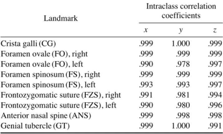
Table 3 reveals that the reproducibility of landmark tracing was excellent according to ICC (r›.978),10 and Table 4 shows that DCs and DFs at both jaws were statis- tically different by paired t-test (P⁄.001). In addition, the mean values of DCs at both jaws were higher than that of DFs. DCmxwas 24.865±21.263 mm which was 17 times higher than DFmx. On the other hand, DCmdwas 40.881±
34.325 mm which was 9.8 times higher than DFmd. This
result indicated that the maxillary deviation was more likely to be changeable depending on the determination of MSP.
Bland-Altman plot showed that the differences between DCs and DFs were not normally distributed (Fig. 2). A low correlation and wide spread plots in Bland-Altman imply good agreement of the variables, while a high cor- relation automatically imply that there is no good agree- ment between two variables.11The plot demonstrated that DCs were statistically higher than DFs depending on the distance from the midpoints to MSPs.
Discussion
The aim of this study was to verify that the determina- tion of different MSPs could result in different 3D cephalo- metric analysis especially for the evaluation of facial asym- metry and therefore imply that appropriate landmarks would be prerequisite for the 3D cephalometric analysis.
Asymmetry of cranium and face was not an uncommon finding.13-16However, there have been a few studies stating whether facial asymmetry was concurrent with cranial asymmetry or not.17In addition there was no study on a possible error according to the determination of MSP in 3D analysis. Therefore, this study could be helpful in expanding clinicians’ scope about 3D analysis for the selection of references.
Proffit et al stated that the main reason seeking a surgi- cal-orthodontic treatment was facial asymmetry and also a long face and skeletal class III deformity.18Accordingly, focusing on class III malocclusion was meaningful to aid in cephalometric analysis for patients with class III dento- facial deformity.
CG was generally accepted as the midpoint of cranium.
The anterior cranial base was closely related with facial skeleton19 and also CG resulted from the sophisticated growth pattern of the anterior cranial base.20Therefore, CG was regarded as linked to facial skeleton as well as cranium.
FO and FS were introduced as reproducible landmarks in both 2D and 3D analysis.4-6,21-24The use of these land- marks was justified by the findings that the growth of cra-
Table 3.Reproducibility of landmark tracing by intraclass correla- tion (ICC) coefficients
Intraclass correlation
Landmark coefficients
x y z
Crista galli (CG) .999 1.000 .999
Foramen ovale (FO), right .999 .999 .999
Foramen ovale (FO), left .990 .978 .997
Foramen spinosum (FS), right .999 .999 .999 Foramen spinosum (FS), left .993 .993 .997 Frontozygomatic suture (FZS), right .991 .981 .994 Frontozygomatic suture (FZS), left .990 .980 .996 Anterior nasal spine (ANS) .999 .998 .998
Genial tubercle (GT) .999 1.000 .991
Table 4.Comparison of jaw deviation between the facial and the cranial midsagittal planes by paired t-test (2-tailed)
Landmark for jaw midpoint DC DF P value
Anterior nasal spine (maxilla) 24.865±21.263 (mm) 1.404±1.116 (mm) P⁄.001
Genial tubercle (mandible) 40.881±34.325 (mm) 4.152±3.199 (mm) P⁄.001
DC: a perpendicular distance from the midpoint to the cranial midsagittal plane, DF: a perpendicular distance from the midpoint to the facial midsagittal plane, Statistical significance is determined at P⁄.05.
nial base was largely complete (¤85%) by 5 years of age and therefore neural or vascular foramina on cranial base can be used as stable landmarks.25-27As stated by Enlow et al, the anterior cranial base showed the different growth pattern and timing compared with the middle cranial base27 which encompassed FO and FS in the sphenoid bone.
Therefore, FO and FS appeared to have different nature compared with CG and FZS. However, FO and FS were suitable for the cephalometric analysis because of their stability in growth pattern. In the present study, apFO and apFS were used as the landmarks of the middle cranial base.
In performing the cephalometric analysis of the face, the reference points or lines were recommended on the non-movable or stable parts. From this regard, the land- mark, such as ANS,3on the jaws was not a suitable refer- ence for evaluation of facial asymmetry because it was possible to be altered during the surgical treatment. Some researchers have used the FZS in both 2D and 3D analy- sis.3,24,28FZS showed the good possibility for the reference in the assessment of facial asymmetry,3which confirmed the usage of FZS in the present study.
In spite of the lower absolute values of DCs and DFs on the maxilla than the mandible, the resultant variations of jaw deviation according to two MSPs were more pro- minent on the maxilla (Table 4). The reference determina- tion probably affected the maxillary asymmetry to a greater extent, which was consistent with finding from Haraguchi et al.16 Although, theoretically, mandibular deviation would be likely to be affected by the different MSPs because of the longer distance from cranium, this result was contrary to that. Further study is needed to explain this finding.
Especially, when facial asymmetry was evaluated, the reference points or lines should be selected with caution.3 From the results of the present study, when the reference points on the middle cranial base with CG were used, the midpoints of jaws were more deviated compared with the reference points on the anterior cranial base and facial skeleton. One should consider the exaggerated results of facial asymmetry using the reference points distant from the facial skeletons. In contrast, jaw deviation relative to the facial MSP might result in unreasonably decreased amounts compared with the cranial MSP. For example, the surgical correction of facial asymmetry in relation to the cranial MSP may lead to the asymmetric facial skele- tons in relation to the facial MSP. A corrective surgery possibly causes a different kind of asymmetry on facial structure depending on the MSP determination. Therefore,
by using 3D CT, the appropriate reference for MSP should be assessed for the individual patient in treatment of facial asymmetry. Further study are needed to support a study on the discrepancy between the surgical outcomes from the different MSPs.
The reference points should be chosen according to the aim of study, the anatomical structure of interest, and methodology. Therefore, if the reference on the middle cranial base was selected with a specific need, it could be a reasonable landmark for cephalometric analysis. One example of this landmark is the mandibular condyle which is considerably consistent with the middle cranial base in growth pattern.22,27 However, one should consider the possible discrepancy originated from the reference points on the anterior and the middle cranial bases relatively.
Confining the subjects to patients with class III dento- facial deformity was the limitation of the present study.
There has been no finding regarding the discrepancy of MSPs according to different skeletal types. Therefore the results of the present study cannot be applied to class I or II skeletal malocclusion. Although it was difficult to col- lect CT data of class I or II skeletal malocclusion because patients with class III dentofacial deformity were the ones who have mainly undergone the surgical treatment in our hospital, those skeletal types should be included.
The close structural connection and proximity between the cranial base and the facial skeleton were suggested.19,27 However, it was implied that the cranial center may not be corresponding to the facial center after the completion of growth by accepting the results of the present study in a broad sense from the anthropometric perspective.
In conclusion, the facial MSP was not in agreement with the cranial MSP in class III dentofacial deformity using three-dimensional computed tomography. The mid- points of the jaws were more deviated using the cranial MSP than the facial MSP. Therefore using the cranial MSP could exaggerate the result of the jaw deviation.
The cephalometric analyses can be discrepant according to the reference determination of MSP in evaluating facial asymmetry of the same patient. With this regard the refer- ence points on the cranial base and facial skeleton should be carefully selected for the cephalometric analysis of facial asymmetry in patients with class III dentofacial deformity.
References
1. Finlay LM. Craniometry and cephalometry: a history prior to the advent of radiography. Angle Orthod 1980; 50 : 312-21.
2. The American Heritage medical dictionary. Boston: Houghton
Mifflin; 2008.
3. Trpkova B, Prasad NG, Lam EW, Raboud D, Glover KE, Major PW. Assessment of facial asymmetries from posteroanterior cephalograms: validity of reference lines. Am J Orthod Dento- facial Orthop 2003; 123 : 512-20.
4. Suri S, Utreja A, Khandelwal N, Mago SK. Craniofacial com- puterized tomography analysis of the midface of patients with repaired complete unilateral cleft lip and palate. Am J Orthod Dentofacial Orthop 2008; 134 : 418-29.
5. Lagravere MO, Hansen L, Harzer W, Major PW. Plane orien- tation for standardization in 3-dimensional cephalometric anal- ysis with computerized tomography imaging. Am J Orthod Dentofacial Orthop 2006; 129 : 601-4.
6. Lagravere MO, Major PW. Proposed reference point for 3- dimensional cephalometric analysis with cone-beam comput- erized tomography. Am J Orthod Dentofacial Orthop 2005;
128 : 657-60.
7. Olszewski R, Cosnard G, Macq B, Mahy P, Reychler H. 3D CT-based cephalometric analysis: 3D cephalometric theoreti- cal concept and software. Neuroradiology 2006; 48 : 853-62.
8. Deguchi T Sr, Katashiba S, Inami T, Foong KW, Huak CY.
Morphologic quantification of the maxilla and the mandible with cone-beam computed tomography. Am J Orthod Dento- facial Orthop 2010; 137 : 218-22.
9. Muramatsu A, Nawa H, Kimura M, Yoshida K, Maeda M, Katsumata A, et al. Reproducibility of maxillofacial anatomic landmarks on 3-dimensional computed tomographic images determined with the 95% confidence ellipse method. Angle Orthod 2008; 78 : 396-402.
10. Fleiss JL, Chilton NW. The measurement of interexaminer agreement on periodontal disease. J Periodontal Res 1983; 18 : 601-6.
11. Bland JM, Altman DG. Statistical methods for assessing agree- ment between two methods of clinical measurement. Lancet 1986; 1 : 307-10.
12. Adams GL, Gansky SA, Miller AJ, Harrell WE Jr, Hatcher DC.
Comparison between traditional 2-dimensional cephalometry and a 3-dimensional approach on human dry skulls. Am J Orthod Dentofacial Orthop 2004; 126 : 397-409.
13. Pirttiniemi PM. Associations of mandibular and facial asym- metries - a review. Am J Orthod Dentofacial Orthop 1994;
106 : 191-200.
14. Galaburda AM, LeMay M, Kemper TL, Geschwind N. Right- left asymmetrics in the brain. Science 1978; 199 : 852-6.
15. Severt TR, Proffit WR. The prevalence of facial asymmetry in the dentofacial deformities population at the University of North Carolina. Int J Adult Orthodon Orthognath Surg 1997;
12 : 171-6.
16. Haraguchi S, Takada K, Yasuda Y. Facial asymmetry in sub- jects with skeletal Class III deformity. Angle Orthod 2002; 72 : 28-35.
17. Baek SH, Cho IS, Chang YI, Kim MJ. Skeletodental factors affecting chin point deviation in female patients with class III malocclusion and facial asymmetry: a three-dimensional analy- sis using computed tomography. Oral Surg Oral Med Oral Pathol Oral Radiol Endod 2007; 104 : 628-39.
18. Proffit WR, Phillips C, Dann C 4th. Who seeks surgical-ortho- dontic treatment? Int J Adult Orthodon Orthognath Surg 1990;
5 : 153-60.
19. Enlow DH, McNamara JA Jr. The neurocranial basis for facial form and pattern. Angle Orthod 1973; 43 : 256-70.
20. Friede H. Normal development and growth of the human neu- rocranium and cranial base. Scand J Plast Reconstr Surg 1981;
15 : 163-9.
21. Janson G, de Lima KJ, Woodside DG, Metaxas A, de Freitas MR, Henriques JF. Class II subdivision malocclusion types and evaluation of their asymmetries. Am J Orthod Dentofacial Orthop 2007; 131 : 57-66.
22. Kanomi R, Hidaka O, Yamada C, Takada K. Asymmetry in the condylar long axis and first molar rotation. J Dent Res 2004;
83 : 109-14.
23. Williamson PC, Major PW, Nebbe B, Glover KE. Landmark identification error in submentovertex cephalometrics. A computerized method for determining the condylar long axis.
Oral Surg Oral Med Oral Pathol Oral Radiol Endod 1998; 86 : 360-9.
24. Lascala CA, Panella J, Marques MM. Analysis of the accuracy of linear measurements obtained by cone beam computed tomography (CBCT-NewTom). Dentomaxillofac Radiol 2004;
33 : 291-4.
25. Waitzman AA, Posnick JC, Armstrong DC, Pron GE. Cranio- facial skeletal measurements based on computed tomography:
Part II. Normal values and growth trends. Cleft Palate Craniofac J 1992; 29 : 118-28.
26. Sgouros S, Natarajan K, Hockley AD, Goldin JH, Wake M.
Skull base growth in childhood. Pediatr Neurosurg 1999; 31 : 259-68.
27. Enlow DH, Hans MG. Essentials of facial growth. Philadelphia:
WB Saunders; 1996.
28. Lee KH, Hwang HS, Curry S, Boyd RL, Norris K, Baumrind S. Effect of cephalometer misalignment on calculations of facial asymmetry. Am J Orthod Dentofacial Orthop 2007; 132 : 15-27.