www.i-mri.org 127
Ductal Carcinoma in situ with
Multicystic Changes in a Patient with Interstitial Mammoplasty via Paraffin Injection: MRI and Pathological
Findings
This is an Open Access article distributed under the terms of the Creative Commons Attribution Non-Commercial License (http://creativecommons.org/licenses/
by-nc/3.0/) which permits unrestricted non-commercial use, distribution, and reproduction in any medium, provided the original work is properly cited.
Received: May 18, 2015 Revised: May 28, 2015 Accepted: May 29, 2015 Correspondence to:
Ok Hee Woo, M.D.
Department of Radiology, Korea University Guro Hospital, 148, Gurodong-ro, Guro-gu, Seoul 152-703, Korea.
Tel. +82-2-2626-1339 Fax. +82-2-863-9282 Email: [email protected]
Copyright © 2015 Korean Society of Magnetic Resonance in Medicine (KSMRM)
iMRI 2015;19:127-130 http://dx.doi.org/10.13104/imri.2015.19.2.127
Case Report
Direct injection of foreign material, such as liquid paraffin and silicone, into the breast can induce a foreign body granulomatous reaction and fibrosis, resulting in hard, nodular breast masses and architectural distortion that can mimic neoplasm.
Conventional methods, including physical examination, mammography, and ultrasonography are of little use to differentiate between foreign body-induced mastopathy and breast cancer. In patients with foreign body injection such as breast augmentation, dynamic contrast enhanced MR imaging is an excellent imaging modality. Here, the authors report the MR imaging and pathological findings of ductal carcinoma in situ (DCIS) with multicystic changes in a 41-year-old woman with a previous history of interstitial mammoplasty by paraffin injection.
Keywords: DCIS; Breast augmentation via Paraffin; Magnetic resonance imaging (MRI)
INTRODUCTION
In patients with foreign body injection into the breasts, conventional methods including physical examination, mammography, and ultrasonography are of little use to differentiate between foreign body-induced mastopathy and breast cancer (1). Dynamic contrast-enhanced MR imaging is an excellent imaging modality in such cases (2-4). In this case report, we present a 41-year-old woman with ductal carcinoma in situ (DCIS) with multicystic changes in the right breast and previous interstitial mammoplasty due to paraffin injection in both breasts. We used MR imaging as our diagnostic tool.
CASE REPORT
A 41-year-old woman was referred to our hospital with a palpable right breast mass, which she had felt for 6 months. She had a history of free paraffin injection in
pISSN 2384-1095 eISSN 2384-1109
Jiyoon Park1, Ok Hee Woo1, Chungyeul Kim2, Kyu Ran Cho3, Bo Kyoung Seo4
1Department of Radiology, Korea University Guro Hospital, Seoul, Korea
2Department of Pathology, Korea University Guro Hospital, Seoul, Korea
3Department of Radiology, Korea University Anam Hospital, Seoul, Korea
4Department of Radiology, Korea University Ansan Hospital, Gyeonggi-do, Korea
www.i-mri.org 128
DCIS with Multicystic Change | Jiyoon Park, et al.
a b
c d
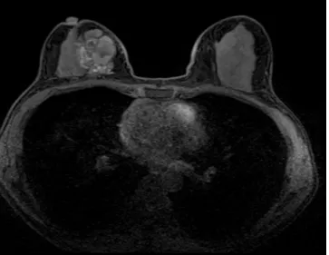
Fig. 1. A 41-year-old woman with DCIS and a previous interstitial mammoplasty via paraffin injection. (a) Axial pre-contrast enhanced T1-weighted, (b) fat-suppressed T1- weighted, (c) T2-weighted MR images of the right breast showing an approximate 4.0 × 4.9 cm sized, multicystic mass. Multifocal, well-circumscribed nodular lesions were observed (arrows) in the subcutaneous layer of both breasts on (d) axial pre-contrast enhanced T1-weighted, and (e) fat-suppressed T1-weighted images.
e
www.i-mri.org 129
http://dx.doi.org/10.13104/imri.2015.19.2.127
both breasts five years previously. Physical examination revealed an approximate 5-cm-sized, hard, mobile mass in the right breast without axillary lymphadenopathy. Due to the difficulty in localization of the palpable mass from artifacts of paraffinomas on conventional US, the patient underwent MRI of both breasts using a 3-Tesla MRI system (Skyra; Siemens, Erlangen, Germany) and a dedicated breast coil. On pre-contrast enhanced axial T1-weighted (Fig. 1a), fat suppressed T1-weighted (Fig. 1b), and T2-weighted (Fig.
1c) MR images, an approximate 4.0 × 4.9 × 4.2 cm sized, multicystic mass was noted in the right breast in the 3-6 o'clock region. In addition, multifocal, non-enhancing, well- circumscribed lesions were observed in the subcutaneous layer of both breasts, suggesting injected foreign bodies (Fig.
1d, e). A post-contrast enhanced T1-weighted MR image
showed a multicystic mass containing several enhancing mural nodules and septa (Fig. 1f). These enhancing nodules showed a type III enhancement kinetic curve, suggesting moderate concern for malignancy. The patient underwent a modified radical mastectomy, sentinel lymph node dissection, and reconstruction with an autologous tissue flap. The pathological diagnosis was ductal carcinoma in situ (DCIS) with lipogranuloma (Fig. 1g, h), without lymph node metastasis. Several nodular, enhancing portions within the multicystic mass on contrast enhancement MRI correlated with DCIS upon pathological findings. Regarding the pathology, multifocal nodular DCIS components were observed and the summation of these was approximately 2.5 x 2.4 cm in extent.
f g
Fig. 1. (f) Well-enhancing mural nodules and septa are seen on an axial post-contrast enhanced T1- weighted fat-suppressed MR image of the right breast.
(g) Photomicrograph of the histopathological specimen showing ductal carcinoma in situ, with a micropapillary pattern and epithelial projection into the duct lumen, lacking a fibrovascular core (Hematoxylin & Eosin staining,
× 100; total extent of DCIS on pathology, roughly 2.5 × 2.4 cm). (h) Photomicrograph of the histopathological specimen showing lipogranuloma (Hematoxylin & Eosin staining, × 100).
h
www.i-mri.org 130
DCIS with Multicystic Change | Jiyoon Park, et al.
DISCUSSION
Breast augmentation using liquid paraffin was used in the early 1900s until the introduction of silicon in 1964 by Cronin and Gerow (5). However, these methods were quickly abandoned due to the serious complications associated with the procedure, including local granulomatous reactions, material-induced mastitis, foreign body reactions, fibrosis, migration of the material, or induction of autoimmune reactions (1). Mammographic findings of paraffinoma include parenchymal distortion, streaky opacities, dystrophic parenchymal calcification around paraffin droplets, and multiple masses (6). Ultrasonography reveals multiple cystic masses in breast parenchyma, axilla, and pectoral muscle (7). Thus, concern about the interference of injected material with physical examination, mammography, and ultrasonography has been raised.
A diagnosis of breast cancer in this setting can be aided by MR imaging (2-4). Distinct MR features of paraffinoma have been reported. Khong et al. (8), hypothesized that paraffinomas have two components: a plaque-like fibrous component that shows intermediate intensity on T1- weighted imaging and hypointensity on T2-weighted images, and a liquid paraffin component that shows hypointensity on both T1- and T2-weighted images. Erguvan-Dogan et al. (7), reported two cases of paraffinomas with a latency period shorter than three years, appearing as low-intensity structures on T1- and high-intensity on T2- and fat- suppressed T2-weighted images, with no enhancement by gadopentetate dimeglumine.
Breast cancer has typical malignant morphological and kinetic features: irregular shape, irregular margins, ductal or segmental distribution, heterogeneous or internal rim, and type 2 or 3 enhancement kinetic patterns (1, 3). DCIS is well-known for its nonmass enhancement with a segmental distribution and early rapid uptake with a plateau curve enhancement on MRI. However, with equal frequency, DCIS can also appear as mass lesions with homogeneous or heterogeneous internal enhancement, but rarely with rim enhancement (9, 10).
Our patient had received interstitial mammoplasty via paraffin injection and presented with a palpable mass in the right breast. Due to the known inability to distinguish a foreign body-induced mass from breast cancer by conventional methods, we used contrast-enhanced MR imaging. In the setting of injected breast enhancement, a MR finding of a multicystic mass alone can be difficult to differentiate as a foreign body-induced inflammatory mass
or a malignant condition. However, our patient presented with several enhancing nodules and septa between the cysts, which helped the diagnosis of carcinoma.
In conclusion, MRI plays an important role in the detection of breast cancer in patients with injected breast enhancement and management of DCIS with multicystic change.
REFERENCES
1. Cheung YC, Su MY, Ng SH, Lee KF, Chen SC, Lo YF. Lumpy silicone-injected breasts: enhanced MRI and microscopic correlation. Clin Imaging 2002;26:397-404
2. Gorczyca DP. MR imaging of breast implants. Magn Reson Imaging Clin N Am 1994;2:659-672
3. Youk JH, Son EJ, Kim EK, et al. Diagnosis of breast cancer at dynamic MRI in patients with breast augmentation by paraffin or silicone injection. Clin Radiol 2009;64:1175- 1180
4. Peng HL, Wu CC, Choi WM, Hui MS, Lu TN, Chen LK. Breast cancer detection using magnetic resonance imaging in breasts injected with liquid silicone. Plast Reconstr Surg 1999;104:2116-2120
5. Cronin TD, Gerow FJ. Augmentation mammoplasty: a new “natural feel” prosthesis. In Broadbent TR, American Association of Plastic Surgeons, American Society of Plastic and Reconstructive Surgeons, International Confederation for Plastic Surgery, Transactions of the International Society of Plastic Surgeons. Transactions of the Third International Congress of Plastic and Reconstructive Surgery. Amsterdam; Excerpta Medica Foundation 1963:
41-49
6. Yang WT, Suen M, Ho WS, Metreweli C. Paraffinomas of the breast: mammographic, ultrasonographic and radiographic appearances with clinical and histopathological correlation.
Clin Radiol 1996;51:130-133
7. Erguvan-Dogan B, Yang WT. Direct injection of paraffin into the breast: mammographic, sonographic, and MRI features of early complications. AJR Am J Roentgenol 2006;186:888-894
8. Khong PL, Ho LW, Chan JH, Leong LL. MR imaging of breast paraffinomas. AJR Am J Roentgenol 1999;173:929-932 9. Kim JA, Son EJ, Youk JH, et al. MRI findings of pure
ductal carcinoma in situ: kinetic characteristics compared according to lesion type and histopathologic factors. AJR Am J Roentgenol 2011;196:1450-1456
10. Yamada T, Mori N, Watanabe M, et al. Radiologic- pathologic correlation of ductal carcinoma in situ.
Radiographics 2010;30:1183-1198