Pseudomembranous Colitis: A Complicated Case with Transient Increase of Carcinoembryonic Antigen
Dong In Nam, Chung Kang, Il Hyung Jung, Hyun Gee Moon, Bo Ram Youn, Nam Hun Lee
Division of Gasteroenterology, Department of Internal Medicine, Gwangju Veterans Hospital, Gwangju, Korea
Introduction
Pseudomembranous colitis (PMC) is a frequently severe and sometimes fatal iatrogenic form of inflammatory bowel disease that is associated with antibiotics in the vast majority of cases.
The most common clinical features of PMC include watery diarrhea, abdominal pain and cramping, fever, leukocytosis, hypoalbuminemia, and hypovolemia [1]. Metronidazole and vancomycin were commonly used for PMC in clinical practice.
Ascites associated with PMC is commonly reported, but PMC accompanied by ascites and pleural effusion has been rarely reported worldwide; only one case has been reported in South Korea [2-4]. Elevation in the carcinoembryonic antigen (CEA) level within ascites fluid has also been rarely reported in patients with PMC [4]. We herein report a case involving a patient with sigmoidoscopy-confirmed PMC who showed elevated serum and ascites CEA levels with a normal level in pleural effusion.
Case
An 80-year-old male was admitted to our hospital with complaints of watery diarrhea, dyspnea, and abdominal disten- sion. He had no recent travel history and had a medical history of hypertension. Eight days before admission, he fell down while walking, resulting in a laceration on his leg. He was admitted to a local hospital, and intravenous ceftriaxone of 2.0 g/day was initiated after the laceration was sutured. The patient developed diarrhea after 5 days of treatment. He was suspected to have acute gastroenteritis and was thus treated with scopolamine and continuous intravenous antibiotics. His diarrhea, dyspnea, and abdominal distension were aggravated ever since.
On physical examination, the patient’s body temperature was 37.8oC, arterial pressure was 100/70 mmHg, pulse rate was 110 beats/min, and respiratory rate was 24 breaths/min. Lung auscultation revealed decreased sounds in both lower lobe fields, and heart auscultation revealed no murmur. The patient’s abdo-
Report
pISSN 2234-3180 • eISSN 2234-2591Pseudomembranous colitis (PMC) is a frequent cause of morbidity and mortality among hospitalized patients. Although diarrhea is the most common manifestation, PMC may be associated with intraperitoneal fluid accumulation in the severe cases. And a few cases showing both ascites and pleural effusion have been reported in patients with PMC. We report a case of PMC who showed elevated serum and ascites levels of carcinoembry- onic antigen (CEA) with a normal CEA level in pleural effusion and who successfully recovered after oral administration of metronidazole. After treatment, the serum CEA level returned to the reference range. (Ewha Med J 2015;38(1):54-58)
Received August 20, 2014 Accepted February 10, 2015 Corresponding author Nam Hun Lee
Department of Internal Medicine, Gwangju Veterans Hospital,
99 Chumdanwolbong-ro, Gwangsan-gu, Gwangju 506-705, Korea
Tel: 82-62-602-6280, Fax: 82-62-602-6931 E-mail: [email protected]
Key Words
Pseudomembranous colitis; Ascites; Pleural effusion; Carcinoembryonic antigen
men was markedly distended, and his lower limbs were edema- tous. Other findings were not remarkable.
The laboratory test showed a white blood cell (WBC) count of 15,320/mm3, hemoglobin level of 11.1 g/dL, and platelet count of 231,000/mm3. The C-reactive protein (CRP) level was 115.9 mg/L (normal, approximately 5 mg/L), and the erythrocyte sedimentation rate (ESR) was 46 mm/hr (normal, approxi- mately 15 mm/hr). Serum chemistry revealed a total protein level of 4.8 g/dL (normal range, 6.7 to 8.7 g/dL) and albumin level of 2.1 g/dL (normal range, 3.5 to 5.2 g/dL). The serum CEA level was 15.9 ng/mL (normal, approximately 5 ng/mL).
A Clostridium difficile toxin enzyme-linked immunosorbent as- say (ELISA) was performed twice and showed negative results in both instances.
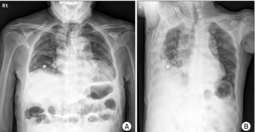
Chest radiographs showed diffuse ground glass opacities, peri- bronchial infiltration in both lungs, and bilateral costophrenic angle blunting. Decubitus chest radiographs showed a moderate amount of bilateral pleural effusion (Fig. 1). Pleural fluid analy-
sis revealed a WBC count of 230/mm3 (neurophils, 3%), total protein level of 2.3 g/dL, albumin level of 0.1 g/dL, adenosine deaminase level of 10 U/L, lactate dehydrogenase level of 161 U/L, and CEA level of 4.3 ng/mL. The fluid was a transudate.
A transthoracic echocardiogram showed no abnormal findings.
Abdominal computed tomography (CT) showed diffuse wall thickening of the colonic loops and a moderate amount of as- cites (Fig. 2A). Ascites fluid analysis revealed a WBC count of 6,880/mm3 (neutrophils, 85%), total protein level of 1.5 g/dL, albumin level of 1.3 g/dL, lactate dehydrogenase level of 185 U/L, adenosine deaminase level of 17.0 IU/L, and CEA level of 19.2 ng/mL. Gram staining and culture results were negative.
Serum-ascites albumin gradient was 0.8 g/dL.
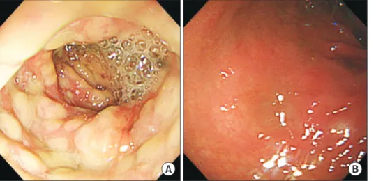
Flexible sigmoidoscopy showed multiple diffuse, yellowish plaques on edematous mucosa (Fig. 3A). The patient was diag- nosed with PMC based on the typical sigmoidoscopic findings, and treatment with metronidazole was initiated (500 mg three times daily for 14 days). The diarrhea, abdominal pain, and
Fig. 1. Chest radiograph upon admis- sion. (A) It shows diffuse ground glass opacities, peribronchial infiltrations in both lungs, and bilateral costophrenic angle blunting. (B) Right decubitus chest radiograph shows a moderate amount of pleural effusion.
Fig. 2. Abdominal computed tomography (CT). (A) It shows diffuse wall thicken- ing of the colonic loops and a moderate amount of ascites. (B) Marked improve- ment in the diffuse wall thickening of the colonic loops and no ascites are present on day 21 of treatment.
A B
A B
fever had resolved by day 5 of treatment. On day 14 of treat- ment, chest radiographs no longer showed diffuse ground glass opacities, peribronchial infiltration, or bilateral costophrenic angle blunting. On day 21 of treatment, the patient underwent a follow-up colonoscopy because of the elevated serum CEA level, and the previous lesions with pseudomembrane disap- peared (Fig. 3B). Esophagogastroduodenoscopy showed no abnormal findings. The repeated abdominal CT showed marked improvement in the diffuse wall thickening of the colonic loops and no ascites (Fig. 2B). The follow-up laboratory test showed a WBC count of 6,770/mm3, hemoglobin level of 10.9 g/dL, platelet count of 245,000/mm3, and CRP level of 0.9 mg/L.
The serum chemistry results showed a total protein level of 7.0 g/dL and albumin level of 3.2 g/dL. The serum CEA level had normalized at 4.6 ng/mL. The patient’s symptoms resolved completely, and he was discharged from the hospital.
Discussion
Disruption of the protective colonic flora by broad-spectrum antibiotics is the most common predisposing factor for the de- velopment of PMC. Various antibiotics may have distinct influ- ences on not only the gut microbiota but also C. difficile. Severe PMC is characterized by abdominal pain, profuse diarrhea, and systemic symptoms such as fever, anorexia, nausea, and malaise.
Leukocytosis, elevated CRP levels, and low albumin levels are frequently seen in patients with severe PMC [5].
C. difficile-associated disease is usually diagnosed follow- ing the demonstration of toxin A and/or B in stool samples. A number of studies have shown that for both assays, the false-
negative rates can be high (10~30%) [6]. Therefore, flexible sigmoidoscopy has been proposed in patients suspected to have C. difficile-associated diarrhea but who have negative stool test results for C. difficile toxins. In our patient, the ELISA results were twice negative for both toxins A and B.
Plain radiographic findings in patients with PMC vary de- pending on the severity and extent of disease. Colonic ileus, small bowel ileus, ascites, and nodular haustral thickening have been identified in patients with PMC. Common CT findings include wall thickening, low-attenuation mural thickening cor- responding to mucosal and submucosal edema, the target sign, pericolonic stranding, and ascites [7].
Ascites is demonstrated by abdominal ultrasonography and CT in approximately 16% to 77% of patients with PMC [2]. Three possible mechanisms have been proposed for the pathogenesis of ascites in patients with PMC: hypoalbuminemia, transmural colonic inflammation with microperforation and infectious peri- tonitis, and toxin-mediated generation of cytokines that enhance vascular permeability [1].
Pleural effusion is rare in patients with PMC, and when pres- ent, seems to be associated with hypoalbuminemia. The pres- ence of pleural effusion also reflects some extent of altered car- diac function, which contributes to the unfavorable outcome [8].
In this patient, the presence of hypoalbuminemia may explain the pathogenesis of the pleural effusion because of the normal N-terminal pro-brain natriuretic peptide level and transthoracic echocardiographic findings.
The serum level of CEA may be increased in patients with malignancies that secrete CEA into the circulation (gastrointesti- nal tract, colorectal, liver, lung, breast, ovarian, pancreatic, and
Fig. 3. Endoscopic findings. (A) Flexible sigmoidoscopy shows multiple diffuse, yellowish plaques on ede matous mucosa.
(B) Colonoscopy shows the absence of both the mucosal edema and pseudo- membrane on day 21 of treatment.
A B
prostate cancers as well as medullary thyroid carcinoma) [9].
But these levels can also increase in smokers and patients with benign disease, including non-malignant liver disease (cirrhosis, hepatitis, alcoholic liver disease), chronic kidney disease, lung disease (pleural inflammation, pneumonia), and bowel inflam- mation (ulcerative colitis, diveticulitis). Benign diseases only rarely give rise to serum values >10 ng/mL [10]. Serum and as- cites CEA levels should be examined in all patients with ascites.
At an ascites CEA cut-off level of 10 ng/mL in one study, 50%
of patients with cancer exhibited significantly elevated serum and ascites CEA levels; however, the serum CEA level was not increased in patients with benign disease [11]. Interestingly, the ascites and serum CEA levels were simultaneously elevated in the present case. Therefore, we evaluated the patient for a hid- den malignancy by esophagogastroduodenoscopy, colonoscopy, and chest and abdominal CT. No findings led us to suspect the presence of malignancy.
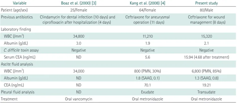
Table 1 provides a summary of previously reported cases of PMC accompanied by ascites and pleural effusion. As in other cases, the present case was also accompanied by ascites and pleural effusion and showed improvement after treatment. Ad- ditionally, elevated serum CEA levels returned to the reference ranges after treatment.
In conclusion, the pleural effusion is synchronous with ascites
in the severe PMC. These abnormalities may be improved by treatment of PMC. Additionally, an elevated CEA level in the patients with severe bowel inflammation such as complicated PMC can be correlated with inflammation.
References
1. Zuckerman E, Kanel G, Ha C, Kahn J, Gottesman BS, Korula J.
Low albumin gradient ascites complicating severe pseudomem- branous colitis. Gastroenterology 1997;112:991-994.
2. Kang CI, Lee HJ, Kim YT, Lee YH, Lee SY, Cho CM, et al. A case of pseudomembraneous colitis associated with ascites. Korean J Gastrointest Endosc 2002;25:466-469.
3. Boaz A, Dan M, Charuzi I, Landau O, Aloni Y, Kyzer S. Pseudo- membranous colitis: report of a severe case with unusual clini- cal signs in a young nurse. Dis Colon Rectum 2000;43:264-266.
4. Kang SJ, Kim KH, Kim KS, Hur JH, Shin YM, Moon HG, et al. A case of pseudomembranous colitis with pleural effusion and ascites. Korean J Med 2008;74(Suppl 1):S53-S57.
5. Monaghan T, Boswell T, Mahida YR. Recent advances in Clos- tridium difficile-associated disease. Gut 2008;57:850-860.
6. Fordtran JS. Colitis due to Clostridium difficile toxins: underdi- agnosed, highly virulent, and nosocomial. Proc (Bayl Univ Med Cent) 2006;19:3-12.
7. Kawamoto S, Horton KM, Fishman EK. Pseudomembranous colitis: spectrum of imaging findings with clinical and pathologic correlation. Radiographics 1999;19:887-897.
8. Valiquette L, Pepin J, Do XV, Nault V, Beaulieu AA, Bedard J, et Table 1. Summary of cases reported previously and the present case of pseudomembranous colitis
Variable Boaz et al. (2000) [3] Kang et al. (2008) [4] Present study
Patient (age/sex) 25/Female 64/Female 80/Male
Previous antibiotics Clindamycin for dental infection (10 days) and ciprofloxacin after hospitalization (4 days)
Ceftriaxone for aneurysmal operation (11 days)
Ceftriaxone for wound management (8 days) Laboratory finding
WBC (/mm3) 34,800 11,210 15,320
Albumin (g/dL) 3.0 1.9 2.1
C. difficile toxin assay Negative Negative Negative
Serum CEA (ng/mL) ND 5.6 15.94 (4.68 after treatment)
Ascite fluid analysis
WBC (/mm3) 34,000 800 (PMN, 30%) 6,800 (PMN, 85%)
Albumin (g/dL) ND 1.8 (SAAG, 0.1) 1.3 (SAAG, 0.8)
CEA (ng/mL) ND 70.1 19.21
Pleural fluid analysis ND Exudate Transudate
Treatment Oral vancomycin Oral metronidazole Oral metronidazole
WBC, white blood cell; CEA, carcinoembryonic antigen; C. difficile, Clostridium difficile; ND, no description; PMN, polymorphonuclear leukocyte;
SAAG, serum ascites albumin gradient.
al. Prediction of complicated Clostridium difficile infection by pleural effusion and increased wall thickness on computed to- mography. Clin Infect Dis 2009;49:554-560.
9. Malati T. Tumour markers: an overview. Indian J Clin Biochem 2007;22:17-31.
10. Duffy MJ. Carcinoembryonic antigen as a marker for colorectal cancer: is it clinically useful? Clin Chem 2001;47:624-630.
11. Loewenstein MS, Rittgers RA, Feinerman AE, Kupchik HZ, Mar- cel BR, Koff RS, et al. Carcinoembryonic antigen assay of ascites and detection of malignancy. Ann Intern Med 1978;88:635-638.