D I A B E T E S & M E T A B O L I S M J O U R N A L
This is an Open Access article distributed under the terms of the Creative Commons At- tribution Non-Commercial License (http://creativecommons.org/licenses/by-nc/3.0/) which permits unrestricted non-commercial use, distribution, and reproduction in any medium, provided the original work is properly cited.
Pancreatic α-Cell Dysfunction in Type 2 Diabetes:
Old Kids on the Block
Jun Sung Moon, Kyu Chang Won
Department of Internal Medicine, Yeungnam University College of Medicine, Daegu, Korea
Type 2 diabetes (T2D) has been known as ‘bi-hormonal disorder’ since decades ago, the role of glucagon from α-cell has lan- guished whereas β-cell taking center stage. Recently, numerous findings indicate that the defects of glucagon secretion get in- volve with development and exacerbation of hyperglycemia in T2D. Aberrant α-cell responses exhibit both fasting and postpran- dial states: hyperglucagonemia contributes to fasting hyperglycemia caused by inappropriate hepatic glucose production, and to postprandial hyperglycemia owing to blunted α-cell suppression. During hypoglycemia, insufficient counter-regulation response is also observed in advanced T2D. Though many debates still remained for exact mechanisms behind the dysregulation of α-cell in T2D, it is clear that the blockade of glucagon receptor or suppression of glucagon secretion from α-cell would be novel thera- peutic targets for control of hyperglycemia. Whereas there have not been remarkable advances in developing new class of drugs, currently available glucagon-like peptide-1 and dipeptidyl peptidase-IV inhibitors could be options for treatment of hypergluca- gonemia. In this review, we focus on α-cell dysfunction and therapeutic potentials of targeting α-cell in T2D.
Keywords: Diabetes mellitus, type 2; Glucagon; Glucagon-secreting cells; Insulin; Insulin-secreting cells
Corresponding author: Kyu Chang Won
Department of Internal Medicine, Yeungnam University College of Medicine, 170 Hyeonchung-ro, Nam-gu, Daegu 705-717, Korea
E-mail: [email protected]
INTRODUCTION
Pancreatic islets, small islands of endocrine cells in the gland, play critical role in blood glucose homeostasis through produc- ing insulin from β-cells and glucagon from α-cells. Under nor- mal physiology, α- and β-cells in the islet regulate each other reciprocally and thereby systemic glucose levels are maintained within narrow range. About 40 years ago, Unger and Orci [1]
suggested “bi-hormonal theory,” which presents that relatively or absolutely hypoinsulinemia and relative hyperglucagonemia raise hyperglycemia in type 2 diabetes (T2D). But the role of α-cell has been neglected for a long time whereas β-cell has been centered in the field of diabetic pathophysiology. Many treatment approaches have been focused on insulin deficiency such as insulin injection, stimulation of endogenous insulin production or by improving insulin sensitivity. Though these strategies have been effective in many type 2 diabetic patients,
diabetes still remains impasse and need to more efforts for overcoming [2,3]. Recently, the role of α-cell was highlighted again that the excessive glucagon from dysfunctional α-cell was recognized an important therapeutic target in diabetes.
This has partly due to the recognition of the glucagon-suppres- sive effect of incretin hormones glucagon-like peptide-1 (GLP- 1) in T2D [4]. In this review, we will focus on the role of α-cell and hyperglucagonemia in the pathogenesis of T2D. Further- more, the clinical relevance and implications for treatments di- rected at targeting glucagon secretion will be discussed.
α-CELL FUNCTION AND GLUCAGON SECRETION
Glucagon is a 29-amino acid, 3485-Da peptide hormone re- leased from pancreatic islet α-cells, cleaved by prohormone convertase-2 from proglucagon molecule. Complex mecha- http://dx.doi.org/10.4093/dmj.2015.39.1.1
pISSN 2233-6079 · eISSN 2233-6087
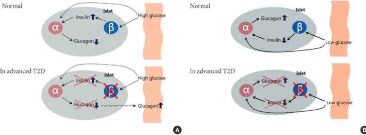
A Normal
In advanced T2D
B Normal
In advanced T2D
Fig. 1. Intra-islet insulin & glucagon secretion. Normal (in nondiabetes) and advanced type 2 diabetes (T2D) of the relationship between the inhibitory effects of pancreatic β-cell insulin secretion on pancreatic α-cell glucagon secretion. Normally, an in- crease in plasma glucose level causes an increase in β-cell insulin secretion that prevents an increase in α-cell glucagon secretion in response to meal. In advanced T2D, however, β-cell failure which is lack of intra-islet signaling result in not only fail to sup- press but also an increase in pancreatic α-cell glucagon secretion (A). A decrease in plasma glucose level causes a decrease in β-cell insulin secretion that signals an increase in α-cell glucagon secretion during hypoglycemia. On the other hand, in the ad- vanced T2D, a decrease in plasma glucose cannot cause a decrease in β-cell insulin secretion, and the absence of that signal re- sults in no increase in pancreatic α-cell glucagon secretion during hypoglycemia (B).
nism are involved in the regulation of pancreatic α-cell, and glucagon secretion is in response to a variety of nutrient, neu- ral, and hormonal factors [5,6]. Glucagon is well known as the counter-regulatory hormone to insulin, and stimulates hepatic glycogenolysis, gluconeogenesis, fatty acid oxidation and keto- genesis. The main stimulus for islet glucagon release is low blood glucose levels, but amino acids such as L-arginine, acti- vation of autonomic nervous system and gastric inhibitory polypeptide (GIP) also stimulate glucagon secretion [7-10]. In contrast, high glucose levels, insulin, somatostatin, amylin, and GLP-1 are suppressor of glucagon release [11-14].
DYSREGULATION OF α-CELL FUNCTION IN TYPE 2 DIABETES
T2D has been considered to as a ‘bihormonal disorder’ [1,15], which is characterized by a progressive pancreatic islet dys- function. Similar to β-cell function in the diabetes, there are a number of characteristic dysfunctions in α-cell. Extremely high plasma glucagon concentrations are observed in states of insulin deficiency, such as type 1 diabetes (T1D), advanced T2D or diabetic ketoacidosis [6,16]. In usual case of T2D, plasma glucagon level is often increased and it suggested con-
tributing to the development of glucose toxicity and exacerba- tion of diabetes [17-21]. Fasting hyperglucagonemia is ob- served in some but not all T2D patients with moderate glyce- mic control [22-24]. ‘Paradoxical’ glucagon response to meal is also observed in patients with T2D, which are elevated glu- cagon levels following a carbohydrate meal and leads to post- prandial hyperglycemia [25-27]. The response of α-cell to hy- perglycemia in diabetes is blunted or vanishing, and plasma glucagon remains inappropriately excessive at comparable blood glucose levels. Moreover, glucagon production is elevat- ed by another stimulus, such as arginine or protein-rich foods, to greater extent in T2D than nondiabetic subjects [23,28].
Even though the mechanisms underlying defects in glucagon secretion are not clear, but the various abnormalities of gluca- gon secretion have separate implications for glucose homeo- stasis and those are summarized at Fig. 1.
Defects of glucagon suppression in T2D exacerbate fasting hyperglycemia
Fasting blood glucose levels is closely linked with abnormal el- evation of hepatic glucose productions in T2D, and the excess glucagon output is contribute to the this pathogenesis [22].
Glucagon accounts for 40% to 50% of basal hepatic glucose
production [22,29-32]. Hepatic insulin resistance or severe in- sulin deficiency mainly causes abnormally high levels of en- dogenous glucose production, inappropriate glucagon level also largely exacerbate fasting hyperglycemia in diabetes. One potential contributor is a relative increase in ratio of α-cells to β-cells in pancreatic islets owing to a decrease in β-cell mass [33,34], but the α-cell mass is similar to that of nondiabetic in- dividuals [35]. Some, but not all, type 2 diabetic patients have increased fasting glucagon levels that can be 50% greater than nondiabetic subjects [22,24]. Interestingly, some type 2 diabetic patients show apparent α-cell dysfunction without changes of their islet cell composition and failure of adjacent β-cells whose defective insulin secretion [36]. Matsuda et al. [37] has been pro- posed the possibility that diabetic patients have enhanced sensi- tivity of hepatic glucose production to glucagon, even though it has not been always consistent [38]. It is important to note that it is in the context of hyperglycemia and hyperinsulinemia (which normally inhibit glucagon production) that the glucagon levels are ‘relatively’ high to the ambient glucose level, as the glucagon levels might not be increased in absolute terms in the early phase of T2D [39,40]. So the concept of glucagon:insulin ratio has been drawn, a term which depicts the overall islet dysfunction in T2D [6,41].
Inverted glucagon secretion in T2D contribute postprandial high glucose
It is well established that glucagon plays a role in postprandial glucose homeostasis. Plasma glucagon levels after oral glucose or carbohydrate meals are not appropriately suppressed in pa- tients with T2D. After meal ingestions, hepatic glucose produc- tion is still remained near fasting levels in diabetic subjects, whereas diminishing abruptly in normal individuals [21], and the failure of glucagon suppression contributes to the postpran- dial hyperglycemia. According to studies, prandial hypergluca- gonemia might be responsible for as much as 50% of the patho- logical increase in plasma glucose excursions following oral glucose intake in T2D [18,24,42]. There is remarkable evidence that insulin reciprocally regulates α-cell glucagon secretion [43], which raise the possibility that β-cell dysfunction could contribute to α-cell dysfunction. Lack of glucagon suppression by high plasma glucose can exaggerate hyperglycemia under situations of insulin deficiency [44]. It could provide simple ex- planation for the aberrant glucagon secretion in diabetes and the reason why this is corrected by exogenous insulin adminis- tration. The significance of paracrine mechanisms has been
highlighted that glucagon secretion is under paracrine control by insulin (the “switch-off” hypothesis) for the architectural proximity between α- and β-cell (Fig. 1). Blockade of insulin signaling with the phosphatidylinositol-3 inhibitor prevents the suppressive effect of high glucose levels to glucagon release from isolated islets [11]. α-Cell specific insulin receptor knock- out (alphaIRKO) mice exhibit mild fasting hyperglycemia and markedly high levels of glucose and glucagon secretion upon feeding [45], providing the pivotal role of insulin signaling in regulation of α-cell. Besides paracrine regulation mediated by insulin as well as other factors (such like γ-aminobutyric acid, Zn2+, somatostatin or γ-hydroxybutyric acid [5,46-48]), it ap- pears that glucagon secretion is also under intrinsic control (exerted within the α-cell itself) [49]. α-Cells obviously possess the ability to respond to glucose at concentrations too low to elicit insulin or somatostatin secretion, and it involves KATP- channels similar to those found in β-cells. Recent study showed that an inverted glucose regulation of glucagon release ob- served in T2D might be a result of a minute increase in α-cell KATP-channel activity [50]. In this context, it has been report- ed that common polymorphism in the KIR 6.2 gene is related to inverted glucagon response, and this gene variant (E23K) is related with an increased risk for diabetes [51]. Some investiga- tors suggested the possibility of that α-cell dysfunction only develops with aging because dysregulated glucagon release is not significant in adolescents with T2D [52,53].
Defective counter regulation in advanced T2D
In contrast with T1D, hypoglycemia is uncommon event—ex- cept iatrogenic—in T2D because the physiological and behav- ioral defenses are valid until late phase. However there is evi- dence that the counter-regulatory effect of glucagon to hypo- glycemia is impaired in subjects with T2D [6,21]. Exact mech- anism is not clear, also ‘paracrine’ and ‘intrinsic’ control are considered to be related with this condition. Given the find- ings that β-cell failure precede defects of α-cell response to lowering glucose levels in advanced T2D. It was reported that the degree of α-cell dysfunction is related with the lack of β-cell function in diabetes [54]. Insulin represses glucagon se- cretion as a pulsatile manner in nondiabetic subject, but this coordination is disrupted in patients with advanced T2D and it could potentially contribute to glucagon dysregulation [55].
In summary, the defect of an increment in glucagon secretion, like the loss of the decrement in insulin secretion, during hy- poglycemia is the result of β-cell failure in advanced T2D [56].
Effects of bariatric surgery on glucagon secretion
Mounting evidence demonstrates bariatric surgery dramati- cally induces remission of T2D, weight reduction, favorable metabolic effects, and can prevent of delay incident T2D.
Roux-en-Y gastric bypass (RYGB) is the most widely used bariatric procedure, and has a significant impact on glucagon secretion. Most of studies, unexpectedly, showed the post- prandial hyperglucagonemia after surgery in patients with T2D as well as nondiabetic subjects [57]. At 1 month after gas- tric bypass, glucagon levels did not change in the fasting state, but rather increased more in response to oral glucose. It ap- pears independent of incretin hormones, and could be associ- ated with acute neuronal changes. Postprandial hyperglucago- nemia was still not suppressed at 6 and 12 months even though fasting glucagon decreased with weight loss [58]. Camastra et al. [59] reported long-term (1 year) effects of RYGB, postpran- dial peaks of glucose and insulin rapidly increase followed by a sharp drop such like “dumping” pattern in both nondiabetic and diabetic subjects. Whereas fasting glucagon levels dropped in both groups after 1 year, postprandial glucagon significantly increased after bypass surgery, which was related with higher rates of endogenous glucose production during the meal chal- lenge [59]. Glucagon and GLP-1 responses to the meal were heightened at early (15 days) after RYGB, but attenuated at 1 year in another study [60]. Sleeve gastrectomy (SLG), in which the pyloric sphincter is intact, induces favorable effects in glu- cose homeostasis similar to that with RYGB. Notwithstanding foregut exclusion does not play a role in SLG, there are no dif- ferences in the change of glucagon levels between RYGB and SLG [60]. It is not determined the exact mechanism about this
‘paradox’; one plausible mechanism is activation of neural pathway by increased glucose levels in the portal vein [57]. Ele- vated portal, compared with systemic, glycemia enhances the early insulin response and hepatic glucose uptake rather than extrahepatic sites uptake. Further potential mechanisms are overstimulation of glucagon by GIP or GLP-2, and cosecretion of glucagon and GLP-1/GIP by intestinal cells.
ROLE OF GLUCAGON AS A THERAPEUTIC TARGET FOR T2D
Even though many advances in fields of diabetes and new hy- poglycemic agents have been developed until recently, most patients with T2D still do not meet the goals. According to above mentioned studies of glucagon metabolism in diabetes,
targeting glucagon would be a reasonable therapeutic strategy in T2D. In fact, we already have been used some effective anti- diabetic drugs that inhibit glucagon secretion. Recent finding showed that the metformin, recommended as the first choice in current guidelines, suppress hepatic glucagon signaling by decreasing production of cyclic AMP [61]. Both exogenous insulin and sulphonylureas partially inhibit glucagon secretion [62,63], but it is not clear how much this contributes to their treatment effect. There are current available agents for sup- pression of glucagon according to primary physiological ac- tions of drug: GLP-1 receptor agonists, DPP-IV inhibitors and amylin mimetics (pramlintide) [64]. However, we still need the development of new agents to reduce or block glucagon and this strategy will require a careful balance between benefi- cial and adverse events.
Glucagon antagonist
There are many experienced studies about the glucagon recep- tor can be targeted for treatment of T2D [5,65]. With regard to glucose homoeostasis can be disrupted by defective in α-cell function as well as β-cell [28], many researchers have attempt- ed to develop potent and selective glucagon receptor antago- nists for over decades [66]. However, there are some concerns about the safety of that approach although antagonism of the glucagon action might be an attractive option [67]. The gluca- gon response to low plasma glucose levels typically lost in pa- tients with absolute insulin deficiency as discussed above.
Similar to this, the blockade of glucagon action or secretion might increase the risk of iatrogenic hypoglycemia in the state of endogenous insulin deficiency such as advanced T2D. Be- sides there are some limitations of development of theses drug, including a lack of specificity or efficacy, toxicity, poten- tial for induction of immune responses and side effects such as pancreatic α-cell hyperplasia [68]. With the recent significant improvements in drug delivery systems, and the overall pa- tient acceptance of subcutaneous administration of other anti- diabetic drugs, such as GLP-1 analogues [69], there is renewed hope for the therapeutic use of peptide-based glucagon recep- tor antagonists [70,71]. Recent pilot study using two peptide- based glucagon receptor antagonists (desHis1Pro4Glu9-gluca- gon or desHis1Pro4Glu9Lys12FA-glucagon) can reverse aspects of genetically and dietary-induced obesity-related diabetes in mice [72].
Glucagon-like peptide-1
There is general consensus that therapy using incretin hor- mone would be effective in part by lowering plasma glucagon levels. GLP-1 is a product of proglucagon that has nearly 50%
homology to glucagon [73]. It is mainly synthesized in the in- testinal L cells whereas minimally in the α-cell. GLP-1 binds a specific receptor that is distinct from, but related to, the GlcaR (glucagon receptor), and there is minimal cross-reactivity among these two ligand-receptor pairs. Contrary to the action of glucagon, GLP-1 regulates hyperglycemia through a variety of mechanisms: enhancing glucose-dependent insulin secre- tion from β-cell and reducing the plasma glucagon. GLP-1 ad- ministration ameliorate plasma glucagon levels, some ques- tions are still remained as to the mechanism by which this oc- curs as most studies have not localized the GLP-1 receptor to α-cells [74]. Thus, alternative explanations, such as indirect control through somatostatin or through neural regulation, have been postulated. GLP-1 receptor antagonist administra- tion leads to induce both fasting hyperglycemia and hyperglu- cagonemia [75,76]. According to these studies, control of α-cell secretion is a physiologic role of GLP-1. Moreover, some study using somatostatin to shut off islet hormone release and exogenous infusions to match the levels seen after GLP-1 ad- ministration suggests that the effects of glucagon suppression and insulin stimulation are similar to maintain fasting glucose in patients with T2D [77]. Remarkably, GLP-1 treatment on T1D patients was effective to control hyperglycemia (3 to 4 mM reduction) coincident with a 40% to 50% decrease in glu- cagon levels [78]. This finding indicate that glucagon suppres- sion is important part of GLP-1 effect on glycemic control. The first clinical trial using a 1-week liraglutide was favorable glu- cose profile, α- and β-cell function in type 2 diabetic patients, and showed significant reduction of plasma glucagon levels [79]. In summary, GLP-1 based treatment in T2D is optimal choice in the context of islet dysfunction [80].
Dipeptidyl peptidase-IV inhibitor
Dipeptidyl peptidase-IV (DPP-IV) inhibitor is orally available drug that prolong the activity of GLP-1, as well as other pep- tides as substrates metabolized by this enzyme. So beneficial effects of DPP-IV inhibitors on glycemic control in T2D have been attributed to GLP-1 and these drugs stimulate insulin se- cretion and reduce plasma glucagon similar to exogenous GLP-1 receptor agonists. Because changes in plasma glucagon have frequently been more pronounced than changes in insu-
lin during chronic use of DPP-IV inhibitors [67], there has been some tendency to attribute the predominant pharmaco- logic effect of these drugs to inhibition of α-cells. Recent evi- dence supports an effect of some DPP-IV inhibitor to improve insulin secretion as well as reduce plasma glucagon [68], which does not support the concept that DPP-IV inhibitors are pri- marily targeting glucagon in treatment of T2D. Vildagliptin improves glucagon dynamics in patients with T2D: reduced glucagon levels during meal, preserves glucagon counter regu- lation during hypoglycemia after 4-week treatment in insulin- treated subjects [81].
Taken together, while there has not been significant progress in developing drugs specifically targeting glucagon pathway for use in T2D, there are some evidences that lowering plasma glu- cagon has beneficial effects on glucose control in patients with T2D. Understanding the mechanisms of the glucagon-sup- pressing effects of GLP-1 receptor agonists and DPP-IV inhibi- tors on glucose lowering would provide the potential for target- ing glucagon in therapeutics of T2D.
CONCLUSIONS
There are many evidences support inadequate glucagon secre- tion in the regulation of both fasting and postprandial glucose homeostasis. Owing to α-cell dysfunction, the defects in glu- cagon secretion is appear that over-secretion when it is not needed and poor production when it is needed. Plasma gluca- gon concentrations would be abnormally high (even within normal ranges) in patients with T2D, and not normally re- sponsive to usual regulation. Studies in animals and humans implicate inappropriate glucagon signaling in hyperglycemia, and we suggest that normalization of glucagon secretion, or even pharmacologic suppression, could have potent effects on glycemic controls in patients with T2D. Currently available therapies that inhibit glucagon secretion, GLP-1 receptor ago- nists and DPP-IV inhibitors could be chosen for this reason.
However, targeting the specific blockade of glucagon in hu- mans is limited available information. Thus, we suggest that more understanding from basic and clinical research is needed to advance this potentially clinical uses.
CONFLICTS OF INTEREST
No potential conflict of interest relevant to this article was re- ported.
ACKNOWLEDGMENTS
This work was supported by the 2014 Yeungnam University Research Grant.
REFERENCES
1. Unger RH, Orci L. The essential role of glucagon in the patho- genesis of diabetes mellitus. Lancet 1975;1:14-6.
2. Moon JS, Ha KS, Yoon JS, Lee HW, Lee HC, Won KC; BETA study group. The effect of glargine versus glimepiride on pan- creatic beta-cell function in patients with type 2 diabetes un- controlled on metformin monotherapy: open-label, random- ized, controlled study. Acta Diabetol 2014;51:277-85.
3. Defronzo RA. Banting Lecture. From the triumvirate to the ominous octet: a new paradigm for the treatment of type 2 dia- betes mellitus. Diabetes 2009;58:773-95.
4. Hare KJ, Knop FK, Asmar M, Madsbad S, Deacon CF, Holst JJ, Vilsboll T. Preserved inhibitory potency of GLP-1 on glucagon secretion in type 2 diabetes mellitus. J Clin Endocrinol Metab 2009;94:4679-87.
5. Gromada J, Franklin I, Wollheim CB. Alpha-cells of the endo- crine pancreas: 35 years of research but the enigma remains.
Endocr Rev 2007;28:84-116.
6. Dunning BE, Gerich JE. The role of alpha-cell dysregulation in fasting and postprandial hyperglycemia in type 2 diabetes and therapeutic implications. Endocr Rev 2007;28:253-83.
7. Mitrakou A, Ryan C, Veneman T, Mokan M, Jenssen T, Kiss I, Durrant J, Cryer P, Gerich J. Hierarchy of glycemic thresholds for counterregulatory hormone secretion, symptoms, and ce- rebral dysfunction. Am J Physiol 1991;260(1 Pt 1):E67-74.
8. Meier JJ, Gallwitz B, Siepmann N, Holst JJ, Deacon CF, Schmidt WE, Nauck MA. Gastric inhibitory polypeptide (GIP) dose- dependently stimulates glucagon secretion in healthy human subjects at euglycaemia. Diabetologia 2003;46:798-801.
9. Rocha DM, Faloona GR, Unger RH. Glucagon-stimulating ac- tivity of 20 amino acids in dogs. J Clin Invest 1972;51:2346-51.
10. Ahren B. Autonomic regulation of islet hormone secretion:
implications for health and disease. Diabetologia 2000;43:393- 410.
11. Xu E, Kumar M, Zhang Y, Ju W, Obata T, Zhang N, Liu S, Wendt A, Deng S, Ebina Y, Wheeler MB, Braun M, Wang Q.
Intra-islet insulin suppresses glucagon release via GABA-GA- BAA receptor system. Cell Metab 2006;3:47-58.
12. Gedulin BR, Rink TJ, Young AA. Dose-response for glucago-
nostatic effect of amylin in rats. Metabolism 1997;46:67-70.
13. de Heer J, Rasmussen C, Coy DH, Holst JJ. Glucagon-like pep- tide-1, but not glucose-dependent insulinotropic peptide, in- hibits glucagon secretion via somatostatin (receptor subtype 2) in the perfused rat pancreas. Diabetologia 2008;51:2263-70.
14. Luft R, Efendic S, Hokfelt T. Somatostatin: both hormone and neurotransmitter? Diabetologia 1978;14:1-13.
15. Unger RH. Glucagon and insulin: a bihormonal system. Com- pr Ther 1976;2:20-6.
16. Muller WA, Faloona GR, Unger RH. Hyperglucagonemia in diabetic ketoacidosis. Its prevalence and significance. Am J Med 1973;54:52-7.
17. Orci L, Baetens D, Rufener C, Amherdt M, Ravazzola M, Studer P, Malaisse-Lagae F, Unger RH. Hypertrophy and hy- perplasia of somatostatin-containing D-cells in diabetes. Proc Natl Acad Sci U S A 1976;73:1338-42.
18. Dinneen S, Alzaid A, Turk D, Rizza R. Failure of glucagon sup- pression contributes to postprandial hyperglycaemia in IDDM. Diabetologia 1995;38:337-43.
19. Larsson H, Ahren B. Islet dysfunction in insulin resistance in- volves impaired insulin secretion and increased glucagon se- cretion in postmenopausal women with impaired glucose tol- erance. Diabetes Care 2000;23:650-7.
20. Ahren B, Larsson H. Impaired glucose tolerance (IGT) is asso- ciated with reduced insulin-induced suppression of glucagon concentrations. Diabetologia 2001;44:1998-2003.
21. Rizza RA. Pathogenesis of fasting and postprandial hypergly- cemia in type 2 diabetes: implications for therapy. Diabetes 2010;59:2697-707.
22. Baron AD, Schaeffer L, Shragg P, Kolterman OG. Role of hy- perglucagonemia in maintenance of increased rates of hepatic glucose output in type II diabetics. Diabetes 1987;36:274-83.
23. Unger RH, Aguilar-Parada E, Muller WA, Eisentraut AM.
Studies of pancreatic alpha cell function in normal and diabet- ic subjects. J Clin Invest 1970;49:837-48.
24. Reaven GM, Chen YD, Golay A, Swislocki AL, Jaspan JB.
Documentation of hyperglucagonemia throughout the day in nonobese and obese patients with noninsulin-dependent dia- betes mellitus. J Clin Endocrinol Metab 1987;64:106-10.
25. Raskin P, Unger RH. Hyperglucagonemia and its suppression.
Importance in the metabolic control of diabetes. N Engl J Med 1978;299:433-6.
26. Sherwin RS, Fisher M, Hendler R, Felig P. Hyperglucagonemia and blood glucose regulation in normal, obese and diabetic subjects. N Engl J Med 1976;294:455-61.
27. Mitrakou A, Kelley D, Mokan M, Veneman T, Pangburn T, Reilly J, Gerich J. Role of reduced suppression of glucose pro- duction and diminished early insulin release in impaired glu- cose tolerance. N Engl J Med 1992;326:22-9.
28. Unger RH. Glucagon physiology and pathophysiology in the light of new advances. Diabetologia 1985;28:574-8.
29. Del Prato S, Castellino P, Simonson DC, DeFronzo RA. Hy- perglucagonemia and insulin-mediated glucose metabolism. J Clin Invest 1987;79:547-56.
30. Liljenquist JE, Mueller GL, Cherrington AD, Keller U, Chias- son JL, Perry JM, Lacy WW, Rabinowitz D. Evidence for an important role of glucagon in the regulation of hepatic glucose production in normal man. J Clin Invest 1977;59:369-74.
31. Brand CL, Jorgensen PN, Knigge U, Warberg J, Svendsen I, Kristensen JS, Holst JJ. Role of glucagon in maintenance of eu- glycemia in fed and fasted rats. Am J Physiol 1995;269(3 Pt 1):E469-77.
32. Gelling RW, Du XQ, Dichmann DS, Romer J, Huang H, Cui L, Obici S, Tang B, Holst JJ, Fledelius C, Johansen PB, Rossetti L, Jelicks LA, Serup P, Nishimura E, Charron MJ. Lower blood glucose, hyperglucagonemia, and pancreatic alpha cell hyper- plasia in glucagon receptor knockout mice. Proc Natl Acad Sci U S A 2003;100:1438-43.
33. Yoon KH, Ko SH, Cho JH, Lee JM, Ahn YB, Song KH, Yoo SJ, Kang MI, Cha BY, Lee KW, Son HY, Kang SK, Kim HS, Lee IK, Bonner-Weir S. Selective beta-cell loss and alpha-cell expan- sion in patients with type 2 diabetes mellitus in Korea. J Clin Endocrinol Metab 2003;88:2300-8.
34. Rahier J, Goebbels RM, Henquin JC. Cellular composition of the human diabetic pancreas. Diabetologia 1983;24:366-71.
35. Henquin JC, Rahier J. Pancreatic alpha cell mass in European subjects with type 2 diabetes. Diabetologia 2011;54:1720-5.
36. Unger RH, Orci L. Paracrinology of islets and the paracrinopa- thy of diabetes. Proc Natl Acad Sci U S A 2010;107:16009-12.
37. Matsuda M, Defronzo RA, Glass L, Consoli A, Giordano M, Bressler P, Delprato S. Glucagon dose-response curve for he- patic glucose production and glucose disposal in type 2 diabet- ic patients and normal individuals. Metabolism 2002;51:1111-9.
38. Nielsen MF, Wise S, Dinneen SF, Schwenk WF, Basu A, Rizza RA. Assessment of hepatic sensitivity to glucagon in NIDDM:
use as a tool to estimate the contribution of the indirect path- way to nocturnal glycogen synthesis. Diabetes 1997;46:2007-16.
39. Dunning BE, Foley JE, Ahren B. Alpha cell function in health and disease: influence of glucagon-like peptide-1. Diabetolo- gia 2005;48:1700-13.
40. Lund A, Bagger JI, Christensen M, Knop FK, Vilsboll T. Glu- cagon and type 2 diabetes: the return of the alpha cell. Curr Diab Rep 2014;14:555.
41. Unger RH. Role of glucagon in the pathogenesis of diabetes:
the status of the controversy. Metabolism 1978;27:1691-709.
42. Shah P, Vella A, Basu A, Basu R, Schwenk WF, Rizza RA. Lack of suppression of glucagon contributes to postprandial hyper- glycemia in subjects with type 2 diabetes mellitus. J Clin Endo- crinol Metab 2000;85:4053-9.
43. Bansal P, Wang Q. Insulin as a physiological modulator of gluca- gon secretion. Am J Physiol Endocrinol Metab 2008;295:E751-61.
44. Shah P, Basu A, Basu R, Rizza R. Impact of lack of suppression of glucagon on glucose tolerance in humans. Am J Physiol 1999;277(2 Pt 1):E283-90.
45. Kawamori D, Kurpad AJ, Hu J, Liew CW, Shih JL, Ford EL, Herrera PL, Polonsky KS, McGuinness OP, Kulkarni RN. In- sulin signaling in alpha cells modulates glucagon secretion in vivo. Cell Metab 2009;9:350-61.
46. Wendt A, Birnir B, Buschard K, Gromada J, Salehi A, Sewing S, Rorsman P, Braun M. Glucose inhibition of glucagon secretion from rat alpha-cells is mediated by GABA released from neigh- boring beta-cells. Diabetes 2004;53:1038-45.
47. Ishihara H, Maechler P, Gjinovci A, Herrera PL, Wollheim CB.
Islet beta-cell secretion determines glucagon release from neigh- bouring alpha-cells. Nat Cell Biol 2003;5:330-5.
48. Li C, Liu C, Nissim I, Chen J, Chen P, Doliba N, Zhang T, Nis- sim I, Daikhin Y, Stokes D, Yudkoff M, Bennett MJ, Stanley CA, Matschinsky FM, Naji A. Regulation of glucagon secre- tion in normal and diabetic human islets by gamma-hydroxy- butyrate and glycine. J Biol Chem 2013;288:3938-51.
49. Rorsman P, Salehi SA, Abdulkader F, Braun M, MacDonald PE. K(ATP)-channels and glucose-regulated glucagon secre- tion. Trends Endocrinol Metab 2008;19:277-84.
50. Zhang Q, Ramracheya R, Lahmann C, Tarasov A, Bengtsson M, Braha O, Braun M, Brereton M, Collins S, Galvanovskis J, Gonzalez A, Groschner LN, Rorsman NJ, Salehi A, Travers ME, Walker JN, Gloyn AL, Gribble F, Johnson PR, Reimann F, Ashcroft FM, Rorsman P. Role of KATP channels in glucose- regulated glucagon secretion and impaired counterregulation in type 2 diabetes. Cell Metab 2013;18:871-82.
51. Tschritter O, Stumvoll M, Machicao F, Holzwarth M, Weisser M, Maerker E, Teigeler A, Haring H, Fritsche A. The prevalent Glu23Lys polymorphism in the potassium inward rectifier 6.2 (KIR6.2) gene is associated with impaired glucagon suppres- sion in response to hyperglycemia. Diabetes 2002;51:2854-60.
52. Elder DA, Prigeon RL, Wadwa RP, Dolan LM, D’Alessio DA.
Beta-cell function, insulin sensitivity, and glucose tolerance in obese diabetic and nondiabetic adolescents and young adults.
J Clin Endocrinol Metab 2006;91:185-91.
53. Tfayli H, Bacha F, Gungor N, Arslanian S. Islet cell antibody- positive versus-negative phenotypic type 2 diabetes in youth:
does the oral glucose tolerance test distinguish between the two? Diabetes Care 2010;33:632-8.
54. Fukuda M, Tanaka A, Tahara Y, Ikegami H, Yamamoto Y, Kumahara Y, Shima K. Correlation between minimal secreto- ry capacity of pancreatic beta-cells and stability of diabetic control. Diabetes 1988;37:81-8.
55. Menge BA, Gruber L, Jorgensen SM, Deacon CF, Schmidt WE, Veldhuis JD, Holst JJ, Meier JJ. Loss of inverse relationship be- tween pulsatile insulin and glucagon secretion in patients with type 2 diabetes. Diabetes 2011;60:2160-8.
56. Segel SA, Paramore DS, Cryer PE. Hypoglycemia-associated au- tonomic failure in advanced type 2 diabetes. Diabetes 2002;51:
724-33.
57. Salehi M, D’Alessio DA. Going with the flow: adaptation of be- ta-cell function to glucose fluxes after bariatric surgery. Diabe- tes 2013;62:3671-3.
58. Bose M, Teixeira J, Olivan B, Bawa B, Arias S, Machineni S, Pi- Sunyer FX, Scherer PE, Laferrere B. Weight loss and incretin responsiveness improve glucose control independently after gastric bypass surgery. J Diabetes 2010;2:47-55.
59. Camastra S, Muscelli E, Gastaldelli A, Holst JJ, Astiarraga B, Baldi S, Nannipieri M, Ciociaro D, Anselmino M, Mari A, Fer- rannini E. Long-term effects of bariatric surgery on meal dis- posal and beta-cell function in diabetic and nondiabetic pa- tients. Diabetes 2013;62:3709-17.
60. Nannipieri M, Baldi S, Mari A, Colligiani D, Guarino D, Cam- astra S, Barsotti E, Berta R, Moriconi D, Bellini R, Anselmino M, Ferrannini E. Roux-en-Y gastric bypass and sleeve gastrec- tomy: mechanisms of diabetes remission and role of gut hor- mones. J Clin Endocrinol Metab 2013;98:4391-9.
61. Miller RA, Chu Q, Xie J, Foretz M, Viollet B, Birnbaum MJ. Bi- guanides suppress hepatic glucagon signalling by decreasing production of cyclic AMP. Nature 2013;494:256-60.
62. Raskin P, Aydin I, Unger RH. Effect of insulin on the exagger- ated glucagon response to arginine stimulation in diabetes mellitus. Diabetes 1976;25:227-9.
63. Pfeifer MA, Halter JB, Judzewitsch RG, Beard JC, Best JD, Ward WK, Porte D Jr. Acute and chronic effects of sulfonyl- urea drugs on pancreatic islet function in man. Diabetes Care
1984;7 Suppl 1:25-34.
64. Inzucchi SE, Bergenstal RM, Buse JB, Diamant M, Ferrannini E, Nauck M, Peters AL, Tsapas A, Wender R, Matthews DR.
Management of hyperglycemia in type 2 diabetes, 2015: a pa- tient-centered approach: update to a position statement of the American Diabetes Association and the European Association for the Study of Diabetes. Diabetes Care 2015;38:140-9.
65. Jiang G, Zhang BB. Glucagon and regulation of glucose me- tabolism. Am J Physiol Endocrinol Metab 2003;284:E671-8.
66. Johnson DG, Goebel CU, Hruby VJ, Bregman MD, Trivedi D.
Hyperglycemia of diabetic rats decreased by a glucagon recep- tor antagonist. Science 1982;215:1115-6.
67. Cryer PE. Minireview: glucagon in the pathogenesis of hypo- glycemia and hyperglycemia in diabetes. Endocrinology 2012;
153:1039-48.
68. Bagger JI, Knop FK, Holst JJ, Vilsboll T. Glucagon antagonism as a potential therapeutic target in type 2 diabetes. Diabetes Obes Metab 2011;13:965-71.
69. Russell S. Incretin-based therapies for type 2 diabetes mellitus:
a review of direct comparisons of efficacy, safety and patient satisfaction. Int J Clin Pharm 2013;35:159-72.
70. Irwin N, Franklin ZJ, O’Harte FP. desHis(1)Glu(9)-glucagon- [mPEG] and desHis(1)Glu(9)(Lys(3)(0)PAL)-glucagon: long- acting peptide-based PEGylated and acylated glucagon recep- tor antagonists with potential antidiabetic activity. Eur J Phar- macol 2013;709:43-51.
71. O’Harte FP, Franklin ZJ, Rafferty EP, Irwin N. Characterisation of structurally modified analogues of glucagon as potential glu- cagon receptor antagonists. Mol Cell Endocrinol 2013;381:26-34.
72. O’Harte FP, Franklin ZJ, Irwin N. Two novel glucagon receptor antagonists prove effective therapeutic agents in high-fat-fed and obese diabetic mice. Diabetes Obes Metab 2014;16:1214-22.
73. Drucker DJ. The biology of incretin hormones. Cell Metab 2006;3:153-65.
74. Gromada J, Rorsman P. New insights into the regulation of glucagon secretion by glucagon-like peptide-1. Horm Metab Res 2004;36:822-9.
75. D’Alessio DA, Vogel R, Prigeon R, Laschansky E, Koerker D, Eng J, Ensinck JW. Elimination of the action of glucagon-like peptide 1 causes an impairment of glucose tolerance after nutri- ent ingestion by healthy baboons. J Clin Invest 1996;97:133-8.
76. Schirra J, Sturm K, Leicht P, Arnold R, Goke B, Katschinski M.
Exendin(9-39)amide is an antagonist of glucagon-like pep- tide-1(7-36)amide in humans. J Clin Invest 1998;101:1421-30.
77. Hare KJ, Vilsboll T, Asmar M, Deacon CF, Knop FK, Holst JJ.
The glucagonostatic and insulinotropic effects of glucagon-like peptide 1 contribute equally to its glucose-lowering action. Di- abetes 2010;59:1765-70.
78. Creutzfeldt WO, Kleine N, Willms B, Orskov C, Holst JJ, Nauck MA. Glucagonostatic actions and reduction of fasting hyperglycemia by exogenous glucagon-like peptide I(7-36) amide in type I diabetic patients. Diabetes Care 1996;19:580-6.
79. Degn KB, Juhl CB, Sturis J, Jakobsen G, Brock B, Chandra- mouli V, Rungby J, Landau BR, Schmitz O. One week’s treat- ment with the long-acting glucagon-like peptide 1 derivative
liraglutide (NN2211) markedly improves 24-h glycemia and alpha- and beta-cell function and reduces endogenous glucose release in patients with type 2 diabetes. Diabetes 2004;53:1187- 94.
80. Meier JJ. GLP-1 receptor agonists for individualized treatment of type 2 diabetes mellitus. Nat Rev Endocrinol 2012;8:728-42.
81. Farngren J, Persson M, Schweizer A, Foley JE, Ahren B. Gluca- gon dynamics during hypoglycaemia and food-re-challenge following treatment with vildagliptin in insulin-treated patients with type 2 diabetes. Diabetes Obes Metab 2014;16:812-8.