Relapsed Intravascular Large B-cell Lymphoma in the Lungs
Jung Yong Hong, M.D., Moon Ki Choi, M.D., Kyung Hee Kim, M.D., Eun Jeong Joo, M.D., Jun Ho Jang, M.D., Kyung Soo Lee, M.D.1,
Young Hyeh Ko, M.D.2 and Won Seog Kim, M.D.
Departments of Medicine, 1Radiology, 2Pathology, Samsung Medical Center, Sungkunkwan University School of Medicine, Seoul, Korea
Intravascular lymphoma (IVL) is a rare form of non-Hodgkin's lymphoma that is characterized by the preferential growth of malignant lymphocytes within blood vessels. Pulmonary presentation of IVL is uncommon, and only a few cases have been reported in Korea. Here, we report on a 59-year-old woman with relapsed intravascular large B-cell lymphoma in the lungs. She had been treated with 6 cycles of rituximab, cyclophosphamide, adriamycin, vincristine, and prednisolone (R-CHOP) combination chemo- therapy for intravascular large B-cell lymphoma in the nasal cavity, and was followed up regularly with no evidence of disease recurrence. About 1 year later, her chest computed tomography showed extensive ground-glass opacity, suggesting interstitial lung disease and, interestingly, diffuse pulmonary fluorodeox- yglucose (FDG) uptake was observed in positron emission tomography (PET). We performed broncho- scopy, bronchoalveolar lavage, and transbronchial lung biopsy. Pathology revealed relapsed intravascular large B-cell lymphoma in the lungs, and she commenced ifosfamide, methotrexate, etoposide, prednisolone (IMVP-16/PD) salvage chemotherapy. After 3 cycles of chemotherapy, PET showed no abnormal FDG uptake. We suggest that a primary or relapsed pulmonary IVL should be considered in the differential diagnosis of unexplained interstitial lung disease and that PET appears be useful in evaluating pulmonary IVL. (Korean J Hematol 2008;43:113-117.)
Key Words: Intravascular lymphoma, Interstitial lung disease, Positron emission tomography
113 접수:2008년 2월 12일, 수정:2008년 5월 26일
승인:2008년 6월 2일
교신저자:김원석, 서울시 강남구 일원동 50번지
135-710, 성균관대학교 의과대학 삼성서울병 원 혈액종양내과
Tel: 02-3410-0295, Fax: 02-3410-1757 E-mail: [email protected]
Correspondence to:Won Seog Kim, M.D.
Division of Hematology-Oncology, Department of Medicine, Samsung Cancer Center, Sungkyunkwan University School of Medicine
50, Ilwon-dong, Gangnam-gu, Seoul 135-710, Korea Tel: +82-2-3410-0295, Fax: +82-2-3410-1757 E-mail: [email protected]
INTRODUCTION
Intravascular lymphoma (IVL) is a rare form of non-Hodgkin's lymphoma that is characterized by proliferation of malignant lymphocytes within the lumina of small to medium-sized vessels.1) The sites most commonly involved are the central nervous system, skin, and bone marrow.2,3) Pul- monary presentation of IVL has been docu-
mented in a few cases. The major clinical symp- toms include fever, cough, dyspnea, and loss of body weight, but these are not diagnostic. Most patients with pulmonary IVL show nonspecific radiological findings, including diffuse inter- stitial infiltrates, pleural effusion, signs of pulmo- nary hypertension, and tumor-like consolidation in the lungs.4-8) The usefulness of positron emis- sion tomography (PET) in diagnosing IVL has not been established, but Odawara et al. and
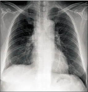
Fig. 1. Linear atelectasis in the right lower lung zone.
Fig. 2. Lung window of a coronal reformatted CT scan shows extensive ground-glass opacity in both lungs except for some areas (arrows). Ground-glass opacity le- sions suggest the presence of interstitial disease process.
CASE REPORT
A 59-year-old woman presented with pro- gressive dyspnea and cough lasting several weeks.
Two years previously, she had been diagnosed with IVL in the nasal cavity (Ann Arbor stage III). After 6 cycles of rituximab, cyclophospha- mide, adriamycin, vincristine, and prednisolone (R-CHOP) combination chemotherapy, she had shown a complete response in her follow-up head and neck CT, and subsequent periodic head and neck CT, chest CT, and abdomen-pelvis CT showed no evidence of recurrent disease.
She was admitted for further evaluation of pro- gressive dyspnea. On admission, her blood pres- sure was 120/74 mmHg, pulse 108/min, and body temperature 36.4oC. Physical examination showed the patient to be acutely ill. There were no pal- pable cervical lymph nodes. Her breathing sound was coarse, and dry crackle was audible at both
no abnormal findings, but arterial blood gas drawn in room air showed PaO2, 39.2mmHg;
PaCO2, 21.1mm/Hg; pH 7.50; bicarbonate, 16.2 mmol/L; and O2 saturation, 81%. Tests for pneu- mococcal and legionella antigen in the urine were negative, and tests for mycoplasma and cytomega- lovirus (CMV) serology in the serum were ne- gative. Chest X-ray showed linear atelectasis in the right lower lung zone (Fig. 1), and chest CT revealed extensive ground-glass opacity, suggest- ing interstitial lung disease process in both lungs (Fig. 2).
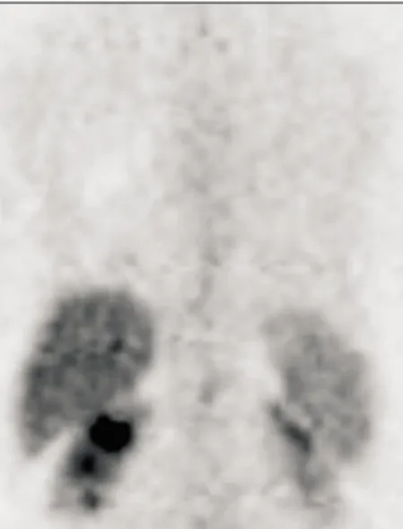
Supplemental oxygen therapy and broad-spec- trum antibiotic treatment was started on suspi- cion of pneumonia. We planned bronchoscopy, bronchoalveolar lavage (BAL), transbronchial lung biopsy (TBLB), and PET to evaluate further the diffuse interstitial infiltrates in both lungs. Inter- estingly, PET demonstrated diffuse FDG uptake
Fig. 3. Maximum intensity projection image of PET demon- strates diffuse FDG uptake in both lungs (arrows).
Abbreviation: L, liver.
Fig. 6. Follow-up PET scan shows reduced lung lesions with nearly complete disappearance of FDG.
Fig. 4. Small and medium-sized blood vessels of the lung filled with intravascular lymphoma cells. Hematoxylin and eosin staining, ×400.
Fig. 5. Immunohistochemical staining for CD20 in neo- plastic cells, ×400.
in both lungs (Fig. 3). She was transferred to the medical ICU for bronchoscopy, BAL, and TBLB.
The bronchoscopic examination showed no endo- bronchial lesion, and BAL was performed in the right middle lobe and TBLB in the right lower lobe, laterobasal segment. Gram stain and cul- ture, Acid-fast bacilli stain and culture, and viral cultures for herpes simplex virus, cytomegalovi- rus, respiratory syncytial virus, adenovirus, and influenza virus in the BAL fluid were negative.
Finally, TBLB pathology showed atypical lym- phocyte proliferation in the small and me- dium-sized blood vessels (Fig. 4); the lympho-
cytes were positive for CD20 in immunohisto- chemistry (Fig. 5). The final diagnosis was con- sistent with relapsed IVL in the lungs. Bone mar- row examination also showed intravascular large B-cell lymphoma involvement.
Ifosfamide, methotrexate, etoposide, and pre- dnisolone (IMVP-16/PD) salvage chemotherapy was started, and her dyspnea was gradually alle- viated. The patient was discharged after chemo- therapy and supportive care of neutropenia. At the time of discharge, her dyspnea had been dra- matically reduced and there was no need for sup- plemental oxygen therapy. We performed 3 cycles of IMVP-16/PD chemotherapy and reevaluated
that is characterized by the presence of lympho- ma cells within the lumina of small to medium- sized vessels, and it has a highly variable end-or- gan involvement and clinical presentation.1) IVL is aggressive and often takes a fatal clinical course.11,12) The B-cell immunotype is most com- mon, although cases with T-cell lineage have been reported.13,14)
IVL patients diagnosed in Western countries show a relatively high frequency of central nerv- ous system and skin involvement,2) whereas pa- tients in Asian countries are more likely to show hemophagocytic syndrome, bone marrow involve- ment, fever, hepatosplenomegaly, and thrombocy- topenia.3) Pulmonary involvement of IVL is also rare. Only a few cases have been reported in Korea, and these have involved nonspecific radio- logical findings, including diffuse interstitial in- filtrates, pleural effusion, signs of pulmonary hy- pertension, and tumor-like consolidation in the lungs.4-8) No PET images of pulmonary IVL have been reported. Interestingly, two recent reports suggest that PET is useful for evaluating IVL.9,10) Odawara et al. reported a patient with dis- seminated IVL and high FDG uptake throughout the entire body on PET. Their patient received R-CHOP combination chemotherapy and showed a complete metabolic response on the follow-up PET.9) Hofman et al. also showed that PET was useful in the early diagnosis of relapsed me- ningeal IVL, and the early diagnosis allowed prompt salvage chemotherapy.10) Our patient is the first reported case of relapsed pulmonary IVL showing diffuse pulmonary FDG uptake on PET and a complete metabolic response after IMVP- 16/PD combination chemotherapy.
In conclusion, we suggest that primary or re-
혈관내 림프종은 비호지킨 림프종의 드문 한 종 류로 혈관내 악성 림프구의 증식을 특징으로 한다.
현재까지 한국에서 혈관내 림프종의 폐 침범은 매 우 드물게 보고되었다. 저자들은 혈관내 B 대세포 림프종이 폐로 재발한 59세 여자환자의 증례를 보 고한다. 환자는 비강에 발생한 혈관내 B 대세포 림 프종에 대하여 6차례의 cyclophosphamide, adriamy- cin, vincristine, prednisolone (R-CHOP) 복합항암요 법 후 완전관해로 추적관찰 중이었다. 항암치료 1년 경과 후 시행한 추적관찰 흉부 전산화 단층촬영에 서 간질성 폐질환을 시사하는 간유리 혼탁화 소견 이 보이고, 양전자방출단층촬영술에서 미만성 폐섭 취가 나타났다. 기관지 내시경, 폐 세척술, 경 기관 지 폐생검을 시행하였다. 폐조직검사 결과에서 재 발성 혈관내 B 대세포 림프종이 진단되었으며, 3차 례의 ifosfamide, methotrexate, etoposide, pre- dnisolone (IMVP-16/PD) 구제 항암요법을 시행하 였다. 구제항암요법 후 양전자방출단층촬영술에서 관찰되었던 미만성 폐섭취는 완전히 소실되었다.
저자들은 원인불명의 간질성 폐질환의 감별진단으 로 원발성 혹은 재발성 혈관내 B 대세포 림프종이 포함되어야 하며, 폐에 발생하는 혈관내 림프종을 평가하는데 양전자방출단층촬영술이 유용할 수 있 음을 제안한다.
REFERENCES
1) Ponzoni M, Ferreri AJ, Campo E, et al. Definition, diagnosis, and management of intravascular large B-cell lymphoma: proposals and perspectives from an international consensus meeting. J Clin Oncol 2007;25:3168-73.
2) Ferreri AJ, Campo E, Seymour JF, et al. Intravas- cular lymphoma: clinical presentation, natural his- tory, management and prognostic factors in a series of 38 cases, with special emphasis on the 'cutaneous variant'. Br J Haematol 2004;127:173-83.
3) Murase T, Nakamura S, Kawauchi K, et al. An Asian variant of intravascular large B-cell lymphoma: clin- ical, pathological and cytogenetic approaches to dif- fuse large B-cell lymphoma associated with haemo- phagocytic syndrome. Br J Haematol 2000;111:826- 34.
4) Aouba A, Diop S, Saadoun D, et al. Severe pulmo- nary arterial hypertension as initial manifestation of intravascular lymphoma: case report. Am J Hematol 2005;79:46-9.
5) Chan VL, Lee CK, Leung WS, Lin SY, Chu CM. A 57-year-old woman with fever and abnormal chest CT findings. Chest 2006;130:924-7.
6) Gabor EP, Sherwood T, Mercola KE. Intravascular lymphomatosis presenting as adult respiratory dis- tress syndrome. Am J Hematol 1997;56:155-60.
7) Ko YH, Han JH, Go JH, et al. Intravascular lympho- matosis: a clinicopathological study of two cases pre- senting as an interstitial lung disease. Histopathology 1997;31:555-62.
8) Yousem SA, Colby TV. Intravascular lymphomatosis presenting in the lung. Cancer 1990;65:349-53.
9) Odawara J, Asada N, Aoki T, et al. 18F-Fluorodeoxy-
glucose positron emission tomography for evaluation of intravascular large B-cell lymphoma. Br J Haematol 2007;136:684.
10) Hofman MS, Fields P, Yung L, Mikhaeel NG, Ireland R, Nunan T. Meningeal recurrence of intra- vascular large B-cell lymphoma: early diagnosis with PET-CT. Br J Haematol 2007;137:386.
11) Bouzani M, Karmiris T, Rontogianni D, et al.
Disseminated intravascular B-cell lymphoma: clin- icopathological features and outcome of three cases treated with anthracycline-based immunochemo- therapy. Oncologist 2006;11:923-8.
12) Ferreri AJ, Campo E, Ambrosetti A, et al. Anthracy- cline-based chemotherapy as primary treatment for intravascular lymphoma. Ann Oncol 2004;15:1215-21.
13) Mori S, Itoyama S, Mohri N, et al. Cellular charac- teristics of neoplastic angioendotheliosis. An im- munohistological marker study of 6 cases. Virchows Arch A Pathol Anat Histopathol 1985;407:167-75.
14) Zuckerman D, Seliem R, Hochberg E. Intravascular lymphoma: the oncologist's "great imitator". Oncologist 2006;11:496-502.