INTRODUCTION
Meningioma has the second highest incidence rate among primary tumors occurring in the central nervous system (CNS), comprising 13–16% of intracranial tumors [1,2]. Based on the fourth classification of World Health Organization (WHO) in 2007, 92% of meningiomas are divided into be-
Recurred Intracranial Meningioma: A Retrospective Analysis for Treatment Outcome and Prognostic Factor
Hyun-Seung Ryu1, Kyung-Sub Moon1, Kyung-Hwa Lee2, Woo-Youl Jang1, Tae-Young Jung1, In-Young Kim1, Shin Jung1
Departments of 1Neurosurgery, 2Pathology, Chonnam National University Research Institute of Medical Sciences, Chonnam National University Hwasun Hospital & Medical School, Hwasun, Korea
Received July 13, 2017 Revised August 15, 2017 Accepted August 24, 2017 Correspondence Kyung-Sub Moon
Department of Neurosurgery, Chonnam National University Hwasun Hospital and Medical School, 322 Seoyang-ro, Hwasun-eup, Hwasun 58128, Korea Tel: +82-61-379-7666 Fax: +82-61-379-7673 E-mail: moonks@chonnam.ac.kr
Background In this study, we aimed to compare repeated resection and radiation treatment, such as Gamma knife radiosurgery (GKRS) or conventional radiotherapy (RT), and investigate the factors in- fluencing treatment outcome, including overall survival (OS), progression-free survival (PFS), and com- plication rates.
Methods We retrospectively reviewed 67 cases of recurred intracranial meningiomas (repeated resection: 36 cases, radiation treatment: 31 cases) with 56 months of the median follow-up duration (range, 13-294 months).
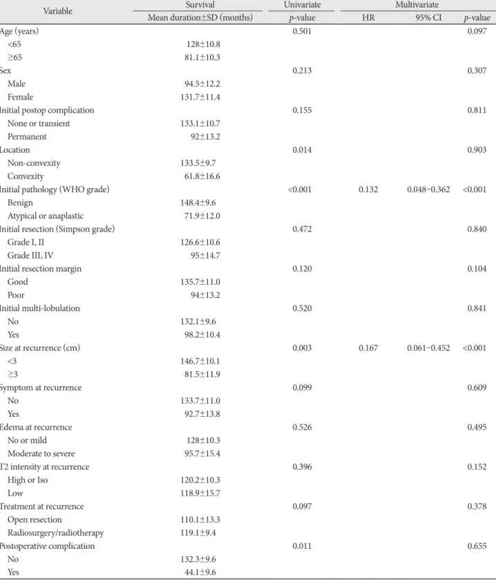
Results The incidence of death rate was 29.9% over follow-up period after treatment for re- curred meningiomas (20/67). As independent predictable factors for OS, benign pathology [hazard ra- tio (HR) 0.132, 95% confidence interval (CI) 0.048-0.362, p<0.001] and tumor size <3 cm (HR 0.167, 95% CI 0.061-0.452, p<0.001) were significantly associated with a longer OS. The incidence of pro- gression rate was 23.9% (16/67). Only treatment modality was important for PFS as an independent predictable factor (GKRS/RT vs. open resection; HR 0.117, 95% CI 0.027-0.518, p<0.005). The com- plication rate was 14.9% in our study (10/67). Larger tumor size (≥3 cm, HR 0.060, 95% CI 0.007- 0.509, p=0.010) was significant as an independent prognostic factor for development of complications.
Although treatment modality was not included for multivariate analysis, it should be considered as a predictable factor for complications (p=0.001 in univariate analysis).
Conclusion The role of repeated resection is questionable for recurred intracranial meningiomas, considering high progression and complication rates. Frequent and regular imaging follow-up is re- quired to detect recurred tumor sized as small as possible, and radiation treatment can be a preferred treatment.
Key Words Intracranial meningioma; Radiosurgery; Recurrent brain tumor; Reoperation;
Treatment outcome.
nign tumors (WHO grade I) that grows comparatively slowly [3-5]. They arise from the arachnoid “cap” cells of the arach- noid villi in the meninges and typically attached to the dural broad base [6]. Some of them invade skull or cause hyperos- tosis to subjacent bone. Gamma knife radiosurgery (GKRS) is recognized as a good treatment option for relatively small me- ningioma with actuarial tumor control rate of 97.9% at 5 years post-GKRS [7]. However, complete surgical removal (Simpson grade I–III) remains the first treatment option of meningioma despite recurrence can be occur in histologically benign menin- giomas on macroscopic total resection and removal of dural at- tachment and abnormal bone (Simpson grade I). Nanda et al.
This is an Open Access article distributed under the terms of the Creative Commons Attribution Non-Commercial License (http://creativecommons.org/licenses/by-nc/4.0) which permits unrestricted non-commercial use, distribution, and reproduction in any medium, provided the original work is properly cited.
Copyright © 2017 The Korean Brain Tumor Society, The Korean Society for Neuro- Oncology, and The Korean Society for Pediatric Neuro-Oncology
[8] revealed that, in benign meningioma, the recurrence rate in Simpson grades 0/I group was 2.9%, otherwise in Simpson grades II–IV group, the recurrence rate was 31%. Further- more, the pathological grade is also important for prediction of recurrence. In atypical meningioma, the overall recurrence rate in Simpson grades I/II resection was 31%. In grades III/IV, the overall recurrence rate was 73%, and this high recurrence rate in these groups was confined within 5 years.
Despite high clinical relevance of recurrence, the proper strategy for the diagnosis and treatment of recurred meningi- omas has not been fully established. In the present study, we analyzed treatment outcomes of patients with recurred me- ningiomas to determine the predictors for survival, progres- sion and postoperative complications.
MATERIALS AND METHODS
PatientsThis study was approved by the Institutional Review Board of the hospital (CNUHH-2017-97) and written informed consent was obtained from patients or their legal surrogates for using clinical data. A retrospective study was performed on 884 patients who received surgery and were histopatho- logically confirmed as a meningioma in our hospital from June 1993 to November 2011. Of the 884 meningioma pa- tients, 75 patients (8.5%) were treated as recurrent meningio- mas. Some patients who had partial uncertain or omitted re- cords were excluded and finally 67 cases were enrolled in the study; open resection (n=36), radiation treatment [GKRS or conventional radiotherapy (RT), n=31].
Treatment
The location and size of the craniotomy was determined for each case, and navigation system was introduced to cor- rectly localize the tumors. In some cases, the feeding vessels and tumor vascularity were evaluated by cerebral angiogra- phy before surgery. In the cases with high bleeding risk, pre- operative embolization was conducted to minimize the hem- orrhage during surgery and ease resection of the tumor.
Whenever possible, gross total resection (GTR) with removal or coagulation of involved dura mater was attempted. How- ever, remnant tumor was inevitable in case in which it had in- vaded the venous sinus or when it was very adherent to the cranial nerves, cerebral arteries and functionally important cortex. GKRS was performed with the Leksell Gamma Knife (model C or Perfexion, Elekta AB, Stockholm, Sweden). The median maximal dose was 26.5 Gy (range, 18–36 Gy), with a median marginal tumor dose of 13.2 Gy (range, 9–18 Gy) at the 45–50% isodose line.
Analysis variables
The clinical and radiological data of the enrolled patients were retrospectively reviewed. Clinicoradiological factors in- cluding, age, sex, location, size, perilesional edema, calcifica- tion, magnetic resonance (MR) imaging characteristics, Simp- son grade, initial neurologic symptoms, and pathology were retrospectively analyzed. The size of the tumor indicates the maximal diameter on 3-dimensional T1-weighted MR images with enhancement. Peritumoral edema, evaluated by axial T2- weighted MR images, were used to classify the patients into 2 groups [none or mild (<5 mm in edema thickness) vs. mod- erate to severe (≥5 mm in edema thickness)]. The extent of tumor resection was classified in accordance with the Simp- son’s classification. Simpson’s grade I or II resection without remnant tumor on follow-up MR imaging was defined as GTR.
WHO classification system was applied for pathological grade for all tumors. Newly-developed neurological deficits or ag- gravated pre-existing deficits after treatment were regarded as treatment-related complications. In this study, we defined the complication as permanent neurologic deficit or sequelae, not transient. If the problem persisted longer than 6 months or needed subsequent surgical operation, it was regarded as a permanent complication. To evaluate tumor recurrence, post- operative follow-up MR imaging was obtained from all pa- tients 6 months after surgery and then annual imaging for the rest of the patient’s life. On follow-up MR imaging, a newly-en- hanced mass in a completely resected case or a re-growing symptomatic mass in an incompletely resected case was regard- ed as progression.
Statistical analysis
Overall survival (OS) was calculated from the date of treat- ment of recurrence until death, or until the date of the last vis- iting for patients who were still alive. Progression-free survival (PFS) rates calculated from date of treatment for recurrence to date of first recurrence in complete resection case or regrowth in remnant case, or last follow-up in patients with no recurrence or regrowth were estimated with the Kaplan-Meier method and compared with the log-rank test. For the multivariate analysis, independent prognostic factors were determined us- ing the Cox’s proportional hazards model. The comparison of a permanent complication occurrence and a categorical variable was done by a Chi-square test or Fisher-exact probability test.
Furthermore, binary logistic regression test was applied for multivariate analysis. All statistical analyses were performed using SPSS version 20.0 for Windows (IBM Corp., Armonk, NY, USA); p<0.05 was considered statistically significant.
RESULTS
Patient’s characteristics
Clinicoradiological characteristics of the enrolled 67 pa- tients with recurred meningiomas are summarized in Table 1.
Several features at initial operation were also analyzed. Poor resection margin between tumor and normal arachnoid was noted in 27 patients (40.2%) and GTR (Simpson grade I/II) was performed in 48 patients (71.7%). In WHO classifica- tion, majority of the patients (42/67, 62.7%) were diagnosed as a WHO grade I. Multi-lobulated tumor was documented in 31 patients (46.2%).
On 2nd treatment, the mean tumor size was 2.1 cm and 29 cases were larger than 3 cm. Most commonly, parasagittal (20.8%), convexity (17.9%), and sphenoidal ridge (11.9%) meningiomas were accounted. Thirty-one patients showed symptoms at recurrence, with headache or dizziness in 8 pa- tients, and cranial nerve symptoms in 13 patients. Median fol- low-up duration was 56 months (range, 13–294 months). On T2-weighted MR images, 11 cases showed low signal intensity and 11 cases revealed moderate to severe peritumoral edema formation.
Overall survival
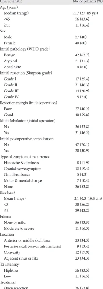
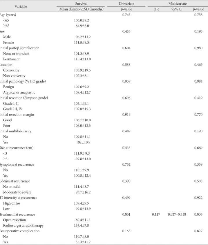
The incidence of death rate was 29.9% over follow-up peri- od after treatment for recurred meningiomas (20/67). The 5-, 10-, and 15-year survival rates were 73.1, 70.1, and 70.1%, re- spectively (Fig. 1A). The results of analyses of the variables correlated with OS are shown in Fig. 1B-F and Table 2.
On univariate analysis, initial pathology, location, size, and postoperative complication showed statistical significance.
Table 1. Clinicoradiological characteristics of 67 patients with re- curred meningiomas
Characteristic No. of patients (%)
Age (years)
Median (range) 55.7 (27–89 yrs)
<65 56 (83.6)
≥65 11 (16.4)
Sex
Male 27 (40)
Female 40 (60)
Initial pathology (WHO grade)
Benign 42 (62.7)
Atypical 21 (31.3)
Anaplastic 4 (6.0)
Initial resection (Simpson grade)
Grade I 17 (25.4)
Grade II 31 (46.3)
Grade III 14 (20.9)
Grade IV 5 (7.4)
Resection margin (initial operation)
Poor 27 (40.2)
Good 40 (59.8)
Multi-lobulation (initial operation)
No 36 (53.8)
Yes 31 (46.2)
Initial postoperative complication
No 47 (70.1)
Yes 20 (30.9)
Type of symptom at recurrence
Headache & dizziness 8 (11.9)
Cranial nerve symptom 13 (19.4)
Gait disturbance 3 (4.5)
Motor & mental change 7 (10.4)
None 36 (53.8)
Size (cm)
Mean (range) 2.1 (0.3–10.8 cm)
<3 38 (56.2)
≥3 29 (43.2)
Edema
None or mild 56 (83.5)
Moderate to severe 11 (16.5)
Location
Anterior or middle skull base 23 (34.3) Posterior skull base or infratentorial 9 (13.4)
Convexity 12 (17.9)
Adjacent sinus or falx 23 (34.3)
T2 intensity
High/Iso 56 (83.5)
Low 11 (16.5)
Treatment
Open resection 36 (53.8)
Table 1. Clinicoradiological characteristics of 67 patients with re- curred meningiomas (continued)
Characteristic No. of patients (%)
Radiosurgery/radiotherapy 31 (46.2) Follow up duration (months)
Median (range) 56 (13–294 months)
Progression after treatment for recurred lesion
No 51 (76.1)
Yes 16 (23.9)
Postoperative complication
No 57 (85.0)
Panhypopituitarysm 1 (1.5)
Mental deepening 1 (1.5)
Tremor 1 (1.5)
Visual defect 1 (1.5)
Hemorrhage 2 (3.0)
Hydrocephalus 2 (3.0)
Wound infection 2 (3.0)
The more aggressive pathology group (WHO grade II/III) manifested a shorter survival time than the benign group (71.9
±12.0 months vs. 148.4±9.6 months, p<0.001). The tumor lo- cation was influential factor for survival time [61.8±16.6 months (convexity) vs. 133.5±9.7 months (non-convexity), p=0.014].
The size of tumor was also an important variable in survival time [146.7±10.1 months (group <3 cm) vs. 81.5±11.9 months (group ≥3 cm), p=0.003]. With regard to permanent compli- cation after treatment for recurrence, the presence of compli- cation had a significantly correlation with shorter survival time (44.1±9.6 months vs. 132.3±9.6 months, p=0.011). Our study showed that the patient treated with open resection sur- vived for a shorter period than patients treated with GKRS/RT, with marginal significance (p=0.097). Pretreatment symptom also showed a marginal significance (p=0.099). On multivari- ate analysis, initial pathology and tumor size were indepen- dent predicting factors for survival rate. Benign pathology [hazard ratio (HR) 0.132, 95% confidence interval (CI) 0.048–
0.362, p<0.001] and tumor size <3 cm HR 0.167, 95% CI 0.061–
0.452, p<0.001) were significantly associated with longer sur-
vival rate.
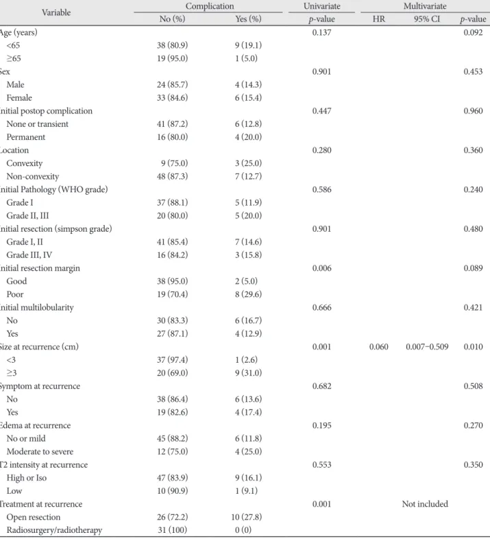
Progression-free survival
The incidence of progression rate was 23.9% over follow- up period after treatment for recurred meningiomas (16/67).
The 5-, 10-, and 15-year PFS were 79.1, 76.1, and 76.1%, re- spectively (Fig. 2A). The results of analyses of the factors cor- related with survival time are shown in Fig. 2B and Table 3. On univariate analysis, only treatment modality for recurred lesion was important for PFS [80.4±11.1 months (open resection) vs. 133.4±7.8 months (GKRS/RT), p=0.001]. This parameter was an independent predictable factor for PFS (GKRS/ RT;
HR 0.117, 95% CI 0.027–0.518, p<0.005). However, factors such as resection degree, pathological grade, tumor size, and location did not show statistical significance in the statistical analyses.
Complication
Permanent complication occurred in 10 patients (14.9%).
The most common complication was hemorrhagic change
0 60 120 180 OS (months)
Cumulative survival
1.0
0.8
0.6
0.4
0.2
0.0
0 60 120 180 OS (months)
p=0.001 WHO Gr I
WHO Gr II & III
Cumulative survival
1.0
0.8
0.6
0.4
0.2
0.0
0 60 120 180 OS (months)
p=0.014 Con
Non-con
Cumulative survival
1.0
0.8
0.6
0.4
0.2
0.0
0 60 120 180 OS (months)
p=0.011 Complication (+)
Complication (-)
Cumulative survival
1.0
0.8
0.6
0.4
0.2
0.0 0 60 120 180
OS (months)
p=0.097 Open resection
GKRS/RT
Cumulative survival
1.0
0.8
0.6
0.4
0.2
0.0 0 60 120 180
OS (months)
p=0.003
<3 cm
≥3 cm
Cumulative survival
1.0
0.8
0.6
0.4
0.2
0.0
A
D
B
E
C
F
Fig. 1. Kaplan-Meier curves showing OS of 67 study patients based on different predictors (overall comparison was estimated using a log- rank test). The number on right lower in each curve represents the p-value. A: Entire patients. B: WHO grade. C: Location. D: Size of tumor.
E: Treatment modality for recurrence. F: Postoperative complication. OS, overall survival; GKRS, Gamma knife radiosurgery; RT, radiotherapy.
(n=2, 3.0%), followed by hydrocephalus (n=2, 3.0%), and in- fection (n=2, 3.0%). The results of analyses of the variables that could be correlated with permanent complications are shown in Table 4.
On univariate analysis, tumor size and treatment modality showed statistical significance. Permanent complications were more prevalent in patient with tumor ≥3 cm than those with tumors <3 cm (2.6% vs. 31.0%, p=0.001). Patients treat-
Table 2. Univariate and multivariate analyses for overall survival from the treatment for recurred meningiomas
Variable Survival Univariate Multivariate
Mean duration±SD (months) p-value HR 95% CI p-value
Age (years) 0.501 0.097
<65 128±10.8
≥65 81.1±10.3
Sex 0.213 0.307
Male 94.5±12.2
Female 131.7±11.4
Initial postop complication 0.155 0.811
None or transient 133.1±10.7
Permanent 92±13.2
Location 0.014 0.903
Non-convexity 133.5±9.7
Convexity 61.8±16.6
Initial pathology (WHO grade) <0.001 0.132 0.048–0.362 <0.001
Benign 148.4±9.6
Atypical or anaplastic 71.9±12.0
Initial resection (Simpson grade) 0.472 0.840
Grade I, II 126.6±10.6
Grade III, IV 95±14.7
Initial resection margin 0.120 0.104
Good 135.7±11.0
Poor 94±13.2
Initial multi-lobulation 0.520 0.841
No 132.1±9.6
Yes 98.2±10.4
Size at recurrence (cm) 0.003 0.167 0.061–0.452 <0.001
<3 146.7±10.1
≥3 81.5±11.9
Symptom at recurrence 0.099 0.609
No 133.7±11.0
Yes 92.7±13.8
Edema at recurrence 0.526 0.495
No or mild 128±10.3
Moderate to severe 95.7±15.4
T2 intensity at recurrence 0.396 0.152
High or Iso 120.2±10.3
Low 118.9±15.7
Treatment at recurrence 0.097 0.378
Open resection 110.1±13.3
Radiosurgery/radiotherapy 119.1±9.4
Postoperative complication 0.011 0.655
No 132.3±9.6
Yes 44.1±9.6
HR, hazard ratio; CI, confidence interval
ed with open resection had a higher complication rate, as compared to the patients treated with GKRS/RT (27.8% vs.
0%, p=0.001). Multivariate analysis confirmed that larger tu- mor size (HR 0.060, 95% CI 0.007–0.509, p=0.010) was sig- nificant as an independent prognostic factor for development of complications. Although treatment modality was not in- cluded for multivariate analysis, it should be considered as one of the predictable factor for complication. Age, sex, pre- operative symptoms, WHO grade, resection degree, tumor consistency, and location were not statistically significant.
Open resection vs. radiation treatment (GKRS/RT) On comparing result from treatment modalities for recurred meningiomas, few showed significant differences, including size of tumors, re-progression rate, and complication rate. In GKRS/RT, mean size of tumors was 2.2 cm, but 4.3 cm in open resection (p<0.001). Based on larger than 3 cm, the number of open resection was over two-fold higher than those of GKRS/
RT (p<0.015). Re-progression rate was 6.5% in GKRS/RT and 38.9% in open resection (p<0.002). Complication rate was 27.8% when treated by open resection; however, there was no complication after GKRS/RT (p<0.001). The Simpson grade or WHO grade were statistically not significant (Table 5).
DISCUSSION
Meningiomas account for approximately one-third of pri- mary CNS tumors [9,10]. In the classification of meningiomas based on the WHO grades, benign meningiomas reportedly comprise 92%, atypical meningioma 5–7%, and malignant meningioma 1–3% of the total [11-15]. Although not all me-
ningiomas warrant treatment, surgery and/or radiation thera- py constitutes the initial therapeutic approach [16].
As meningiomas are histologically benign tumors, macro- scopic total resection is expected to be curative. However, several meningiomas recur after macroscopic total resection.
In WHO grade I meningiomas, 5-year recurrence or pro- gression rates are reported as approximately 10% after com- plete resection and 45% after non-radical resection [17,18].
Van Alkemade et al. [19] showed that OS at 5, 10, 15, and 20 years was 91.5, 81.4, 62.5, and 53.4%, respectively. After 13 years, the observed survival is positioned significantly under the expected survival curve due to excess mortality related to brain tumor or stroke. Recurrence rates at 5, 10, and 15 years were also reported as 18, 26, and 32%, respectively.
Atypical and malignant meningiomas are associated with an increased risk of local recurrence and decreased OS, as compared with WHO grade I meningiomas. Klinger et al.
[20] reported that 44% of atypical meningiomas developed clinical-radiologic evidence of recurrence at an average of 32.4 months after surgical resection. In our previous study, overall recurrence rate of atypical meningioma was 25.5% (14% in Simpson’s grade I and 37% in Simpson’s grade II/III/IV) [21].
In malignant meningiomas, the recurrence rate after com- plete extirpation is 20–40% by the decade and increases to 40–60% in partial extirpation patients [22]. Statistical signifi- cances varied by studies, nevertheless, histologically higher grade meninigioma showed higher recurrence or progress rate.
According to original Simpson’s grade, recurrent rates were 9% for grade I, 16% for grade II, and 29% for grade III [23].
This is comparable with results from another cohort and it suggests the remnant tumor cells left behind removed space
0 30 60 90 120 150 PFS (months)
Cumulative survival
1.0
0.8
0.6
0.4
0.2
0.0
0 30 60 90 120 150 p=0.001 PFS (months)
Open resection GKRS/RT
Cumulative survival
1.0
0.8
0.6
0.4
0.2
0.0
A B
Fig. 2. Kaplan-Meier curves showing PFS of 67 study patients based on different predictors (overall comparison was estimated using a log- rank test). The number on right lower in each curve represents the p-value. A: Entire patients. B: Treatment modality for recurrence. PFS, progression-free survival; GKRS, Gamma knife radiosurgery; RT, radiotherapy.
and that cells could be in continuous proliferation [24]. Some authors propose that a Simpson grade 0 resection can decrease recurrence by additional removal a 2–4 cm outwards each du- ral margin [25,26].
Although the majority occur within the first two to three years after resection, late recurrences are common. Recent studies indicate that adjuvant RT or GKRS after surgery show good outcomes for preventing recurrence, but guidelines of
Table 3. Univariate and multivariate analyses for progression-free survival from the treatment for recurred meningiomas
Variable Survival Univariate Multivariate
Mean duration±SD (months) p-value HR 95% CI p-value
Age (years) 0.745 0.758
<65 106.0±9.2
≥65 84.9±8.0
Sex 0.455 0.193
Male 96.2±13.2
Female 111.8±9.5
Initial postop complication 0.604 0.980
None or transient 101.3±8.9
Permanent 115.4±13.0
Location 0.588 0.469
Convexitiy 103.9±19.5
Non-convexity 107.3±8.1
Initial pathology (WHO grade) 0.938 0.984
Benign 107.6±9.2
Atypical or anaplastic 109.4±12.7
Initial resection (Simpson grade) 0.695 0.419
Grade I, II 105.1±9.1
Grade III, IV 109.0±15.3
Initial resection margin 0.914 0.770
Good 106.7±10.0
Poor 106.0±12.3
Initial multilobularity 0.489 0.190
No 109.8±11.1
Yes 102±10.9
Size at recurrence (cm) 0.433 0.669
<3 111.9± 9.3
≥3 97.8±13.0
Symptom at recurrence 0.752 0.359
No 110.1±9.9
Yes 100.8±12.4
Edema at recurrence 0.390 0.503
No or mild 111.4±8.7
Moderate to severe 93.7±16.2
T2 intensity at recurrence 0.499 0.922
High or Iso 109.4±9.5
Low 99.8±13.9
Treatment at recurrence 0.001 0.117 0.027–0.518 0.005
Open resection 80.4±11.1
Radiosurgery/radiotherapy 133.4±7.8
Postoperative complication 0.165 0.827
No 110.7±8.0
Yes 55.3±11.7
HR, hazard ratio; CI, confidence interval
treatment for already recurred meningioma are still not estab- lished. In our study, independent predictive factor for longer survival were benign pathology (HR 0.132, 95% CI 0.048–
0.362, p<0.001) and tumor size <3 cm (HR 0.167, 95% CI 0.061–
0.452, p<0.001). In univariate statistics, tumor location and complication at the first operation showed statistical signifi- cance. Treatment modality (open resection vs. radiation
treatment) showed marginal significance. In PFS, only radia- tion treatment was an independent predictive factor (HR 0.117, 95% CI 0.027–0.518, p<0.005). Regarding complication rates, larger than 3 cm size (HR 0.060, 95% CI 0.007–0.509, p=
0.010) was a significant independent predictive factor for de- velopment of complications. Although treatment modality was not a significant factor, it should be considered as a pre-
Table 4. Univariate and multivariate analyses for postoperative complication from the treatment for recurred meningiomas
Variable Complication Univariate Multivariate
No (%) Yes (%) p-value HR 95% CI p-value
Age (years) 0.137 0.092
<65 38 (80.9) 9 (19.1)
≥65 19 (95.0) 1 (5.0)
Sex 0.901 0.453
Male 24 (85.7) 4 (14.3)
Female 33 (84.6) 6 (15.4)
Initial postop complication 0.447 0.960
None or transient 41 (87.2) 6 (12.8)
Permanent 16 (80.0) 4 (20.0)
Location 0.280 0.360
Convexity 9 (75.0) 3 (25.0)
Non-convexity 48 (87.3) 7 (12.7)
Initial Pathology (WHO grade) 0.586 0.240
Grade I 37 (88.1) 5 (11.9)
Grade II, III 20 (80.0) 5 (20.0)
Initial resection (simpson grade) 0.901 0.480
Grade I, II 41 (85.4) 7 (14.6)
Grade III, IV 16 (84.2) 3 (15.8)
Initial resection margin 0.006 0.089
Good 38 (95.0) 2 (5.0)
Poor 19 (70.4) 8 (29.6)
Initial multilobularity 0.666 0.421
No 30 (83.3) 6 (16.7)
Yes 27 (87.1) 4 (12.9)
Size at recurrence (cm) 0.001 0.060 0.007–0.509 0.010
<3 37 (97.4) 1 (2.6)
≥3 20 (69.0) 9 (31.0)
Symptom at recurrence 0.682 0.508
No 38 (86.4) 6 (13.6)
Yes 19 (82.6) 4 (17.4)
Edema at recurrence 0.195 0.270
No or mild 45 (88.2) 6 (11.8)
Moderate to severe 12 (75.0) 4 (25.0)
T2 intensity at recurrence 0.553 0.350
High or Iso 47 (83.9) 9 (16.1)
Low 10 (90.9) 1 (9.1)
Treatment at recurrence 0.001 Not included
Open resection 26 (72.2) 10 (27.8)
Radiosurgery/radiotherapy 31 (100) 0 (0)
HR, hazard ratio; CI, confidence interval
dictable factor for complication. As shown in Table 5, radia- tion treatment was better in all parameters than open resec- tion as the treatment modality for recurred lesion.
In addition to surgical resection or radiation treatment, pharmacologic therapy is in use for recurred meningiomas [27]. Mainly antiangiogenic agents such as bevacizumab, vat- alanib, and sunitinib have made promising results but they are still in issue to be used clinically [28,29]. Other agents in- cluding hydroxyurea, epidermal growth factor inhibitor, hor- monal therapeutics, α-interferon, and somatostatin receptor agonists have been tried for recurred meningioma [30-32]. Up to now, pharmacologic therapy is not first therapeutic option for recurred meningioma and used narrowly for refractory re- current tumor. However, the solidity of the evidence might soon advance with identification of the pharmacologic target effect.
Major limitation of this study is its retrospective design, which makes many types of bias. Another limitation is inclu- sion of all WHO grades for studied patients, possibly leading a selection bias for treatment. In addition, the size of recurred tumor may cause another selection bias for treatment modal-
ity. The relatively small number of patients and short follow- up duration can be other limitations in current study. Further studies need to be performed prospectively with large numbers of benign pathology for establishing treatment strategy of re- curred meningiomas.
In conclusion, although open surgical resection is the treat- ment choice for symptomatic or large sized meningiomas, it is questionable for recurred tumors, considering high progres- sion and complication rates after repeated open resection. Thus, frequent and regular imaging follow-up is required to detect recurred tumor sized as small as possible, and radiation treat- ment can be considered as a preferential treatment.
Conflicts of Interest
The authors have no financial conflicts of interest.
Acknowledgments
This study was supported by a grant of Chonnam National University Hospital Research Institute of Clinical Medicine.
REFERENCES
1. Hoessly GF, Olivecrona H. Report on 280 cases of verified parasagittal Table 5. Comparison of clinicoradiological characteristics according to treatment modalities for recurred meningiomas
Characteristics Radiosurgery/RT Open resection p-value
Characteristics related with 2nd treatments Age (years)
Mean±SD 57.6±13.7 54.1±13.6 0.864
≥65 (%) 35.5 25.0 0.505
Sex
Male (%) 45.2 38.9 0.787
Location
Convexity (%) 12.9 22.2 0.321
Size (cm)
Mean±SD 2.2±1.4 4.3±2.7 <0.001
≥3 (%) 25.8 58.3 0.015
Low in T2 (%) 22.6 11.1 0.206
Moderate/high peritumoral edema (%) 22.6 25 1.000
Severe symptom (%) 35.5 33.3 1.000
Interval between 1st and 2nd treatments (months) 74.0±56.6 42.6±37.0 0.170
Re-progression rate (%) 6.5 38.9 0.002
Complication rate (%) 0 27.8 0.001
Expire rate (%) 19.4 38.9 0.140
Follow-up duration (mean±SD, months) 68.4±45.5 61.7±46.5 0.747
Characteristics related with initial treatment (%)
Multilobulation 51.6 41.7 0.570
Poor resection plane 29.0 50.0 0.135
Simpson’s grade ≥III 38.7 19.4 0.141
WHO grade II/III 29.0 44.4 0.295
Complication after 1st treatment 29.0 30.6 1.000
RT, radiotherapy; SD, standard deviation
meningioma. J Neurosurg 1955;12:614-26.
2. Mirimanoff RO, Dosoretz DE, Linggood RM, Ojemann RG, Martuza RL. Meningioma: analysis of recurrence and progression following neu- rosurgical resection. J Neurosurg 1985;62:18-24.
3. Louis DN, Ohgaki H, Wiestler OD, et al. The 2007 WHO classification of tumours of the central nervous system. Acta Neuropathol 2007;114:
97-109.
4. Willis J, Smith C, Ironside JW, Erridge S, Whittle IR, Everington D. The accuracy of meningioma grading: a 10-year retrospective audit. Neuro- pathol Appl Neurobiol 2005;31:141-9.
5. Ojemann SG, Sneed PK, Larson DA, et al. Radiosurgery for malignant meningioma: results in 22 patients. J Neurosurg 2000;93 Suppl 3:62-7.
6. Buetow MP, Buetow PC, Smirniotopoulos JG. Typical, atypical, and misleading features in meningioma. Radiographics 1991;11:1087-106.
7. Kollová A, Liscák R, Novotný J Jr, Vladyka V, Simonová G, Janousková L.
Gamma Knife surgery for benign meningioma. J Neurosurg 2007;107:
325-36.
8. Nanda A, Bir SC, Konar S, et al. Outcome of resection of WHO Grade II meningioma and correlation of pathological and radiological predic- tive factors for recurrence. J Clin Neurosci 2016;31:112-21.
9. Ostrom QT, Gittleman H, Fulop J, et al. CBTRUS Statistical Report:
Primary Brain and Central Nervous System Tumors Diagnosed in the United States in 2008-2012. Neuro Oncol 2015;17 Suppl 4:iv1-iv62.
10. Wrensch M, Minn Y, Chew T, Bondy M, Berger MS. Epidemiology of primary brain tumors: current concepts and review of the literature.
Neuro Oncol 2002;4:278-99.
11. Al-Mefty O, Kadri PA, Pravdenkova S, Sawyer JR, Stangeby C, Husain M. Malignant progression in meningioma: documentation of a series and analysis of cytogenetic findings. J Neurosurg 2004;101:210-8.
12. Lim YS, Kim MK, Park BJ, Kim TS, Lim YJ. Long term clinical out- comes of malignant meningiomas. Brain Tumor Res Treat 2013;1:85-90.
13. Harris AE, Lee JY, Omalu B, Flickinger JC, Kondziolka D, Lunsford LD.
The effect of radiosurgery during management of aggressive meningio- mas. Surg Neurol 2003;60:298-305; discussion 305.
14. Jung HW, Yoo H, Paek SH, Choi KS. Long-term outcome and growth rate of subtotally resected petroclival meningiomas: experience with 38 cases. Neurosurgery 2000;46:567-74; discussion 574-5.
15. Kallio M, Sankila R, Hakulinen T, Jääskeläinen J. Factors affecting opera- tive and excess long-term mortality in 935 patients with intracranial me- ningioma. Neurosurgery 1992;31:2-12.
16. Chamberlain MC. Treatment of meningioma, including in cases with no further surgical or radiotherapy options. Oncology (Williston Park) 2015;29:369-71.
17. Marosi C, Hassler M, Roessler K, et al. Meningioma. Crit Rev Oncol Hematol 2008;67:153-71.
18. Gondi V, Tome WA, Mehta MP. Fractionated radiotherapy for intracra- nial meningiomas. J Neurooncol 2010;99:349-56.
19. van Alkemade H, de Leau M, Dieleman EM. Impaired survival and long-term neurological problems in benign meningioma. Neuro Oncol 2012;14:658-66.
20. Klinger DR, Flores BC, Lewis JJ, et al. Atypical meningiomas: recur- rence, reoperation, and radiotherapy. World Neurosurg 2015;84:839-45.
21. Moon HS, Jung S, Jang WY, Jung TY, Moon KS, Kim IY. Intracranial Meningiomas, WHO Grade II: Prognostic Implications of Clinicopath- ologic Features. J Korean Neurosurg Soc 2012;52:14-20.
22. Adegbite AB, Khan MI, Paine KW, Tan LK. The recurrence of intracra- nial meningiomas after surgical treatment. J Neurosurg 1983;58:51-6.
23. Simpson D. The recurrence of intracranial meningiomas after surgical treatment. J Neurol Neurosurg Psychiatry 1957;20:22-39.
24. Giombini S, Solero CL, Morello G. Late outcome of operations for su- pratentorial convexity meningiomas. Report on 207 cases. Surg Neurol 1984;22:588-94.
25. de Vries J, Wakhloo AK. Repeated multifocal recurrence of grade I, grade II, and grade III meningiomas: regional multicentricity (primary new growth) or metastases? Surg Neurol 1994;41:299-305.
26. Borovich B, Doron Y, Braun J, et al. Recurrence of intracranial menin- giomas: the role played by regional multicentricity. Part 2: Clinical and radiological aspects. J Neurosurg 1986;65:168-71.
27. Nabors LB, Portnow J, Ammirati M, et al. Central nervous system cancers, version 2.2014. Featured updates to the NCCN Guidelines. J Natl Compr Canc Netw 2014;12:1517-23.
28. Kaley TJ, Wen P, Schiff D, et al. Phase II trial of sunitinib for recurrent and progressive atypical and anaplastic meningioma. Neuro Oncol 2015;
17:116-21.
29. Lou E, Sumrall AL, Turner S, et al. Bevacizumab therapy for adults with recurrent/progressive meningioma: a retrospective series. J Neurooncol 2012;109:63-70.
30. Chamberlain MC, Glantz MJ. Interferon-alpha for recurrent World Health Organization grade 1 intracranial meningiomas. Cancer 2008;113:2146- 31. Chamberlain MC, Glantz MJ, Fadul CE. Recurrent meningioma: salvage 51.
therapy with long-acting somatostatin analogue. Neurology 2007;69:
969-73.
32. Kaley T, Barani I, Chamberlain M, et al. Historical benchmarks for medical therapy trials in surgery- and radiation-refractory meningioma:
a RANO review. Neuro Oncol 2014;16:829-40.