대한치과재료학회지 46(2) : 75-88, 2019 Korean Journal of Dental Materials (
Korean J Dent Mater
) ISSN:2384-4434 (Print); 2384-3268 (Online) Available online at http://www.kadm.org http://dx.doi.org/10.14815/kjdm.2019.46.2.75근단 협착부 직경, 세척액 주입속도, 주사침 말단의 형상이 치근단에 작용하는 압력에 미치는 영향
이창하
1,3
, 조설아1,3
, 임범순2,3
, 이인복1,3,*
1서울대학교 치의학대학원 치과보존학교실, 2치과생체재료과학교실, 3치학연구소
Effect of apical constriction diameter, irrigant flow rate, and needle tip design on apical pressure
Chang-Ha Lee
1,3, Seol-Ah Jo
1,3, Bum-Soon Lim
2,3, In-Bog Lee
1,3,*Departmen of Conservative Dentistry1, Dept. of Dental Biomaterials Science2, School of Dentistry and Dental Research Institute3, Seoul National University, Seoul, Korea
The purpose of this study was to evaluate the effects of apical constriction (AC) diameter, irrigant flow rate, and needle tip design on apical pressure (AP) during the root canal irrigation. Five extracted human mandibular premolars were instrumented up to #35 (0.06 taper) using nickel-titanium rotary instruments. AC was determined at 1 mm from the apical foramen. Three needles with different tip designs (notched, side-vented, and flat) were placed 3 mm from AC. APs were measured with varying flow rates of 0.05, 0.1, 0.2, and 0.3 mL/s. The AC diameter of the teeth was enlarged to #40 and #45 (0.06 taper) successively, and the aforementioned measurement procedure was repeated (n=5). When the other conditions were controlled, AP increased with decreasing AC diameter or increasing irrigant flow rate, and the AP of flat needle was the highest, followed by notched, and side-vented needle (p<0.05). The APs with 0.05 mL/s flow rate were lower than central venous pressure (5.88 mmHg) for all conditions. Under the conditions of the present study, flat needle is not recommended in narrow AC (0.35 mm), as the value of the AP with a flow rate of 0.05 mL/s is similar to the central venous pressure. However, in AC diameter of
>0.35 mm, open-end (notched or flat) needles can be used to improve irrigant replacement in the apical portion using a flow rate of 0.05 mL/s.
Key words :Apical constriction diameter, Apical pressure, Irrigant flow rate, Needle tip design
Chang-Ha Lee (ORCID: 0000-0002-4623-1475) Seol-Ah Jo (ORCID: 0000-0001-7158-1306) Bum-Soon Lim (ORCID: 0000-0003-3112-0227)
Correspondence: In-Bog Lee (ORCID: 0000-0001-8083-1530) 101, Daehak-ro, Jongro-gu, Seoul, 03080, Republic of Korea
Affiliation: Department of Conservative Dentistry, School of Dentistry, Seoul National University, Seoul, Republic of Korea
Tel: +82-2-2072-3953, Fax: +82-2-2072-3859 E-mail: [email protected]
Received: May
. 06, 2019; Revised: Jun. 01, 2019; Accepted: Jun. 03, 2019서 론
치아의 근관 치료는 우식이나 외상으로 인한 치수 노출 또는 치수염, 치수괴사, 치근단염 등, 치수 및 치근단 질환으 로 인해 시행된다. 근관 치료의 성공을 위해서는 잔존 치수 또는 세균에 감염된 조직을 제거하기 위한 과정이 필수적이 다(1). 수동형 및 회전식 기구를 사용한 기계적 근관 형성과 항균작용을 가지는 세척액 적용을 통해 근관계의 멸균을 도모할 수 있으며, 근관 세척과정은 기계적 근관 형성의 한계점을 보완해 준다는 측면에서 중요한 의의를 가진다 (2-5).
근관 세척액의 적용 시 치근단 조직으로 가해지는 압력 (apical pressure, AP)이 평균 중심정맥압(5.88 mmHg)을 초과하는 경우 근관세척액(1-5.25% NaOCl 용액)이 근첨 밖으로 나가 치근단 조직으로 침투할 위험이 있다(6, 7).
NaOCl 용액을 근관 내에 국한하여 사용할 경우 안전하고 효율적으로 항균작용을 도모할 수 있지만 근첨공을 통해 치근단 주위 조직으로 NaOCl 용액이 유출될 경우 강한 산화제로 작용하여 용혈, 궤양, 세포 파괴 등 독성 작용을 야기할 가능성이 있다(8). 이러한 NaOCl 용액의 독성은 물과 반응하여 생성되는 활성 산소, 아민과 반응하여 생성되 는 클로라민, 산과 반응하여 생성되는 염소 가스에 의해 야기된다(9). 근관 세척액에 의한 압력이 치근단 조직에 가해지거나 세척액이 근첨 밖으로 유출될 경우 기계적 자극, 화학매개물, 면역계 반응, 환자의 심리적 요인 등에 의해 술 후 통증이 발생하게 되는데(10), 술 후 통증의 빈도는 3-58%의 다양한 정도로 보고되었다(11). 음압 방식으로 근 관 세척을 시행한 경우 AP가 감소하였고 술 후 통증 발생 정도와 진통제 복용 빈도도 더 낮은 결과를 보였다(10, 12, 13). 따라서 양압 방식으로 근관 세척 시 과도한 압력이 치근단 조직에 작용하지 않도록 하는 것이 중요하다.
근관 세척 시 세척액의 교환 효과는 세척액이 근첨 방향으 로 침투하는 정도와 세척액의 흐름 패턴에 의해 결정된다 (14-17). 세척액의 주입속도는 근관 내에서 흐름 패턴을 결정하는데 중요한 역할을 하고 주사침 말단을 근첨에 가깝 게 위치시켜야 높은 세척액 교환 효과를 얻을 수 있으며, 주사침의 종류에 따라서도 세척액 교환 효과의 차이가 발생 한다. 또한 세척액의 근첨 방향으로의 침투 효과는 근관의
확대 정도와 관계되며(18), 근관 형성 시 근단부 직경이 커지거나 근관의 taper 정도가 커지면 근단부 세척 효율은 증가한다(19, 20).
근단부 세척 효율을 증가시키기 위해 세척액 주입 속도를 증가시키고 주사침 말단을 근첨 방향으로 위치시키거나 주사침 말단의 형상 또는 근단부 직경을 변화시킬 경우 AP가 증가하거나 세척액 유출량이 증가할 수 있으므로 이에 대한 고려가 필요하다. AP는 세척액 주입속도가 증가할수록 증가하였고(21, 22), 근단 부위에서 유출된 세척액의 양은 말단 개방형 주사침일수록, 주사침이 근첨 부위에 가까이 위치할수록, 근단부 직경이 감소할수록 증가하였다(23, 24).
최근 연구에서 발치된 사람 치아에서 주사침 말단의 형상과 위치, 세척액 주입속도를 변화시키며 AP를 측정한 결과 AP는 주사침이 근단에 가깝게 위치할수록, 세척액 주입속도 가 증가할수록 증가하였고 말단 개방형 주사침에서 AP가 높았다(25).
이와 같이 AP와 세척액 유출량은 다양한 요소들에 의해 영향을 받는다는 여러 연구결과들이 있지만 근단부 확대 정도가 근관 세척에 미치는 영향에 대한 연구는 주로 근관 내 세균 및 잔사의 제거 효과에 관한 연구이며(26, 27), 발치된 사람 치아에서 다양한 요소를 함께 변화시키며 AP를 측정한 연구는 드물다. 따라서 본 연구에서는 근단 협착부 직경, 세척액 주입속도, 주사침 말단의 형상을 변화시킬 때 발생하는 AP를 발치된 사람 치아에서 측정하여 비교 및 분석하고자 하였다.
귀무가설은 “형성된 근관의 근단 협착부 직경, 근관 세척 액의 주입속도, 주사침 말단의 형상에 따라 AP의 차이가 없을 것이다.”로 설정하였다.
재료 및 방법
1. Apical pressure (AP) 측정 장비
근관 세척 시 치근단에 작용하는 압력을 측정하기 위해
Figure 1(a)와 같은 장비(Bio-Pressure, IB Systems, Seoul,
Korea)를 이용하였다. 스텝 모터로 동작하는 시린지 펌프에
의해 원하는 주입 속도로 설정된 세척액이 플라스틱 튜브와
(a)
(b)
Figure 1. (a) Schematic diagram of a custom-made apical pressure measuring instrument (Bio-Pressure, IB Systems, Seoul, Korea).
(b) Magnified view of tooth model.
주사침을 통해 치근단 부위로 전달되며 플라스틱 튜브를 통해 치근단 부위와 연결된 압력 센서에 의해 AP가 측정 된다.
2. 치아 시편 및 근관 세척용 주사침 제작
교정 치료 목적으로 발치된 우식 및 수복물이 없는 사람
하악 소구치(IRB 승인번호: CRI09005)를 현미경(OPMI
pico, Carl Zeiss Meditec AG, Jena, Germany)으로 검사하여
균열이 없는 5개의 치아를 선택하였다. 각 치아에 근관
와동 형성 후 #15 k-file로 치근단공의 개방성을 확인하고
치근단공으로부터 1 mm 짧은 지점을 근단 협착부로 정하였
다. 이후 #35(0.06 taper) 니켈-티타늄 회전식 기구(K3,
Sybron Dental Specialties, Orange, CA)로 근관을 형성하고
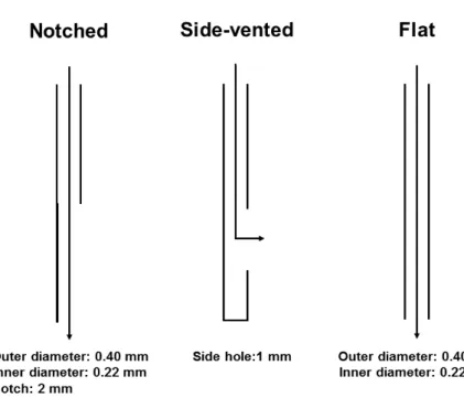
Figure 2. Three different needle tip designs.
3.5% NaOCl (Sodium Hypochlorite solution, Duksan Pure Chemicals Co., Ansan, Korea)로 근관세척 시행하였다.
원통형(직경 3 mm × 길이 8 mm) 알루미늄 관을 50 µm 알루미나(Al
2O
3) 입자(BasicMobil, Renfert, Hilzingen, Germany)로 샌드블라스팅하고, 금속 프라이머(Reliance, Ortho Prod. Inc. Itasca, IL, USA)로 처리한 후 건조하였다.
치근면은 인산(Scotchbond Universal Etchant, 3M Deutschland GmbH, Neuss, Germany)으로 산-부식 처리하 였고, 상아질 접착제 적용 후 알루미늄 관에 치근단 부위가 잠기도록 위치시켜 유동형 복합레진(DenFil Flow, Vericom Co., Anyang, Korea)으로 치아와 알루미늄 관을 접착하였 다. 복합레진(Filtek Z250 Universal Restorative, 3M ESPE, St. Paul, MN, USA)과 nail varnish로 경계면을 봉쇄하였다 (Figure 1b).
외경 0.40 mm, 내경 0.22 mm, 길이 25 mm의 근관 세척용 notched 주사침(Endo-Eze Irrigator tips, Ultradent Product, Inc., South Jordan, UT, USA)의 notch 부분을 제거하여 flat 주사침을 제작하였고, flat 주사침의 말단을 접착제 (cyanoacrylate glue)로 밀봉하고 말단으로 부터 1 mm 상방 위치의 측면에 1 mm 길이의 구멍을 형성하여 side-vented 주사침을 제작하였다(Figure 2).
3. Apical pressure (AP) 측정
AP 측정을 위해 제작한 치아 시편은 스탠드에 고정하고
치아와 접착된 알루미늄 관 하부를 멸균증류수가 담긴 플라
스틱 튜브를 이용하여 압력 센서와 연결하였다. Notched,
side-vented, flat의 3가지 유형의 주사침을 플라스틱 튜브로
시린지와 연결하여 세척액(멸균증류수)이 주입될 수 있도록
하였다. 각각의 주사침 말단은 근단 협착부(apical constric-
tion, AC)로부터 치관 방향으로 3 mm 거리가 되는 지점(주사
침 말단의 위치)에 연결하였다. 이 때 수동형 근관치료용
줄(MANI K-FILES, Mani Inc., Utsunomiya, Tochigi, Japan)
의 구성품인 rubber stop을 치아 각각의 치관부 참고점에
연결하여 근관 내에서 주사침 말단의 위치가 일정하도록
하였다. 각 군에 대하여 세척액 주입속도는 0.05, 0.1, 0.2,
0.3 mL/s로 변화시키면서 AP를 측정하였다(n=5). #40, #45
(0.06 taper) 니켈-티타늄 회전식 기구(K3, Sybron Dental
Specialties)를 이용한 근관 확대를 통해 근단 협착부 직경을
0.40, 0.45 mm로 변화시킨 후 앞서 언급한 측정과정을
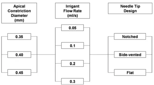
각각 반복하였다. 근단 협착부 직경, 세척액 주입속도, 주사
침 말단의 형상 변화의 세부사항은 Figure 3과 같다.
Figure 3. Details of the experimental groups.
4. 통계 분석
근단 협착부 직경, 세척액 주입속도, 주사침 말단의 형상 에 따른 AP의 비교를 위해 삼원배치 분산분석(three-way ANOVA)를 시행하였다(n=5). 분산분석의 결과는 Tukey's post-hoc test를 이용하여 사후검정 하였다. 상관분석은 Pearson correlation analysis를 이용하였다. 모든 통계분석 은 SPSS software (Version 21.0, SPSS Inc., Chicago, IL, USA)를 이용하여 이루어졌으며, 검정을 위한 유의수준은 5%로 설정하였다.
결 과
직경 0.35 mm의 근단 협착부로부터 치관 방향으로 3 mm 거리에 위치시킨 notched 주사침을 이용하여 세척액 주입속도를 변화시키면서 측정한 AP의 시간의 흐름에 따른 대표적 곡선은 Figure 4와 같다. 근단 협착부 직경, 세척액 주입속도 및 주사침 말단의 형상 변화에 따른 AP 측정값은 Table 1에 제시하였다.
1. 근단 협착부 직경에 따른 apical pressure (AP)의 변화
세척액 주입속도와 주사침 말단의 형상이 동일한 조건
하에서 근단 협착부 직경이 감소할수록 AP는 유의하게 증가 하였다(p<0.05, Table 1, Figure 5). 상관분석 결과 근단 협착부 직경과 AP 사이에는 약한 음의 상관관계를 가짐을 알 수 있었다(Pearson coefficient = -0.157).
2. 세척액 주입속도에 따른 apical pressure (AP)의 변화
근단 협착부 직경과 주사침 말단의 형상이 동일한 조건 하에서 세척액 주입속도가 증가할수록 AP는 유의하게 증가 하였다(p<0.05, Table 1, Figure 5). 상관분석 결과 세척액 주입속도와 AP 사이에는 강한 양의 상관관계를 보였다 (Pearson coefficient = 0.904). 세척액 주입속도가 0.05 mL/s인 경우는 근단 협착부 직경과 주사침 말단의 형상에 관계없이 모든 군에서 중심정맥압보다 낮은 AP를 나타냈다.
세척액 주입속도가 0.1 mL/s인 경우는 근단 협착부 직경이 0.45 mm이고, side-vented 주사침 적용 시에만 중심정맥압 보다 낮은 AP를 나타냈다. 세척액 주입속도가 0.2 mL/s 이상인 경우는 모든 경우에서 AP가 중심정맥압보다 높았다.
3. 주사침 말단의 형상에 따른 apical pressure (AP)의 변화
근단 협착부 직경과 세척액 주입속도가 동일한 조건 하에
서 side-vented 주사침이 가장 낮은 AP를 보였고, notched,
Figure 4. Representative curve of apical pressure change vs. flow rate over time with a notched needle at a tip position of 3 mm from the apical constriction of 0.35 mm diameter.
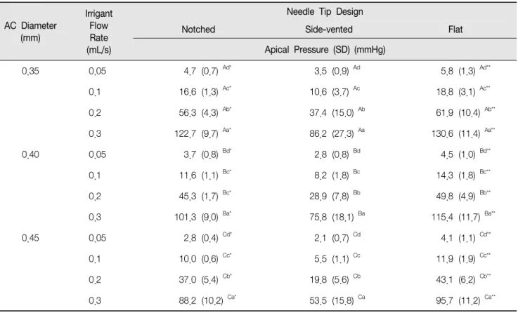
Table 1. Apical pressure with varying apical constriction (AC) diameter, irrigant flow rate, and needle tip design
AC Diameter (mm)
Irrigant Flow Rate (mL/s)
Needle Tip Design
Notched Side-vented Flat
Apical Pressure (SD) (mmHg)
0.35 0.05 4.7 (0.7) Ad* 3.5 (0.9) Ad 5.8 (1.3) Ad**
0.1 16.6 (1.3) Ac* 10.6 (3.7) Ac 18.8 (3.1) Ac**
0.2 56.3 (4.3) Ab* 37.4 (15.0) Ab 61.9 (10.4) Ab**
0.3 122.7 (9.7) Aa* 86.2 (27.3) Aa 130.6 (11.4) Aa**
0.40 0.05 3.7 (0.8) Bd* 2.8 (0.8) Bd 4.5 (1.0) Bd**
0.1 11.6 (1.1) Bc* 8.2 (1.8) Bc 14.3 (1.8) Bc**
0.2 45.3 (1.7) Bc* 28.9 (7.8) Bb 49.8 (4.9) Bb**
0.3 101.3 (9.0) Ba* 75.8 (18.1) Ba 115.4 (11.7) Ba**
0.45 0.05 2.8 (0.4) Cd* 2.1 (0.7) Cd 4.1 (1.1) Cd**
0.1 10.0 (0.6) Cc* 5.5 (1.1) Cc 11.9 (1.9) Cc**
0.2 37.0 (5.4) Cb* 19.8 (5.6) Cb 43.1 (6.2) Cb**
0.3 88.2 (10.2) Ca* 53.5 (15.8) Ca 95.7 (11.2) Ca**
Different superscript upper-case letters indicate statistically significant differences among the groups with different AC diameter.
Different superscript lower-case letters indicate statistically significant differences among the groups with different irrigant flow rate.
The superscript “⁕” and “⁕⁕” indicate statistically significant differences among the groups with different needle tip design.
(a) (b)
(c)
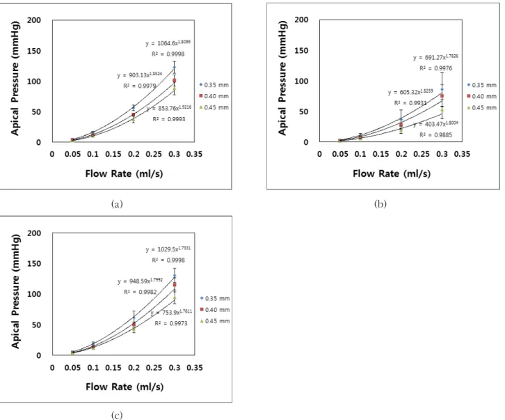
Figure 5. Apical pressure as a function of irrigant flow rate (0.05, 0.1, 0.2 and 0.3 mL/s) with varying apical constriction diameter (0.35, 0.40, and 0.45 mm). (a) Notched, (b) side-vented, and (c) flat needle.
flat 주사침 순서로 AP가 유의하게 증가하였다(p<0.05, Table 1, Figure 5).
4. 교호작용
근단 협착부 직경과 세척액 주입속도 간에 교호작용이 관찰되었고, 세척액 주입속도가 증가할수록 근단 협착부 직경에 따른 AP 변화가 컸으며, 근단 협착부 직경이 감소할 수록 세척액 주입속도에 따른 AP 변화가 컸다(p<0.05).
세척액 주입속도와 주사침 말단의 형상 사이에도 교호작용 이 관찰되었고 세척액 주입속도가 증가할수록 주사침 말단 의 형상에 따른 AP값의 차이가 증가하였으며, side-vented
주사침은 세척액 주입속도에 의한 AP 변화가 가장 작고 flat 주사침은 세척액 주입속도에 의한 AP 변화가 가장 크게 나타났다(p<0.05). 근단 협착부 직경과 주사침 말단의 형상 간에는 교호작용이 관찰되지 않았다(p>0.05).
고 찰
본 연구에서는 근관 확대 정도와 근관 세척 과정에서
조절 가능한 두 가지 조건을 변화시키며 AP를 측정하여
비교 및 분석하였다. AP는 세척액이 근첨으로 향하는 순방
향 흐름과 근관 입구로 향하는 역방향 흐름의 비율, 세척액 주입속도에 따라 변화할 것으로 가정하고 근단 협착부 직경, 주사침 끝부분의 개구방향, 근관 세척액의 유속을 변화시켰 다. 발거한 사람 치아 근관의 해부학적 형태의 다양성으로 인해 AP 측정값의 편차가 발생할 가능성이 있어 이를 최소화 하기 위해 치아를 선별하기 전에 방사선 사진을 촬영하여 직선 형태의 단근관이며 확대 전 근관의 taper가 크지 않은 치아를 선택하였다.
선별한 단근관의 사람 하악소구치를 대상으로 치근단공 으로부터 1 mm 짧은 지점을 근관장으로 결정하여 근단 협착부를 형성하였다. 사람 하악소구치의 경우 치근단공으 로부터 1 mm 거리에서 협-설 직경이 0.35 mm, 근-원심 직경은 0.28 mm로 보고된 바(28), 근단 협착부 직경을 0.35, 0.40, 0.45 mm로 증가시키면서 AP를 측정하였다. 이전 연구에서 임상 과정에서 안전하고 효율적인 근관 세척을 위해 근단 협착부로부터 3 mm 떨어진 지점에 주사침 말단을 위치시키는 것이 추천되었고(25), 27 gauge 주사침을 사용 한 경우 근단 협착부 직경이 0.30 mm 이상일 경우 주사침 말단을 근단에서 3 mm 떨어뜨려 위치시켜도 세척액이 근단 부위까지 도달하였다(29). 따라서 본 연구에서는 주사침 말단 위치를 근단 협착부로부터 3 mm 떨어진 지점으로 고정하고 나머지 조건을 변화시키면서 실험을 진행하였다.
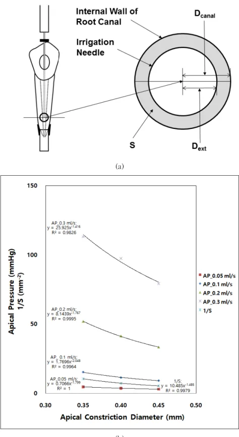
근관 확대를 통한 근단 협착부 직경 증가는 주사침 외경 (D
ext)과 주사침 말단이 위치한 곳의 근관 직경(D
canal= A + (L × T), A: 근단 협착부 직경, L: 근단 협착부와 주사침 말단 사이의 거리, T: 확대된 근관의 경사도(taper)) 사이에 놓인 면적(S)을 변화시키며
와 같이 표현될 수 있다(19) (Figure 6a). 근단 협착부 직경에 따른 세 가지 주사침의 AP 평균값 및 S의 역수 값의 변화를 나타낸 Figure 6(b)에서 근단 협착부 직경이 증가하면 AP 평균값과 S의 역수 값은 멱함수(power function)적으로 감 소한다. 즉 근단 협착부 직경이 증가하면 세척액이 근관 입구 방향으로 향하는 역방향 흐름이 증가하게 되어 AP가 감소한다. 이는 이전 computational fluid dynamics 연구와 일치하는 결과였다(19). 또한 S의 역수 값의 감소 경향은 세척액 주입속도가 0.1 mL/s일 경우의 AP 감소와 유사하게 나타났고, 주입속도가 0.2, 0.3 mL/s인 경우는 AP의 감소가
더욱 급격하게, 0.05 mL/s일 경우는 AP의 감소가 더욱 완만 하게 일어났다. 베르누이의 유체에 관한 방정식에 따른 유체 의 dynamic pressure는
ρν
2=
ρ
(ρ = 주입
유체의 밀도, ν= 단면에서 유체의 주입 평균속도, Q = 유체의 유량, A= 유체가 통과하는 단면적)로 표현되는데 이에 따르면 AP는 주사침 말단에서 나온 세척액 흐름중 역방향 흐름을 제외한 나머지 유량의 제곱에 비례하게 된다.
따라서 동일한 S 면적에서 세척액 주입속도가 감소하면 AP는 주입속도 감소량 보다 더 큰 폭의 감소를 보이게 된다.
즉, AP의 변화는 S와 세척액 주입 속도 두 가지 변수가 함께 작용하는 함수의 결과로 나타남을 알 수 있다.
본 연구에서 세척액 주입속도의 변화에 대한 AP 변화를 그래프로 나타내보면 AP는 세척액 주입속도 증가에 따라
의 멱함수(power function)적 증가를 보였다 (Figure 5). 이는 세척액 주입속도를 변화시키며 AP를 측정 한 이전 연구와도 일치하는 결과이다(21, 25). 이론적으로 AP는 세척액 주입속도의 제곱에 비례하여 증가하지만(25) 본 연구에서는
형태의 멱함수 지수 b의 평균값이 1.81로 이론적 지수 값인 2보다 다소 작게 측정되었는데 이는 유체의 점성에 따른 마찰로 인한 손실 때문이었을 것이다.
근관 세척 시 분당 4 mL의 주입 속도(0.067 mL/s) 보다 빠른 속도로 근관 세척액을 적용할 경우 치근단으로 세척액 유출의 위험이 증가하는 반면 세척액 교환 효과의 개선은 기대하기 어려움이 보고된 바 있다(22). 다른 연구에서는 15 초당 1 mL의 세척액을 적용하는 것을 적정 속도로 추천하 고 있다(30). 본 연구 결과에서는 세척액 주입속도가 0.05 mL/s인 경우는 근단 협착부 직경과 주사침 말단의 형상에 관계없이 중심정맥압(5.88 mmHg)보다 낮은 AP값을 보여 비교적 안전한 근관 세척을 수행할 수 있음을 알 수 있었다.
그러나 이 결과는 주사침 말단을 근단 협착부로부터 3 mm
떨어진 지점에 위치시킨 결과이며, 말단 개방형 주사침의
경우 주사침 말단에서 2-3 mm, 말단 폐쇄형 주사침의 경우
1-1.5 mm 이내에서 세척액 교환 효과가 있다고 보고된
바 있다(16, 19, 29). 이러한 사실들을 종합해 볼 때 근단
협착부 직경이 0.35 mm인 경우 side-vented와 같은 말단
폐쇄형 주사침은 근단부에서 세척액 교환 효과를 얻기 어렵
(a)
(b)
Figure 6. (a) Cross-sectional diagram at the tip of the needle (D
canal: diameter of the root canal, D
ext: external diameter of the needle,
). (b) Mean apical pressure of three different needles with varying irrigant flow rate, and inverse
annulus area (1/S) as a function of the apical constriction diameter.
고 flat 주사침은 AP가 중심정맥압과 유사하므로 notched 주사침 적용을 고려해볼 수 있다. 근단 협착부 직경이 0.45 mm로 증가하게 되면 임상에서 사용이 위험하다고 간주되는 flat 주사침의 경우에도 근단 협착부 직경이 0.35 mm인 경우의 notched, side-vented 주사침과 유사한 AP값을 나타 냈다. 이는 역방향 흐름을 통해 치근단에 작용하는 압력이 상당 부분 해소되기 때문으로 말단 개방형 주사침을 사용하 여 보다 개선된 세척액 교환 효과를 얻는 방법이 고려될 수 있다.
본 연구에서는 발치된 사람 치아에서 측정한 AP가 근단 협착부 직경, 세척액 주입속도, 주사침 말단의 형상에 따라 유의한 차이를 나타내 귀무가설은 기각되었고, 근단 협착부 직경과 주사침 말단의 형상 간의 교호작용을 제외한 각 변수들 간 교호작용도 관찰되었다. 한편 아크릴 블록에 형성 한 인공 근관 모델에서 세척액 유출량을 연구한 결과에서는 형성된 근관의 근단 협착부 직경이 #25로 작거나 세척액 주입속도가 0.3 mL/s와 같이 큰 경우에서만 유출량의 차이 가 관찰되었고(24), 발거된 치아에서 치근단공의 크기는 세척액 유출량과 유의한 관련성이 없음이 보고되기도 하였 다(31). 따라서 추후 연구에서는 발거된 사람 치아에서 근단 협착부 직경, 세척액 주입속도, 주사침 말단의 형상에 따라 근관 세척 시 세척액의 유출량을 측정해보고 AP와 세척액 유출량 사이의 관계를 알아볼 필요가 있을 것이다.
결 론
본 실험실 연구의 한계 내에서 다음과 같은 결론을 얻었 다. 안전한 근관 세척을 위해 주사침을 근단 협착부로부터 3 mm 거리에 위치시키고 0.05 mL/s의 주입속도로 세척액을 적용하는 것이 요구된다. 근관의 확대 정도가 작을 때(근단 협착부 직경: 0.35 mm), flat needle의 경우 0.05 mL/s의 속도로 세척액 주입 시 AP가 중심정맥압과 유사하므로 사용 이 추천되지 않는다. 근관 확대 정도가 증가하여 역방향 흐름이 충분히 허용되는 조건(근단 협착부 직경: 0.40 또는 0.45 mm)에서는 근단부 세척액 교환 효과를 증진시키기 위해 0.05 mL/s의 세척액 주입속도로 notched, flat과 같은 말단 개방형 주사침을 사용할 수 있다.
사 사
이 연구는 한국연구재단의 기초과학연구프로그램에 의한 교육부의 지원 (No.2016R1D1A1B03931827)에 의해 이루 어졌음.
참고문헌
1. Moiseiwitsch JR, Trope M. Nonsurgical root canal therapy treatment with apparent indications for root-end surgery. Oral Surg Oral Med Oral Pathol Oral Radiol Endod. 1998;86(3):335-40.
2. Peters OA, Schönenberger K, Laib A. Effects of four Ni-Ti preparation techniques on root canal geometry assessed by micro computed tomography. Int Endod J. 2001;34(3):221-30.
3. Paqué F, Balmer M, Attin T, Peters OA. Preparation of oval-shaped root canals in mandibular molars using nickel-titanium rotary instruments: a micro-computed tomography study. J Endod. 2010;36(4):703-7.
4. Haapasalo M, Shen Y, Qian W, Gao Y. Irrigation in endodontics. Dent Clin North Am. 2010;54(2):
291-312.
5. Boutsioukis C, Psimma Z, van der Sluis LW. Factors affecting irrigant extrusion during root canal irrigation:
a systematic review. Int Endod J. 2013;46(7):599-618.
6. Baumann UA, Marquis C, Stoupis C, Willenberg TA, Takala J, Jakob SM. Estimation of central venous pressure by ultrasound. Resuscitation. 2005;64(2):
193-9.
7. Zhu WC, Gyamfi J, Niu LN, Schoeffel GJ, Liu SY, Santarcangelo F, et al. Anatomy of sodium hypochlor- ite accidents involving facial ecchymosis - a review.
J Dent. 2013;41(11):935-48.
8. Guivarc'h M, Ordioni U, Ahmed HM, Cohen S, Catherine
JH, Bukiet F. Sodium Hypochlorite Accident: A
Systematic Review. J Endod. 2017;43(1):16-24.
9. Slaughter RJ, Watts M, Vale JA, Grieve JR, Schep LJ.
The clinical toxicology of sodium hypochlorite. Clin Toxicol (Phila). 2019;57(5):303-11.
10. Gondim E Jr, Setzer FC, Dos Carmo CB, Kim S.
Postoperative pain after the application of two different irrigation devices in a prospective randomized clinical trial. J Endod. 2010;36(8):1295- 301.
11. Sathorn C, Parashos P, Messer H. The prevalence of postoperative pain and flare-up in single- and multiple-visit endodontic treatment: a systematic review. Int Endod J. 2008;41(2):91-9.
12. Haapasalo M, Shen Y, Wang Z, Park E, Curtis A, Patel P, et al. Apical pressure created during irrigation with the GentleWave
TMsystem compared to conven- tional syringe irrigation. Clin Oral Investig. 2016;20(7):
1525-34.
13. Topçuoğlu HS, Topçuoğlu G, Arslan H. The Effect of Apical Positive and Negative Pressure Irrigation Methods on Postoperative Pain in Mandibular Molar Teeth with Symptomatic Irreversible Pulpitis: A Randomized Clinical Trial. J Endod. 2018;44(8):1210- 5.
14. Boutsioukis C, Lambrianidis T, Kastrinakis E. Irrigant flow within a prepared root canal using various flow rates: a Computational Fluid Dynamics study. Int Endod J. 2009;42(2):144-55.
15. Boutsioukis C, Verhaagen B, Versluis M, Kastrinakis E, van der Sluis LW. Irrigant flow in the root canal:
experimental validation of an unsteady Computational Fluid Dynamics model using high-speed imaging. Int Endod J. 2010;43(5):393-403.
16. Boutsioukis C, Verhaagen B, Versluis M, Kastrinakis E, Wesselink PR, van der Sluis LW. Evaluation of irrigant flow in the root canal using different needle types by an unsteady computational fluid dynamics model. J Endod. 2010;36(5):875-9.
17. Gulabivala K, Ng YL, Gilbertson M, Eames I. The
fluid mechanics of root canal irrigation. Physiol Meas.
2010;31(12):R49-84.
18. Gulabivala K, Patel B, Evans G, Ng YL. Effects of mechanical and chemical procedures on root canal surfaces. Endodontic Topics. 2005;10(1):103-22.
19. Boutsioukis C, Gogos C, Verhaagen B, Versluis M, Kastrinakis E, van der Sluis LW. The effect of apical preparation size on irrigant flow in root canals evaluated using an unsteady Computational Fluid Dynamics model. Int Endod J. 2010; 43(10):874-81.
20. Boutsioukis C, Gogos C, Verhaagen B, Versluis M, Kastrinakis E, van der Sluis LW. The effect of root canal taper on the irrigant flow: evaluation using an unsteady Computational Fluid Dynamics model. Int Endod J. 2010;43(10):909-16.
21. Khan S, Niu LN, Eid AA, Looney SW, Didato A, Roberts S, et al. Periapical pressures developed by nonbinding irrigation needles at various irrigation delivery rates.
J Endod. 2013;39(4):529-33.
22. Park E, Shen Y, Khakpour M, Haapasalo M. Apical pressure and extent of irrigant flow beyond the needle tip during positive-pressure irrigation in an in vitro root canal model. J Endod. 2013;39(4):511-5.
23. Psimma Z, Boutsioukis C, Kastrinakis E, Vasiliadis L. Effect of needle insertion depth and root canal curvature on irrigant extrusion ex vivo. J Endod. 2013;
39(4):521-4.
24. Chang JW, Cheung AW, Cheung GS. Effect of root canal dimensions, injection rate, and needle design on the apical extrusion of an irrigant: an in vitro study.
J Investig Clin Dent. 2015;6(3):221-7.
25. Lee CH, Jo SA, Lim BS, Lee IB. Effect of needle tip design and position, and irrigant flow rate on apical pressure. Korean J Dent Mater. 2018;45(4):275-86.
26. Usman N, Baumgartner JC, Marshall JG. Influence of instrument size on root canal debridement. J Endod.
2004;30(2):110-2.
27. Falk KW, Sedgley CM. The influence of preparation
size on the mechanical efficacy of root canal irrigation in vitro. J Endod. 2005;31(10):742-5.
28. Wu MK, R'oris A, Barkis D, Wesselink PR. Prevalence and extent of long oval canals in the apical third.
Oral Surg Oral Med Oral Pathol Oral Radiol Endod.
2000;89(6):739-43.
29. Hsieh YD, Gau CH, Kung Wu SF, Shen EC, Hsu PW, Fu E. Dynamic recording of irrigating fluid distribution
in root canals using thermal image analysis. Int Endod J. 2007;40(1):11-7.
30. Darcey J, Jawad S, Taylor C, Roudsari RV, Hunter M. Modern Endodontic Principles Part 4: Irrigation.
Dent Update. 2016;43(1):20-33.
31. Williams CE, Reid JS, Sharkey SW, Saunders WP.
In-vitro measurement of apically extruded irrigant in
primary molars. Int Endod J. 1995;28(4):221-5.
Original Article
근단 협착부 직경, 세척액 주입속도, 주사침 말단의 형상이 치근단에 작용하는 압력에 미치는 영향
이창하
1,3
, 조설아1,3
, 임범순2,3
, 이인복1,3,*
1서울대학교 치의학대학원 치과보존학교실, 2치과생체재료과학교실, 3치학연구소