Abstract :
Key Words :
Departments of Neurology, and Physiology , Keimyung University School of Medicine, Daegu, Korea Department of Neurology, Catholic University of Daegu , School of Medicine
Daegu, Korea
Taurine (2-amino ethanesulfonic acid) is a low- molecular-weight organic cellular constituent that is found in considerable amounts in algae, animal kingdom but absent in bacterial and plant kingdom. It is ubiquitously, present in human tissues particularly at high concentrations in excitable and secretory cells [1].
Taurine has been known to play important roles in cellular osmoregulation [2-4], Ca2+ modulation [5,6], antioxidation [7] and radioprotection [8]. It is characterized by a sulfur-containing zwitterionic-amino acid in normal physiological pH with high hydrophilicity. Higher water solubility thus helps to act as an important organic osmolyte. Taurine readily interacts with neutral phospholipids [9] with both positive and negative charges in their molecules, thereby modifying the functions of membrane proteins embedded in, such as ion channels [10,11], membrane- associated proteins [12,13] as well as those in the membranes of intracellular organelles [14]. It reveals that taurine inhibits the ATP-sensitive potassium (KATP) channels in cardiac [15,16] and skeletal muscles [17].
Ischaemic or hypoxic insults on the excitable cells could affect less by taurine efflux from the cells, thus relieving its inhibition of KATPchannels [15,18]. This in turn hyperpolarizes the membrane potential and lets the cell consume less ATP. However, detailed molecular mechanism of the inhibition remains still unclear.
Moreover, the taurine effect has not yet been evaluated on vascular smooth muscle-and pancreatic beta cell- type KATPchannels.
The type of KATPchannels differs in different tissues.
The KATP channel is an octameric complex with 4 sulfonylurea receptor (SURx) subunits and 4 inwardly rectifying potassium channel (Kir6.2 or 6.1) subunits.
Kir6.2/SUR1 is for the beta cell and some neurons [19,20], the cardiac and skeletal KATP channels of Kir6.2/SUR2A [21,22], and the smooth muscle and some neuronal KATP channels of Kir6.1 or
Kir6.2/SUR2B [23,24]. The Kir forms a pore while the SUR is a regulatory subunit that endows the Kir with sensitivity to drugs such as the inhibitory sulfonylureas [25] like glibenclamide and to K+channel openers [26].
Recently, it is suggested that taurine increases the sensitivity of KATPchannels on skeletal muscle cells, i.e.
Kir6.2/SUR2A, to a sulfonylurea glibenclamide [17], implying taurine inhibits the KATP channel activity interfering with the glibenclamide-binding site of the channel. In contrast to SUR2A or 2B that has only a benzamido-binding site for glibenclamide, it is known that two cytoplasmic loops of SUR1 (one between transmembrane (TM) domain 15 and 16, and the other between TMs 5 and 6) are likely responsible for glibenclamide-binding, which lie in close proximity in the three-dimensional structure of the KATPchannel [27].
The former loop may be shared for the sulfonylureas, such as tolbutamide, gliclazide, etc, which have the sulfonylurea moiety only in the molecules. The latter loop between TMs 5 and 6, despite the residues unidentified as yet, may binds sulfonylureas with the benzamido moiety. Furthermore, it is not clear whether taurine acts on SUR, Kir6.2 or both. Using recombinant KATPchannels expressed in Xenopus oocyte membrane, this study was undertaken to elucidate whether taurine inhibits all three types of KATP channels, and also whether there exists any difference in the potency according to the type of SURs. The molecular mechanism for the taurine action was also explored.
Mouse Kir6.2 (Genbank D50581; 20,28), rat SUR1 (Genbank L40624; 19), rat SUR2A (Genbank D83598;
21) and rat SUR2B (Genbank D86038; 23) cDNAs were cloned in the pBF vector. Mutagenesis of individual amino acids was performed using the altered
sites II System (Promega). Capped mRNA was prepared using the mMESSAGE mMACHINE large scale in vitro transcription kit (Ambion, Austin, TX, USA), as previously described [29].
Female Xenopus laevis were anaesthetised with MS222 (2 g/L added to the water). One ovary was removed via a mini-laparotomy, the incision was sutured and the animal was allowed to recover.
Immature stage V-VI oocytes were incubated for 60 min with 1.0 mg/mL collagenase (type V, Sigma, USA) and manually defolliculated. Oocytes were then coinjected with about 0.1 ng of wild-type or mutant Kir6.2 mRNA and about 2 ng of mRNA encoding either SUR2A or SUR2B. The final injection volume was 50 nL/oocyte. Isolated oocytes were maintained in Barth s solution and studied for 1-4 days after the injection [29].
Patch pipettes were pulled from borosilicate glass and had resistances of 250-500 k when filled with pipette solution. Macroscopic currents were recorded from giant excised inside-out patches at a holding potential of 0 mV and at 20-24 [29]. Currents were evoked by repetitive 3-second voltage ramps from -110 mV to +100 mV and recorded using a GeneClamp 500 patch-clamp amplifier (Axon Instruments, Forster, USA). Currents were filtered at 10 kHz, digitised at 0.4 kHz using a Digidata 1200 Interface and analysed using pClamp8.2 software (Axon Instruments, Forster, USA).
Records were stored on videotape and resampled at 20 Hz for presentation in the figures.
The pipette (external) solution contained (mM): 140 KCl, 1.2 MgCl2, 2.6 CaCl2, 10 HEPES (pH 7.4 with KOH). The intracellular (bath) solution contained (mM): 107 KCl, 2 MgCl2, 1 CaCl2, 10 EGTA, 10
HEPES (pH 7.2 with KOH; final [K+] ~140 mM). Rapid exchange of solutions was achieved by positioning the patch in the mouth of one of a series of adjacent inflow pipes placed in the bath.
In ramp experiments, the slope conductance was measured by fitting a straight line to the current-voltage relation between -20 mV and -100 mV: the average of 5 consecutive ramps was calculated in each solution. The control current was taken as the mean of the current amplitude in control solution immediately before and after drugs application (averaged over 10 seconds in each case) except for glibenclamide irreversible on SUR1. Concentration-response curves were fit to the Hill equation: G/Gc = 1 / (1 + ([drug] / IC50)h), where [drug] is the drug concentration, IC50 is the drug concentration at which inhibition is half-maximal, and h is the slope factor (Hill coefficient). All chemicals were purchased from Sigma Chemical Co. (St Louis, USA) except taurine (Tocris, Ellisville, USA). Drug- containing bath solutions were prepared just before the experiment. Glibenclamide and gliclazide were prepared as 10 and 50 mM stock solutions, respectively in DMSO and diluted immediately before use to the final concentrations indicated. Data were fit using Microcal Origin software and are presented as mean SEM.
The taurine effect on the activities of different types of KATPchannels was explored in the inside-out mode.
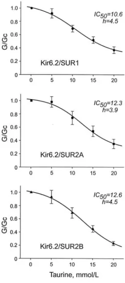
When applied intracellulary, taurine inhibited KATP
channel activity in a dose-dependent manner (Fig. 1 &
2). An IC50 was 10.6 1.34 mM for Kir6.2/SUR1 channel, 12.3 1.9 mM for Kir6.2/SUR2A, and 12.6
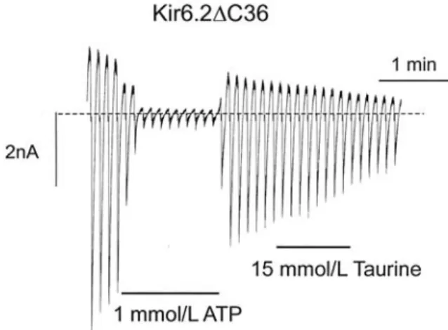
1.3 mM for Kir6.2/SUR2B, indicating that the extent of inhibitory potency was quite similar. The Hill coefficients (h) were 4.5, 3.9, and 4.5, respectively. To examine the site on which taurine acts, taurine (15 mM) was applied onto Kir6.2 C36 channel [30], a truncated form of Kir6.2 in which the last 36 amino acids of the C terminus had been deleted (Fig. 3). As expected, the channel current of Kir6.2 C36 did not respond to the taurine application, implying the site of taurine being on the SUR subunits, not Kir6.2 subunit.
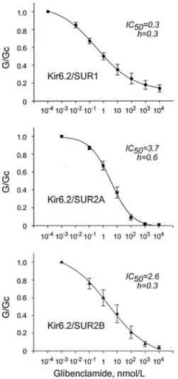
In order to elucidate whether taurine acts on the SURs interfering with the glibenclamide-binding sites, the channels were exposed to serial concentrations of glibenclamide in the presence of 10 mM taurine. In the presence of taurine, the glibenclamide sensitivities of all three recombinant KATP channels were markedly enhanced than those previously reported in the absence of taurine (31,32,33) (Fig. 4). They were fit to the Hill equation and yielded the IC50Sof 0.3 0.05 mM for Kir6.2/SUR1, 3.7 0.67 mM for Kir6.2/SUR2A, and 2.6 0.98 mM for Kir6.2/SUR2B. The Hill coefficients were 0.3 0.02, 0.6 0.05, and 0.3 0.05, respectively (Fig. 5).
As previously described, glibenclamide would bind two-binding sites on SUR1 as opposed to SUR2A or 2B, which has only the benzamido-binding site for glibenclamide. Gliclazide, another drug of sulfonylurea that binds only the sulfonylurea-, but not benzamido- binding site on SUR1, was further tested to ascertain the sites on SUR1 interacting with taurine. Gliclazide with taurine (10 mM), not as in the case of glibenclamide, similarly inhibited the Kir6.2/SUR1
channel compared to that in the absence of taurine (Fig.
6A), suggesting that taurine interacts with only sulfonylurea binding sites on SUR1. The IC50was 2.9 1.47 mM, and the Hill coefficient was 0.5 0.11 (Fig. 6B).
In the present study, we compared the taurine effects on the activities of the three-types of KATP
channels, and demonstrated that taurine nearly equally inhibited the currents of Kir6.2/SUR1, Kir6.2/SUR2A, and Kir6.2/SUR2B channels, suggesting that the types of SUR subunits are not critical for the inhibitory mechanism of taurine. This suggestion was further supported by the fact that the glibenclamide sensitivities of the recombinant KATP channels were also equally accentuated in the presence of taurine. Since the effect of taurine was not observed in Kir6.2 C36, but shown with the glibenclamide sensitivity of the channels, taurine may interact with SURs, in particular, their glibenclamide-binding sites, but not Kir6.2.
A recent study revealed that two nonadjacent cytosolic loops of SUR1, linking TMs 5 and 6 and TMs 15 and 16, are critical for [3H]-glibenclamide binding [34]. Mutation of serine (S) 1237 in the cytoplasmic loop between TMs 15 and 16 to tyrosine (Y) abolishes the high-affinity block of Kir6.2/SUR1 channels by tolbutamide [35], which has only the sulfonylurea moiety in its molecule. It is likely that this residue also interacts with the sulfonylurea moiety of drugs such as glibenclamide, glimepiride, and gliclazide. The fact that glibenclamide blocks Kir6.2/SUR1-S1237Y channels indicates that residues other than S1237 are also critical for binding of this drug. Likewise, the block of Kir6.2/SUR1 by meglitinide and repaglinide, which do not possess a sulfonylurea moiety, is unaltered by the S1237Y mutation [35,36], suggesting that these drugs do not interact with this residue. This raises the
possibility that the TM 5-6 loop may interact with the benzamido moiety of glibenclamide. Because SUR2 has only the benzamido-binding site, glibenclamide could bind to SUR2 at one site, in contrast to SUR1 at two sites. Gliclazide, which has only the sulfonylurea moiety in its molecule, may bind to SUR1 at only the
sulfonylurea site, whereas SUR2 would not bind the drug. In the present study, taurine was found to contribute to the sensitization of Kir6.2/SUR1 channels to glibenclamide, but not gliclazide. This suggests that taurine interacts only with the benzamido-binding site and the interaction with only one binding site could be enough for the taurine action.
Taurine affects many different ion channels in cell membranes, including the Na+, Ca2+, Cl-, and K+ channels. The underlying molecular mechanism seems
Fig. 1. Macroscopic currents recorded from inside-out patches in response to a series of voltage ramps from -110 to +100 mV. Oocytes were injected with mRNAs encoding Kir6.2/SUR1 (upper), Kir6.2/SUR2A (middle), or Kir6.2/SUR2B (lower). Taurine was applied at the bars indicated.
Fig. 2. Mean taurine dose-response relationships for Kir6.2/SUR1 (upper; n=5), Kir6.2/SUR2A (middle; n=5), or Kir6.2/SUR2B (lower; n=6) currents. Date represent Mean SE.
to be related with its zwitterionic nature similar to neutral membrane phospholipids, such as phosphatidylcholine and phosphatidylethanolamine [9].
It also interacts with phosphatidylserine, an acidic phospholipid, rendering higher Ca2+-binding affinity.
High-affinity taurine-binding sites have been identified on Na+-dependent and-independent taurine transporters which are associated with taurine influx [37] and efflux [38], respectively. Low-affinity taurine binding was mainly observed with the neutral membrane phospholipids [9], possibly altering membrane architecture and fluidity. In skeletal muscle fibers, the inhibitory effect of taurine on KATPchannel activity is independent of the functional state of the channel [17], supporting the notion that taurine may allosterically modify the KATPchannel activity by binding to the polar phase of the membrane phospholipids that are functionally related to the SUR, but not SUR itself.
KATPchannels, which couple cellular metabolism to membrane excitability, are found at high density in a variety of cell types, including the pancreatic beta cells, cardiac, smooth, and skeletal muscles, and some brain neurons. In all these tissues, opening of KATPchannels in
response to metabolic stress leads to inhibition of electrical activity. Thus, when muscular or neuronal cells are exposed to ischaemia or hypoxia, taurine moves out of the cells [39], relieving its inhibition of KATPchannels. This can induce an earlier activation of KATPchannel activity to save cellular energy [15,16,17].
KATP channels also play important roles in neuronal regulation of glucose homeostasis [40], seizure protection [41], and control of vascular smooth muscle tone [42]. In the beta cells, inhibited by cellular ATP and sulfonylureas, KATP channels are critical for the glucose-induced insulin secretion. The fact that taurine Fig. 3. A macroscopic current recorded from inside-out
patches in response to a series of voltage ramps from -110 to +100 mV. Oocytes were injected with mRNAs encoding Kir6.2 C36. Taurine or ATP was applied at the bars indicated.
Fig. 4. Macroscopic currents recorded from inside-out patches in response to a series of voltage ramps from -110 to +100 mV. Oocytes were injected with mRNAs encoding Kir6.2/SUR1 (upper), Kir6.2/SUR2A (middle), or Kir6.2/SUR2B (lower). Glibenclamide was applied at the bars indicated.
inhibits all types of KATP channels tested to a similar extent suggests that it plays important roles in the regulation of cellular functions linked to KATPchannel activity. It is well known that taurine is readily moved out of the cell as an osmolyte to prevent cell swelling during hypoosmotic insults. Furthermore, the concentrations of taurine are different among tissues and species; for example, 10-100 mol/L in serum, 30 mol/L/g wet wt in the rat heart, 52 mol/L/g dry wt in the pancreatic islets, 26 mol/L/g dry wt in the exocrine pancreas, 25.6 mol/L/g dry wt in the bovine cerebral
cortex, 60 mol/L/g dry wt in the brain secretory structure [1,43]. Since the difference of SUR subunits could not modify the taurine inhibition of KATPchannel activity, intracellular endogenous taurine concentration seems to be a critical factor to regulate KATPchannel activity.
In conclusion, the inhibitory effect of taurine on KATPchannel activity is not related with different SUR subunits, but rather with intracellular concentration of Fig. 5. Mean glibenclamide dose-response relationships
for Kir6.2/SUR1 (upper; n=5), Kir6.2/SUR2A (middle; n=7), or Kir6.2/SUR2B (lower; n=5) currents. Date represent Mean SE.
Fig. 6. A. A macroscopic current recorded from inside- out patches in response to a series of voltage ramps from -110 to +100 mV. Oocytes were injected with mRNAs encoding Kir6.2/SUR1.
Gliclazide was applied at the bars indicated.
B. Mean gliclazide dose-response relationships for Kir6.2/SUR1 (n=4) currents. Date represent Mean SE.
taurine. Taurine may interfere with the benzamido- binding site on SURs, but not Kir6.2 of KATPchannel.
1. Huxtable RJ. Physiological actions of taurine.
Physiol Rev 1992;72:101-63.
2. Song D, O Regan MH, Phillis JW. Amino acid release during volume regulation by cardiac cells:
cellular mechanisms. Eur J Pharmacol 1998;341:273- 80.
3. Cala PM. Volume regulation by red blood cells.
Mechanisms of ion transport between cells and mechanisms. J Physiol (London) 1983;4:33-52.
4. Fugelli K, Vislie T. Physiological response to acid water in brown trout (Salmo trutta L.): cell volume regulation in heart ventricle tissue. J Exp Biol 1982;101:71-82.
5. Franconi F, Stendardi I, Martini F, Zilletti L, Giotti A. Interaction between organic calcium-channel blockers and taurine in vitro and in vivo. J Pharm Pharmacol 1982;34:329-30.
6. Sawamura A, Sada H, Azuma J, Kishimoto S, Sperelakis N. Taurine modulates ion influx through Ca2+channels. Cell Calcium 1990;11:251-9.
7. Lewis CPL, Haschek WM, Wyatt I, Cohen GM, Smith LL. The accumulation of cystamine and its metabolism to taurine in rat lung slices. Biochem Pharmacol 1989;38:481-8.
8. Dokshina GA, Silaeva TY. Effect of taurineon insulin secretion by isolated pancreatic tissue of intact and irradiated rats. Radiobiologiya 1976;16:446-9.
9. Huxtable RJ, Sebring LA. Towards a unifying theory for the actions of taurine. Trends Pharmacol Sci 1986;7:481-5.
10. De Luca A, Pierno S, Camerino DC. Effect of taurine depletion on excitation-contraction coupling and Cl-conductance of rat skeletal muscle. Eur J Pharmacol 1996;296:215-22.
11. Steele DS, Smith GL, Miller DJ. The effects of taurine on Ca2+uptake by the sarcoplasmic reticulum and Ca2+sensitivity of chemically skinned rat heart.
J Physiol 1990;422:499-511.
12. Igisu H, Izumi K, Goto I, Kina K. Effects of taurine on the ATPase activity in the human erythrocyte membrane. Pharmacology 1976;14:362-6.
13. Sebring LA, Huxtable RJ. Taurine modulation of calcium binding to cardiac sarcolemma. J Pharmacol Exp Ther 1985;232:445-51.
14. Lombardini JB. Effects of taurine and mitochondrial metabolic inhibitors on ATP-dependent Ca2+uptake in synaptosomal and mitochondrial subcellular fractions of rat retina. J Neurochem 1988;51:200-5.
15. Han J, Kim E, Ho WK, Earm YE. Blockade of the ATP-sensitive potassium channel by taurine in rabbit ventricular myocytes. J Mol Cell Cardiol 1996;28:2043-50.
16. Satoh H. Direct inhibition by taurine of the ATP- sensitive K+channel in guinea pig ventricular cardiomyocytes. Gen Pharmacol 1996;27:625-7.
17. Tricarico D, Barbieri M, Camerino DC. Taurine blocks ATP-sensitive potassium channels of rat skeletal muscle fibres interfering with the sulphonylurea receptor. Br J Pharmacol 2000;130 :827-34.
18. Nichols CG, Lederer WJ. Adenosine triphosphate- sensitive potassium channels in the cardiovascular system. Am J Physiol 1991;261:H1675-86.
19. Aguilar-Bryan L, Nichols CG, Wechsler SW, Clement JP 4th, Boyd AE 3rd, Gonzalez G, et al.
Cloning of the beta cell high-affinity sulfonylurea receptor: a regulator of insulin secretion. Science 1995;268:423-6.
20. Inagaki N, Gonoi T, Clement JPt, Namba N, Inazawa J, Gonzalez G, et al. Reconstitution of IKATP: an inward rectifier subunit plus the sulfonylurea receptor. Science 1995;270:1166-70.
21. Inagaki N, Gonoi T, Clement JP, Wang CZ, Aguilar- Bryan L, Bryan J, et al. A family of sulfonylurea
receptors determines the pharmacological properties of ATP-sensitive K+ channels. Neuron 1996;16:1011-7.
22. Chutkow WA, Simon MC, Le Beau MM, Burant CF. Cloning, tissue expression, and chromosomal localization of SUR2, the putative drug-binding subunit of cardiac, skeletal muscle, and vascular KATPchannels. Diabetes 1996;45:1439-45.
23. Isomoto S, Kondo C, Yamada M, Matsumoto S, Higashiguchi O, Horio Y, et al. A novel sulfonylurea receptor forms with BIR (Kir6.2) a smooth muscle type ATP-sensitive K+channel. J Biol Chem 1996;271:24321-4.
24. Yamada M, Isomoto S, Matsumoto S, et al.
Sulphonylurea receptor 2B and Kir6.1 form a sulphonylurea-sensitive but ATP-insensitive K+ channel. J Physiol 1997;499(Pt 3):715-20.
25. Proks P, Reimann F, Green N, Gribble F, Ashcroft F.
Sulfonylurea stimulation of insulin secretion.
Diabetes 2002;51 Suppl 3:S368-76.
26. Garrino MG, Plant TD, Henquin JC. Effects of putative activators of K+ channels in mouse pancreatic beta-cells. Br J Pharmacol 1989;98:957- 65.
27. Schwanstecher M, Schwanstecher C, Dickel C, Chudziak F, Moshiri A, Panten U, et al. Location of the sulphonylurea receptor at the cytoplasmic face of the beta-cell membrane. Br J Pharmacol 1994;113:903-11.
28. Sakura H, Ammala C, Smith PA, Gribble FM, Ashcroft FM. Cloning and functional expression of the cDNA encoding a novel ATP-sensitive potassium channel subunit expressed in pancreatic beta-cells, brain, heart and skeletal muscle. FEBS Lett 1995;377:338-44.
29. Gribble FM, Ashfield R, Ammala C, Ashcroft FM.
Properties of cloned ATP-sensitive K+ currents expressed in Xenopus oocytes. J Physiol 1997;
498(Pt 1):7-98.
30. Tucker SJ, Gribble FM, Zhao C, Trapp S, Ashcroft
FM. Truncation of Kir6.2 produces ATP-sensitive K+ channels in the absence of the sulphonylurea receptor. Nature 1997;387:79-83.
31. Gribble FM, Tucker SJ, Seino S, Ashcroft FM.
Tissue specificity of sulfonylureas: studies on cloned cardiac and beta-cell KATPchannels. Diabetes 1998;47:1412-8.
32. Dorschner H, Brekardin E, Uhde I, Schwanstecher C, Schwanstecher M. Stoichiometry of sulfonylurea-induced ATP-sensitive potassium channel closure. Mol Pharmacol 1999;55:1060-6.
33. Russ U, Hambrock A, Artunc F, Loffler-Walz C, Horio Y, Kurachi Y, et al. Coexpression with the inward rectifier K(+) channel Kir6.1 increases the affinity of the vascular sulfonylurea receptor SUR2B for glibenclamide. Mol Pharmacol 1999;56:955-61.
34. Mikhailov MV, Mikhailova EA, Ashcroft SJ.
Molecular structure of the glibenclamide binding site of the beta-cell KATP channel. FEBS Lett 2001;499:154-60.
35. Ashfield R, Gribble FM, Ashcroft SJ, Ashcroft FM.
Identification of the high-affinity tolbutamide site on the SUR1 subunit of the KATP channel.
Diabetes 1999;48:1341-7.
36. Dabrowski M, Wahl P, Holmes WE, Ashcroft FM.
Effect of repaglinide on cloned beta cell, cardiac and smooth muscle types of ATP-sensitive potassium channels. Diabetologia 2001;44:747-56.
37. Atlas M, Bahl JJ, Roeske W, Bressler R. In vitro osmoregulation of taurine in fetal mouse hearts. J Mol Cell Cardiol 1984;16:311-20.
38. Fugelli K, Thoroed SM. Taurine transport associated with cell volume regulation in flounder erythrocytes under anisosmotic conditions. J Physiol 1986;374:245-61.
39. Phillis JW, O Regan MH. Energy utilization in the ischemic/reperfused brain. Int Rev Neurobiol 2002;51:377-414.
40. Miki T, Liss B, Minami K, Shiuchi T, Saraya A,
Kashima Y, et al. ATP-sensitive K+channels in the hypothalamus are essential for the maintenance of glucose homeostasis. Nat Neurosci 2001;4:507-12.
41. Yamada K, Ji JJ, Yuan H, Miki T, Sato S, Horimoto N, et al. Protective role of ATP-sensitive potassium channels in hypoxia-induced generalized seizure.
Science 2001;292:1543-6.
42. Quayle JM, Nelson MT, Standen NB. ATP-sensitive and inwardly rectifying potassium channels in smooth muscle. Physiol Rev 1997;77:1165-232.
43. Briel G, Gylfe E, Hellman B, Neuhoff V.
Microdetermination of free amino acids in pancreatic islets isolated from obese-hyperglycemic mice. Acta Physiol Scand 1972;84:247-53.