유방초음파 술기
관동대학교 의과대학 제일병원 외과학교실, 1차의과학대학교 강남차병원 외과
고승상, 이지현1
Practical Technique of Breast Sonography
SeungSang Ko, Ji Hyun Lee
1Department of Surgery, Cheil General Hospital & Women's Health Care Center, KwanDong University College of Medicine,
1
Department of Surgery, Kangnam Cha Hospital, Cha University College of Medicine, Seoul, Korea
Received May 3, 2013 Revised October 2, 2013 Accepted October 9, 2013
Sonography has a significant role in diagnostic breast imaging. It is most commonly used as an adjunctive test in characterization of lesions detected using other imaging modalities or by physical examination. Sonography is recognized as the modality of choice in evaluation of women who are symptomatic and younger than 30 years of age, pregnant, or lactating.
Combined mammography and breast sonography appear to have a role in screening high-risk populations. The standard terminology and description, such as Breast Imaging Reporting and Data System for Ultrasound (BI-RADs-US) lexicon is helpful in differential diagnosis between benign and malignancy. Biopsy is indicated for any feature consistent with malignancy. Breast sonography is the modality of choice for interventional procedures. The role of breast sonography is being further investigated.
Keywords: Breast image, Mammography, Ultrasonography, Breast imaging reporting and
data system for ultrasound (BI-RADs-US) lexiconCorrespondence to:
SeungSang Ko
Department of Surgery, Cheil General Hospital, Kwandong University College of Medicine, 1-19 Mukjeong-dong, Jung-gu, Seoul 100-380, Korea Tel: +82-2-2000-7080 Fax: +82-2-2000-7791 E-mail: [email protected]
서 론
유방초음파검사는 유방X선촬영과 함께 유방질환을 진 단하는데 '필수적인 검사'라고 할 수 있다. 유방초음파검 사는 유방X선촬영과 같은 방사선 노출이 없으면서, 간편 한 조작으로, 때에 구애 없이, 짧은 시간 동안에, 신속하게 실시간으로 병변의 실체를 볼 수 있는 장점이 있고, 유방 초음파검사에서 보이는 모든 병변은 유방초음파 유도 하 에 조직학적 진단이 가능하기 때문에 유방초음파검사의 이용 빈도는 날로 증가추세에 있다.
Mammography와 Breast Ultrasonography의 원리
X-ray가 물체를 투과하여 얻어지는 영상은 물질을 이 루고 있는 분자 구조에 의해 영향을 받으나, 물질의 상태, 즉 기체, 액체, 고체에 의해서는 영향을 받지 않는 것으로 되어 있다. 유방을 이루고 있는 구조물은 지방(fat)과 유 기 성분(organic components)으로 되어 있으며, 다시 유 기 성분은 유관유엽구조(ductolobular structures)내에 있는 결체 조직, 상피 조직, 분비물(organic fluid, cells and cellular debris)과 혈액으로 이루어져 있다. 이런 유 기 성분은 약 80%가 물이며, 20%는 유기 물질(organic material)이다. 물은 10%의 수소와 90%의 산소로 구성되
REVIEW ARTICLE
J Surg Ultrasound 2014;1:11-15
Journal of Surgical Ultrasound JSU
어 있고, volumic mass를 1로 볼 때, 유기 물질은 10% 수 소, 12% 탄소, 77% 질소 또는 산소로 이루어져 있어 vol- umic atomic mass가 거의 물과 동일하다. 따라서 유방 내 유기 성분은 유방촬영술(mammography)상 물과 같이 하 얗게 보인다. 한편, 지방은 12% 수소, 12% 질소 또는 산 소, 76% 탄소로 구성되어 있으며, 구성 분자 가운데 탄소 가 차지하는 비율이 크기 때문에, 지방의 volumic mass는 약 0.9로 유기 물질의 volumic mass보다 작으므로 유방 촬영술상 검게 보이게 된다. 다시 말하면, 유방촬영술 volumic atomic mass의 미세한 차이에 의해서 물과 동일 한 유기 성분과 지방을 구별하게 된다. 그러나 유기 성분 을 이루고 있는 각각의 조직들 간의 volumic atomic mass 의 차이가 대단히 작기 때문에 유방촬영술로 각각의 조직 들을 명확히 구별할 수 없으며, 특히 유기 성분에는 지방 이 매우 다양하게 혼재되어 있으므로 유방촬영술로 유방 의 구조물을 정확하게 분석한다는 것은 불가능하다.
반면에, 초음파(ultrasound)는 물질의 상태(액체, 기 체, 고체)와 물질의 역학적 속성인 탄력성(elasticity)과 밀집성(compactness)에 매우 예민하다. 초음파는 solid collagenous fiber로 이루어져 있으며 solid body로 보이 는 ''결체 조직"과 반유동성의 유방 "지방"과 액체 상태의
"관소엽 구조물(ductolobular structures)"을 뚜렷하게 구별하게 된다. 초음파는 관소엽 구조물에 차 있는 액체의 공간적 배열을 보임으로써 유관의 윤곽을 알 수 있게 하 며, 상피 질환이 있으면 관소엽 구조물 모양의 변화를 감 지할 수 있게 된다. 또한, 초음파는 병적인 상피 증식으로 상피의 두께가 증가하면 할수록 더욱 뚜렷하게 결체 조직 과의 감별이 용이하다. 따라서 흐릿하고 모호한 영상을 보 이는 유방촬영술보다 유방에서 종괴의 소견을 찾는 데는 초음파가 뛰어난 것을 알 수 있다.(1)
유방 초음파검사의 적응증(2,3)
ㆍ모든 촉지성 종괴: 종괴가 고형인지 낭성인지를 구별 하고 양성과 악성을 비교적 정확히 진단 할 수 있다.
ㆍ임상적으로 촉지 되나 유방촬영술상 병소가 불분명 한 경우
ㆍ임상적으로 촉지 되지 않으나, 유방촬영술상 병소가 의심되는 경우
ㆍ유방촬영술상 치밀 유방(dense breast)으로 숨어 있
는 병소를 찾기 위한 경우
ㆍ30세 이하의 젊은 여성, 임산부나 수유기 여성에서 병소 유무를 알아보는 경우
ㆍ수술 전 위치고정 및 결정(localization needle bi- opsy)을 하는 경우와 MammotomeⓇ을 시행하는 경우 ㆍ유방암 수술 받은 환자의 추적검사
ㆍ국소적 유방통증과 모호한 임상증상을 호소하는 경우 ㆍ유방 성형수술을 받은 환자
유방 초음파검사 방법
ㆍPerpendicular or Transductal view: 유관을 수직으 로 자른 단면을 보는 방법
ㆍAxial view: 유관 주행방향을 따라 보는 방법으로
"ductal echography"라고 하기도 한다. 이 방법은 종 양이 nipple에서 몇 시 방향에 어느 정도의 거리에 떨 어져 있는지 종양의 위치를 보다 정확히 알 수 있게 하 며, 병변과 유관소엽과의 상관관계를 perpendicular view보다 정확히 알 수 있게 하는 장점이 있다.(4)
유방 초음파상 종괴의 영상의 위치를 명명하는 방법
ㆍBreast laterality: right or left
ㆍBreast quadrant: inner high (A), inner lower (B) outer high (C), outer lower (D), beneath nipple (E)
ㆍAxis: 예) (10', 30 mm) of the right breast
유방초음파상 종괴의 크기를 명명하는 방법(5)
ㆍLine a: LDP (Largest Diameter Plane: section A) ; 초음파 영상에 보이는 종괴 단면 중 가장 긴 직경을
거리
ㆍLine c: LDP의 수직이 되는 거리
ㆍLine b: LDP의 perpendicular view에서 가장 긴 직 경의 거리
Tumor size = a × b × c mm (boundary echoes를 포함)
"Depth/Width ratio"에서는 boundary echoes가 제외
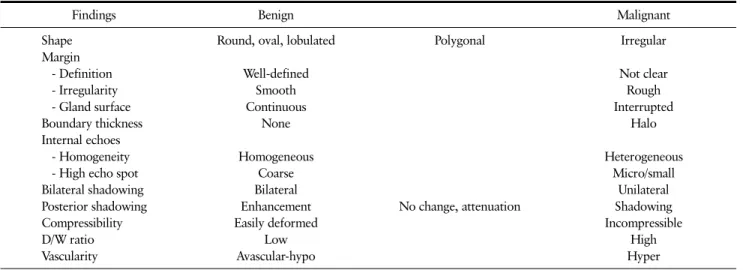
Table 1. Signs for Differential Diagnosis of Breast Lesion
Findings Benign Malignant
Shape Margin - Definition - Irregularity - Gland surface Boundary thickness Internal echoes - Homogeneity - High echo spot Bilateral shadowing Posterior shadowing Compressibility D/W ratio Vascularity
Round, oval, lobulated
Well-defined Smooth Continuous
None
Homogeneous Coarse Bilateral Enhancement Easily deformed
Low Avascular-hypo
Polygonal
No change, attenuation
Irregular
Not clear Rough Interrupted
Halo
Heterogeneous Micro/small
Unilateral Shadowing Incompressible
High Hyper
된 hypoechoic tumor의 길이만으로 측정한다.초음파화상의 정상 유방구조(6)
ㆍSkin
ㆍSubcutaneous fat layer
ㆍSuperficial layer of superficial fascia ㆍCooper's ligament
ㆍMammary gland
ㆍRetromammary fat tissue ㆍDeep layer of superficial fascia ㆍRetromammary space
ㆍFascia of pectoralis major muscle & pectoralis major muscle
ㆍRib
Guidelines of Ultrasonic Diagnosis (7-9)
이와 같은 유방초음파 소견들(Table 1) 가운데 어느 한 가지만으로는 유방암의 진단을 정확히 결정하기는 어렵 다. 그러나 유방암을 예견할 수 있는 가장 중요한 소견은 irregular shape, irregular margin, lateral boundary thickness이나 halo sign, increased vascularity로 유방 암 진단에 큰 도움이 되는 것으로 되어 있다.(10,11) Internal echoes의 기준은 subcutaneous fat layer의
echo를 기준으로 하여 hypoechoic density, isoechoic density, hyperechoic density로 결정되며, high reso- lution ultrasound로는 종양 내의 미세석회화(microcal- cification)는 대부분 관찰할 수 있어 유방암의 진단에 도 움을 주지만, internal echoes의 homogeneity를 기준하 여 유방암을 진단하기는 어렵다. Bilateral shadowing의 소견도 특징적인 섬유선종(fibroadenoma)에서 tadpole tail sign이 보이는 경우 진단에 도움을 주지만, 유방암을 결정적으로 진단하는데 미흡하며, unilateral shadowing 이 보인다고 유방암이라고 속단하는 것 보다는 shape, margin, boundary thickness의 유무를 참고하여 진단하 는 것이 올바른 방법이다. Posterior shadowing의 기준은 종괴 주변 유선조직의 echoic density를 기준하여 en- hancement, no change, attenuation, shadowing으로 구분하며, 이전에는 posterior shadowing이 보이는 경우 유방암을 의심한다고 주장하기도 하였지만 유방암의 세 포가 가득 찬 고형 유형(solid type)의 유방암에서는 얼마 든지 posterior enhancement가 보일 수 있으며 본원에서 진단된 유방암환자의 초음파 소견을 분석 결과에서도 약 50%에서 posterior enhancement의 소견을 보였다.
posterior shadowing에 변화가 없는 종괴에서도 역시 유 방암이 있을 수 있으며 일본의 보고에 의하면 papil- lo-tubular type의 유방암에서 posterior shadowing이 나타나지 않는 것으로 보고하고 있다. posterior shad- owing은 종양내의 섬유화가 많이 진행된 경성 유형 (scirrhous type)의 유방암에서 대개 보이나, 석회화된 섬
유선종(calcified fibroadenoma)은 양성이지만 posterior shadowing이 보인다. 따라서 posterior shadowing은 조 직학적 분류상 고형 유형(solid type), 유두-선형 유형 (papillo-tubular type), 경성 유형(scirrhous type)에 따라 유방암의 초음파 소견이 다양하게 보일 수 있다고 하 는 일본학자들의 주장에 공감하며, posterior shadowing 의 소견 역시 유방암의 진단에 결정적인 단서를 제공하지 는 못하는 것 같다. 작은 크기의 유방암을 진단하는데 D/W ratio 혹은 D/T ratio가 도움을 주는 것으로 보고되 고 있으며, 대부분의 초음파 소견이 해부학적 양상을 나타 내는데 비하여 수치적인 개념을 표시하는 유일한 소견이 다. D/W ratio은 boundary thickening의 소견인 hyper- echoic density를 제외한 hypoechoic tumor의 직경의 길 이만으로 측정하며, D/W ratio가 0.7 이하인 경우에는 양 성 종양을 의심하고, D/W ratio가 1 이상인 경우에는 악성 을 의심하는 것으로 보고되고 있지만, 이것 또한 유방암 진단에 참고할 만한 소견일 뿐이다.(7-10)
이와 같은 일차적인 유방초음파 소견이외에도 유방암 을 진단하는데 함께 고려해야 할 중요한 이차적인 유방 초 음파의 소견은 다음과 같다.(7,12)
ㆍDuctal spreading or extension ㆍSatellite lesions
ㆍLobular extension: thorny, burning bush. hook, tassel, comet, starfish or octopus patterns ㆍC ligament disruption
ㆍT ligament disruption: Cruciform arrangement:
T sign & + sign (plus sign) ㆍC or T ligament convergency ㆍRoundel pattern
ㆍSkin change: swelling, flattening, incisure, dimpling
ㆍAxillary lymph node enlargement
일차적, 이차적 유방 초음파 소견들을 토대로 유방암을 의심하여 진단하는 것은 영상 진단에 의한 추측일 뿐 확진 은 아니며, 확진을 하기 위해서는 반드시 조직검사가 필요 하다는 것을 잊지 말아야 한다.
최근에는 검사자의 주관적인 해석이 많은 유방초음파 검사에 의한 진단을 표준화하여 환자 치료에 도움을 주기 위하여 유방촬영술에 적용되어 사용하고 있는 BI-RADS 의 category 1-5를 유방초음파검사에도 적용하여 사용하
는 것을 시도하고 있으며, 이러한 시도로 인해 category 1-5의 환자군의 구분과 특성이 명확해지면, 환자들에게 불필요한 침습적 처치를 줄이는데 기여할 것으로 사료된 다.(10,13)
유방 초음파검사는 유방촬영술보다 해부적 구조를 분 해하는 해상능력이 뛰어나고, 검사 방법이 신속하고 간편 하기 때문에, 유방병변에 대한 여러 조직검사(core nee- dle biopsy, fine needle aspiration cytology, local- ization needle biopsy, mammotomeⓇ)를 시행할 때 유 방촬영술 유도하보다 초음파 유도하를 이용하는 경우가 전체 검사의 80% 이상을 차지하고 있으며 최근에는 유방 초음파검사를 시행하는 빈도가 계속 증가하고 있다. 그러 나 유방 초음파검사만으로 진단이 쉽지 않은 경우도 적지 않다. 유방 초음파 검사 상 양성 경향 지니고 있는 종괴의 1.6-3%정도는 조직검사 상 악성 종양이며, 유방 초음파검 사만을 시행하는 경우 약 10%에서 오진이 될 가능성이 있다 고 보고하고 있다.(10,14,15) 상피내암(Ductal Carcinoma In Situ; DCIS)로 진단되는 경우 가운데 약 10%는 유방 초 음파 검사 상 종괴의 소견이 없이 정상으로 보이며, 종괴 가 있는 경우에는 진단의 가능성이 높지만 종괴 없이 미세 석회화가 유선 조직에 산포되어 있는 경우에는 세밀한 관 찰이 있어야만 병소를 발견할 수 있다.(14,16) 침윤성 소 엽암(Invasive Lobular Carcinoma; ILC)의 암세포는 유 관 주위 조직이나 유관을 따라 침윤하여 유방조직에 암세 포가 흩어져 있어 종괴를 형성하지 않으며, 침윤성 유관암 (Invasive Ductal Carcinoma; IDC)과는 달리, 정상조직 의 면을 따라 수평방향으로 퍼지는 경향이 있다. 침윤성 소엽암의 경우에도 유방초음파 검사 상 병소가 보이지 않 는 경우가 있으며, 이런 경우를 Butler 등은 12%로 보고하 고 있다.(17,18) 전형적인 수질암(medullary carcinoma) 은 internal echo의 중심부분이 hyperechoic한 경우가 많 은데 이를 "target sign"이라고 하여 진단에 도움을 주는 것으로 보고하고 있지만(7) 전체적인 초음파검사의 소견 들이 양성과 감별이 어려운 경우도 빈번하며(7,19,20), 순 수한 점액암(mucinous carcinoma)인 경우도 양성 종양 의 특징을 보이는 경우가 빈번하기 때문에 감별에 세밀한 주의가 요구된다.(21) 섬유선종의 크기가 크면 클수록 미 세소엽(microlobulation)의 소견이 보이는 경우가 많기 때문에 악성 종양으로 오인이 되는 경우도 있으며(10), 엽 상 종양(Phyllodes tumor)인 경우에는 진단이 용이할 수
있지만 초음파검사만으로는 악성인지 양성인지 구별은 불가능한 것으로 되어 있다.(22) 유방 초음파검사로 악성 과 양성을 감별하는 데는 다소의 어려운 점이 있지만, 특 별한 경우를 제외하고는 대부분 양, 악성의 구별은 가능하 리라고 생각한다. 그러나 유방 초음파의 소견만으로 병리 학적 분류에 따른 특수 유형(special type)의 조직형까지 정확히 알기에는 각각의 조직형의 유방초음파 소견들이 비특이적이고 서로 중첩되는 부분이 많기 때문에 어렵다 고 생각된다. 따라서 이런 문제는 현재 사용하고 있는 초 음파기계보다 성능이 더 나은 컬러 초음파(color ultra- sound)와 같은 기계들이 상용화되어 생체와 똑 같은 화상 을 얻을 수 있는 미래에는 해결될 수 있을 것으로 전망한다.
결 론
유방초음파검사는 "유방 속을 투시하는 눈"과 같은 역 할을 하고 있다. 유방초음파검사를 통하여 유방내의 실질 들을 정확히 관찰하고 진단하고자 하는 학문적인 노력이 뒤따른다면, 또 다른 각도에서 유방질환을 평가할 수 도 있을 것이며, 더 나아가서는 유방초음파에 대해도 기존과 는 다른 학문적인 진전도 기대해 볼 수 있다고 생각한다.
REFERENCES
1. Teboul M, Halliwell M. Atlas of ultrasound and ductal echography of the breast: The introduction of anatomic intelligence into breast imaging. Oxford, England; Cam- bridge, Mass., USA: Blackwell Science; 1995. p.18-20.
2. Oh KK. Breast Image. 1996;33-9.
3. Yonsei Univeristy School of Medicine, Department of Radiology. Diagnostic approach of Early Breast Cancer.
1996;61-4.
4. Teboul M, Halliwell M. Atlas of ultrasound and ductal echography of the breast: The introduction of anatomic intelligence into breast imaging. Oxford, England; Cam- bridge, Mass., USA: Blackwell Science; 1995. p.83-151.
5. Mizutani M. 13th ICUEB 2003;146-7.
6. Oh KK. Breast Image. 1996;13-27.
7. Oh KK. Breast Image. 1996;44-133.
8. Mizutani M. 13th ICUEB 2003;142-57.
9. Ei Ueno. Real-time breast ultrasound. 1991.
10. Stavros AT, Thickman D, Rapp CL, Dennis MA, Parker SH, Sisney GA. Solid breast nodules: use of sonog- raphy to distinguish between benign and malignant lesions. Radiology 1995;196:123-34.
11. Milz P, Lienemann A, Kessler M, Reiser M. Evaluation of breast lesions by power Doppler sonography. Eur Radiol 2001;11:547-54.
12. Teboul M, Halliwell M. Atlas of ultrasound and ductal echography of the breast: The introduction of anatomic intelligence into breast imaging. Oxford, England; Cam- bridge, Mass., USA: Blackwell Science; 1995. p.204-343.
13. Mendelson EB. BI-RADS for ultrasound. 13th ICUEB 2003:41, 141.
14. Skaane P, Sauer T. Ultrasonography of malignant breast neoplasms. Analysis of carcinomas missed as tumor.
Acta Radiol 1999;40:376-82.
15. Durfee SM, Selland DL, Smith DN, Lester SC, Kaelin CM, Meyer JE. Sonographic evaluation of clinically palpable breast cancers invisible on mammography.
Breast J 2000;6:247-51.
16. Kasumi F et al. Ultrasonic Image of Noninvasive Car- cinomas: Breast Ultrasound Update 1994;168-73.
17. Butler RS, Venta LA, Wiley EL, Ellis RL, Dempsey PJ, Rubin E. Sonographic evaluation of infiltrating lobu- lar carcinoma. AJR Am J Roentgenol 1999;172:325-30.
18. Cawson JN, Law EM, Kavanagh AM. Invasive lobular carcinoma: sonographic features of cancers detected in a BreastScreen Program. Australas Radiol 2001;45:
25-30.
19. Yilmaz E, Lebe B, Balci P, Sal S, Canda T. Compar- ison of mammographic and sonographic findings in typical and atypical medullary carcinomas of the breast. Clin Radiol 2002;57:640-5.
20. Cheung YC, Chen SC, Lee KF, Wan YL, Ng SH. Sono- graphic and pathologic findings in typical and atyp- ical medullary carcinomas of the breast. J Clin Ultrasound 2000;28:325-31.
21. Memis A, Ozdemir N, Parildar M, Ustun EE, Erhan Y.
Mucinous (colloid) breast cancer: mammographic and US features with histologic correlation. Eur J Radiol 2000;35:39-43.
22. Lifshitz OH, Whitman GJ, Sahin AA, Yang WT.
Radiologic-pathologic conferences of the University of Texas M.D. Anderson Cancer Center. Phyllodes tumor of the breast. AJR Am J Roentgenol 2003;180:332.