52 354
This is an Open Access article distributed under the terms of the Creative Commons Attribution Non-Commercial License (http://creativecommons.org/licenses/by-nc/4.0/) which permits unrestricted non-commercial use, distribution, and reproduction in any medium, provided the original work is properly cited.
Copyright © 2019. Anatomy & Cell Biology
Introduction
The Stafne bone cavity (SBC), first reported by Edward Stafne in 1942 [1], also called the static bone cavity, salivary inclusion cyst [2], latent cyst [3], and lingual bone defect [4], is an asymptomatic bony defect that is commonly located in- ferior to the mandibular canal and slightly above the inferior border of the mandible [1, 5]. The reported incidence of the SBC ranges from 0.1% to 6.06% [5, 6]. It is usually found on one side but occasionally, on both sides of the mandible [7].
The submandibular gland, fat tissue, connective tissue, lym- phatic tissue, muscle or vessels have all been considered as potential contents of the SBC [4]. In the clinical setting, most SBC is found incidentally during panoramic radiography.
Since the SBC is an asymptomatic bone cavity and does not require treatment, it is relatively rare to see the actual bony defect without radiographic images. Therefore, herein, we re-
port a case of unilateral SBC in a cadaver.
Case Report
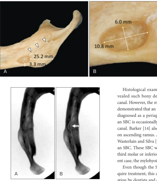
A 76-year-old at death male Caucasian cadaveric head (fresh-frozen) was dissected. During routine dissection of the head, the mandible was dissected, and the soft tissues of the mandible were removed. After the mandible was separated from the head, a large bony defect was found on the lingual surface of the right mandible, which was located 25.2 mm anterior to the angle of the mandible and 3.3 mm superior to the inferior border of the mandible. The shape and size of the bony defect was oval and 10.8×6.0 mm, respectively (Figs. 1, 2). The bony defect had a smooth surface and did not show any pathological condition. Therefore, it was determined to be an SBC. The SBC had continuity with the right mylohyoid groove. The depth of the SBC and width of the mandible at the SBC was 4.15 mm, 3.14 mm, respectively. The SBC was not observed on the left side.
Discussion
Studies on the SBC using dry mandibles were reported by Harvey and Noble in 1968 [7] and Kay in 1974 [8]. Since
Case Report
https://doi.org/10.5115/acb.19.019 pISSN 2093-3665 eISSN 2093-3673
Corresponding author:
Joe Iwanaga
Seattle Science Foundation, 550 17th Ave, James Tower, Suite 600, Seattle, WA 98122, USA
Tel: +1-2067326500, Fax: +1-2067326599, E-mail: [email protected]
Stafne bone cavity: a rare cadaveric case report
Joe Iwanaga
1,2, T. L. Wong
1, Shogo Kikuta
1,2, R. Shane Tubbs
1,31Seattle Science Foundation, Seattle, WA, USA, 2Dental and Oral Medical Center, Kurume University School of Medicine, Kurume, Japan, 3Department of Anatomical Sciences, St. George’s University, St. George’s, Grenada, West Indies
Abstract: The Stafne bone cavity (SBC), also called the static bone cavity, salivary inclusion cyst, latent cyst, and lingual bone defect is an asymptomatic bony defect that is commonly located inferior to the mandibular canal and slightly above the inferior border of the mandible. It is rare to see the actual bony defect in the cadaver because of its relatively low incidence of 0.1% to 6.06%. We report a unilateral SBC found in a 76-year-old at death male Caucasian cadaver and involving the right mandible.
The SBC was oval in shape with a smooth surface and measured 10.8×6.0 mm. The SBC was continuous with the right mylohyoid groove. Since actual photographs of the SBC are lacking in the literature, this report might provide additional insight for better understanding the SBC.
Key words: Cadaver, Anatomy, Stafne bone, Mylohyoid groove, Variations Received February 7, 2019; Revised March 15, 2019; Accepted March 25, 2019
Stafne bone cavity cadaveric case report
https://doi.org/10.5115/acb.19.019
Anat Cell Biol 2019;52:354-356
355
www.acbjournal.org
then, most of the reported studies examined either patients’
panoramic radiography or computed tomography. The etiol- ogy of SBC is unclear. Many hypotheses exist regarding the cause of the bony defect, including a hypertrophic lobe of the submandibular gland, erosion by vascular compression [9], or incomplete Meckel cartilage ossification [9]. Minowa et al. [9]
hypothesized that an SBC results from bone resorption or an overlying lipoma. In a case report by Kaya et al. [10], an ec- topic parotid gland was found in the SBC. SBCs are most fre- quently found in patients over 50 years old [9] and there have been no reports of SBCs in patients younger than 10 years old [11]. On conventional panoramic radiographs, the SBC usually resembles a unilocular cystic lesion with well-defined borders. Multiple SBC was also reported by Aguiar et al. [12].
Histological examination by Harvey and Noble [7] re- vealed such bony defects did not invade the mandibular canal. However, the most recent study by Hisatomi et al. [13]
demonstrated that an SBC in the premolar area might be mis- diagnosed as a periapical lesion or odontogenic cyst. Thus, an SBC is occasionally not located inferior to the mandibular canal. Barker [14] also reported a case with an SBC situated on ascending ramus. An anthropological study conducted by Wasterlain and Silva [15] investigated ancient mandibles with an SBC. These SBC were most commonly situated below the third molar or inferior to the mylohyoid groove. In the pres- ent case, the mylohyoid groove had continuity with the SBC.
Even though the SBC is asymptomatic and does not re- quire treatment, this anomaly should be taken into consider- ation by dentists and oral surgeon in order to avoid misdiag- nosis.
Author Contributions
Conceptualization: JI, RST. Data acquisition: JI, TLW, SK.
Data analysis or interpretation: JI, TLW, SK. Drafting of the manuscript: JI, TLW, SK. Approval of the final version of the manuscript: all authors.
Conflicts of Interest
No potential conflict of interest relevant to this article was reported.
References
1. Stafne EC. Bone cavities situated near the angle of the mandible.
J Am Dent Assoc 1942;29:1969-72.
2. Fordyce GL. The probable nature of so-called latent haemor- Fig. 2. Fluoroscopy of the right mandible (superiorinferior view)
showing depth of Stafne bone defect. (A) Plain radiography. (B) Pointing the Stafne bone defect (dotted line and arrow).
Fig. 1. Stafne bone cavity (SBC) of the right mandible. (A) SBC located 25.2 mm anterior to the posterior border of the ramus and 3.3 mm superior to the inferior border of the mandible. Note the mylohyoid groove (arrowheads) has con ti nuity with the SBC. (B) Mag ni fied photo of Fig. 1A.
Anat Cell Biol 2019;52:354-356 Joe Iwanaga, et al
356
www.acbjournal.org https://doi.org/10.5115/acb.19.019
rhagic cysts of the mandible. Br Dent J 1956;101:40-2.
3. Rushton MA. Solitary bone cysts in the mandible. Br Dent J 1946;81:37-49.
4. Etoz M, Etoz OA, Sahman H, Sekerci AE, Polat HB. An unusual case of multilocular Stafne bone cavity. Dentomaxillofac Radiol 2012;41:75-8.
5. Quesada-Gómez C, Valmaseda-Castellón E, Berini-Aytés L, Gay-Escoda C. Stafne bone cavity: a retrospective study of 11 cases. Med Oral Patol Oral Cir Bucal 2006;11:E277-80.
6. Li B, Long X, Cheng Y, Wang S. Cone beam CT sialography of Stafne bone cavity. Dentomaxillofac Radiol 2011;40:519-23.
7. Harvey W, Noble HW. Defects on the lngual surface of the man- dible near the angle. Br J Oral Surg 1968;6:75-83.
8. Kay LW. Some anthropologic investigations of interest to oral surgeons. Int J Oral Surg 1974;3:363-79.
9. Minowa K, Inoue N, Sawamura T, Matsuda A, Totsuka Y, Na- kamura M. Evaluation of static bone cavities with CT and MRI.
Dentomaxillofac Radiol 2003;32:2-7.
10. Kaya M, Ugur KS, Dagli E, Kurtaran H, Gunduz M. Stafne bone
cavity containing ectopic parotid gland. Braz J Otorhinolaryngol 2018;84:669-72.
11. Hansson LG. Development of a lingual mandibular bone cavity in an 11-year-old boy. Oral Surg Oral Med Oral Pathol 1980;49:
376-8.
12. Aguiar LB, Neves FS, Bastos LC, Crusoé-Rebello I, Ambrosano GM, Campos PS. Multiple stafne bone defects: a rare entity.
ISRN Dent 2011;2011:792145.
13. Hisatomi M, Munhoz L, Asaumi J, Arita ES. Stafne bone defects radiographic features in panoramic radiographs: assessment of 91 cases. Med Oral Patol Oral Cir Bucal 2019;24:e12-9.
14. Barker GR. A radiolucency of the ascending ramus of the man- dible associated with invested parotid salivary gland material and analogous with a Stafne bone cavity. Br J Oral Maxillofac Surg 1988;26:81-4.
15. Wasterlain SN, Silva AM. Study of stafne's defects in Late Neo- lithic, Late Roman, Medieval and Modern skeletal samples from Portugal. Int J Osteoarchaeol 2012;22:423-34.