Association between Adipokines and Coronary Artery Lesions in Children with Kawasaki Disease
Body fat is an important source of adipokine, which is associated with energy balance and inflammatory and immune responses. However, the role of adipokines in coronary artery complications in Kawasaki disease (KD) has not yet been fully explained. We investigated whether serum adipokine level can be a useful marker for patients with KD who are at higher risk of developing coronary artery lesion (CAL). We measured adipokine levels and other inflammatory parameters in 40 patients with KD, 32 febrile controls, and 15 afebrile controls. Interleukin (IL)-6, tumor necrosis factor (TNF)-α and other laboratory parameters were also measured before and after intravenous immunoglobulin therapy, and in the convalescent phase. At admission, the serum resistin levels in KD children were significantly higher than those in controls (177.56 ng/mL in KD children, 76.48 ng/mL in febrile controls, and 17.95 ng/mL in afebrile controls). In patients with KD, resistin levels were significantly associated with decreased hemoglobin levels (P = 0.049) and increased IL-6 levels (P = 0.014). The serum IL-6 levels were significantly higher and body mass index was significantly lower in the group of KD with CALs than those without CALs (228.26 ng/mL vs. 39.18 ng/mL and 15.09 vs. 16.60, respectively). In conclusion, resistin is significantly elevated in KD patients, although it has no prognostic value of predicting coronary artery lesion in the acute stage.
Keywords: Leptin; Adiponectin; Resistin; Interleukin-6; Tumor Necrosis Factor-alpha;
Mucocutaneous Lymph Node Syndrome; Coronary Artery Bypass Hyun Jung Kim,1 Eun Hye Choi,2
and Hong Ryang Kil3
1Department of Pediatrics, Eulji Universitiy School of Medicine, Daejeon; 2Eulji Medi-Bio Research Institute, Eulji University, Daejeon; 3Department of Pediatrics, Chungnam National University School of Medicine, Daejeon, Korea
Received: 17 March 2014 Accepted: 24 July 2014 Address for Correspondence:
Hong Ryang Kil, MD
Department of Pediatrics, Chungnam National University Hospital, Chungnam National University School of Medicine, 282 Munhwa-ro, Jung-gu, Daejeon 301-726, Korea Tel: +82.42-280-7251, Fax: +82.42-255-3158 E-mail: [email protected]
Funding: This research was supported by EMBRI Grants (2013 EMBRIDJ) from the Eulji University.
http://dx.doi.org/10.3346/jkms.2014.29.10.1385 • J Korean Med Sci 2014; 29: 1385-1390
INTRODUCTION
Kawasaki disease (KD) is an acute febrile disease with coronary and other systemic vasculitis that occurs predominantly in in- fancy and early childhood. Although the pathogenesis of KD has not been clearly identified, serum levels of proinflammato- ry cytokines, such as interleukin (IL)-6 and tumor necrosis fac- tor (TNF)-α, are elevated during the acute phase of KD, sug- gesting that they may be involved in the development of KD (1).
White adipose tissue which used to be simply known as a storage of surplus energy is now perceived as an independent and active endocrine organ. It produces various kinds of adipo- kines such as leptin, adiponectin and resistin, which have ma- jor effects on obesity related metabolic disease by controlling fat metabolism, energy homeostasis and insulin sensitivity. More- over, adipokines participate in the systemic inflammatory re- sponse with strong reciprocal influences on other cytokines such as TNF-α, IL-6 and IL-10, and in regulating the systemic inflammatory response, angiogenesis, and subclinical athero- sclerosis (2).
Recent studies have reported that circulating adiponectin levels in patients with acute KD were significantly lower than
that in convalescent KD patients and controls (3). The role of hypoadiponectinemia or hyperadiponectinemia on the degree of severity of KD remains controversial. Also, previous reports have shown that the serum resistin levels were elevated in KD children than healthy children, but its concentrations were un- likely to help predict the prognosis of disease in acute KD pa- tients (4). There have been contrasting reports on the potential role of leptin in the pathogenesis of KD. Some published stud- ies have suggested that leptin participates in the systemic infla- mmatory response (5), and increased leptin concentration is an independent risk factor for coronary heart disease (6). In contrast, Liu et al. (7) reported that serum leptin levels in pa- tients with KD were lower than those in healthy controls, which suggests that leptin is not involved in the generalized inflam- mation seen in KD patients. Taking this evidence into consider- ation, we hypothesized that adipokines might exert direct or in- direct effects on the progression of KD, and that they may be predictive factors for development of coronary artery lesions (CAL). Therefore, we compared the sereial levels of adipokines, IL-6, and TNF-α in KD patients and control groups to assess whether an association is present between circulating adipo- kine levels and CAL in KD patients.
Cardiovascular Disorders
MATERIALS AND METHODS Patients and data collection
The study included 40 Korean children with KD and 47 age-mat- ched children recruited between May and November 2013. The 40 KD patients were enrolled within 8 days of the onset of ill- ness, with day 1 defined as the first day of fever symptoms. The control groups consisted of 47 subjects (32 febrile patients and 15 afebrile patients) with a mean body mass index (BMI) of 16.15 ± 1.82 kg/m2. The febrile control patients had either pneu- monia, acute tonsillitis, or acute cervical lymphadenitis. The afebrile control patients had acute gastroenteritis, acute urti- caria, or Henoch-Schonlein purpura.
Echocardiography
Echocardiography was performed within 8 days of the onset of fever or before intravenous immunoglobulin (IVIG) adminis- tration. CALs were diagnosed on the basis of the Z scores of the left main coronary artery, proximal left anterior descending cor- onary artery, and proximal right coronary artery, and were de- fined as the Z scores of 2.0 or more. The value of Z scores from a standardized coronary artery dimension was calculated from the body surface area (8) based on formula of Haycock’s et al. (9).
Laboratory analysis
Levels of leptin, adiponectin and resistin were measured in 87 cases, including groups: KD patients with normal coronary ar- teries (n = 28), KD patients with dilated coronary arteries (n = 12), the acute febrile control group (n = 32), and the afebrile control group (n = 15). The serum levels of leptin, adiponectin, resistin, IL-6, and TNF-α were assayed with an enzyme-linked immunosorbent assay (ELISA) kit (abcamR, R&D Systems, Cam- bridge, United Kingdom). White blood cells counts (WBC), he- moglobin, hematocrit, platelet count, alanine aminotransferase (ALT), aspartate aminotransferase (AST), C-reactive protein (CRP), erythrocyte sedimentation rate (ESR), and lipid panels were obtained in all study subjects; blood samples were drawn before IVIG therapy in KD patients.
Serial blood samples were obtained from all KD patients in the acute stage, 2-4 days after IVIG infusion, and in the conva- lescent phase, when ESR was normal (on days 30-35). Samples were prepared at the appropriate dilutions and paired samples were assayed together according to the instructions of the man- ufacturers.
Statistical analysis
Normally distributed continuous data were expressed as mean
± standard deviation. Comparisons of the frequencies between groups were analyzed using chi-square tests. Differences among groups were assessed using the unpaired 2-tailed t-tests and analysis of variance (ANOVA). The significance of difference
was calculated by Scheffe’s test, and a P value less than 0.05 was considered as statistically significant.
Ethics statement
Informed consent was obtained from the parents of all the chil- dren, and the study protocol was approved by the Eulji Univer- sity Hospital institutional review board (No. 2013-03-012). In- formed consent was confirmed by IRB.
RESULTS
Baseline patient characteristics and laboratory findings The KD group included 25 boys and 15 girls with a mean age at diagnosis of 35.18 ± 22.09 months with a range from 5 months to 8 yr. Of the 40 KD patients, 33 patients (82.5%) were diagnos- ed with typical KD and 7 patients (17.5%) with incomplete KD.
The mean time until start of IVIG treatment was 5.43 ± 1.19 days.
The mean age of the control groups was 34.41 ± 21.09 mon ths and 41.07 ± 22.80 months in febrile and afebrile control groups, respectively.
The mean intervals of blood sampling day from the onset of illness before and after IVIG therapy in the KD patients were 5.43 ± 1.19 and 8.00 ± 1.60 days, respectively.
Two patients resisted the initial IVIG treatment, but respond- ed to the second IVIG treatment.
At admission, the levels of WBC, neutrophil count, ALT, ESR, CRP, and N-terminal fragment of B-type natriuretic peptide (NT- proBNP) were significantly higher in KD patients compared with the control groups. The level of HDL-C was lower in KD patients compared with the control groups (P = 0.008 in febrile controls, P = 0.004 in afebrile controls) (Table 1).
Levels of TNF-α, IL-6, adipokines and other parameters in KD patients
The serum resistin levels at baseline in KD children were signif- icantly higher than those in the febrile controls (177.56 ng/mL [3.12-546.83] vs. 76.48 ng/mL [0.17-257.6], P = 0.024). There were no significant differences between groups of KD with and without CALs in age, gender, ALT, AST, CRP, and NT-proBNP levels. However, serum IL-6 levels were significantly higher and hemoglobin levels were significantly lower in the KD group with CALs than those without CALs (P = 0.016 and P = 0.018, respectively). There were no significant differences in serum leptin, resistin, adiponectin, and TNF-α levels between groups of KD with and without CALs (Table 2).
Echocardiographic findings
Twelve of the 40 patients (30.0%) showed dilation of coronary arteries in the acute stage of KD. All of the KD patients with CALs at admission showed normal coronary arteries at 1 month after discharge. None of the KD patients developed coronary artery
aneurysms as evaluated by echocardiography before and after IVIG therapy. There was no difference between the KD patients
and the febrile control patients in the degree of left ventricular fractional shortening. The ratio of mitral peak velocity of early filling to early diastolic mitral annular velocity measured by tis- sue Doppler imaging (TDI) was increased in the acute KD group compared with the febrile control group, which did not reach a statistical significance (Table 3).
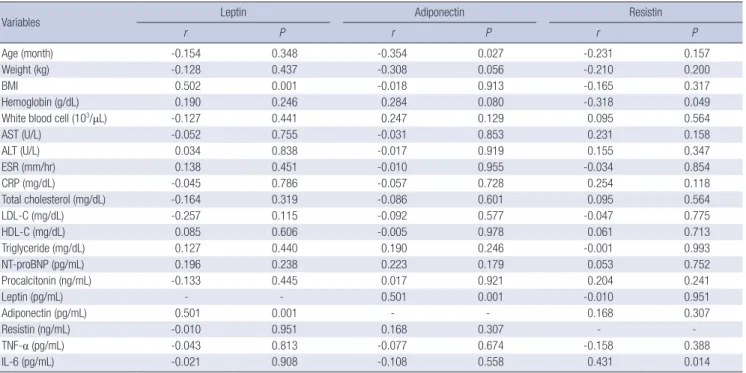
Correlations between adipokines and other parameters Hemoglobin and IL-6 levels were significantly correlated with resistin levels in patients with KD (r = -0.318, P = 0.027; r = 0.431, P = 0.014, respectively). Age in KD patients was negatively as- sociated with adiponectin levels (r = -0.354, P = 0.027). In addi- tion, BMI was positively associated with leptin levels in KD (r = 0.502, P = 0.001). A positive correlation was found between leptin and adiponectin levels before IVIG treatment in acute stage of Table 1. Baseline characteristics of patients with KD and control subjects
Parameters KD patients (n = 40) Febrile controls (n = 32) Afebrile controls (n = 15) P
Age (month) 35.18 ± 22.09 34.41 ± 21.01 41.07 ± 22.80 0.598
Gender (male/female) 25/15 14/18 8/7 0.284
Weight (kg) 14.24 ± 5.39 14.0 ± 3.41 15.24 ± 5.16 0.696
BMI 16.15 ± 1.82 16.1 ± 1.74 16.0 ± 1.91 0.966
Hemoglobin (g/dL) 11.73 ± 0.96 13.15 ± 5.49 12.55 ± 0.89 0.216
White blood cell (103/μL) 13.26 ± 43.55 11.56 ± 5.08 8.52 ± 3.20 0.003
Neutrophil (%) 66.5 ± 13.51 60.43 ± 15.65 47.94 ± 15.68 < 0.001
AST (U/L) 103.55 ± 216.31 37.34 ± 7.57 38.33 ± 13.31 0.122
ALT (U/L) 73.73 ± 103.27 19.34 ± 7.94 19.33 ± 10.62 0.003
ESR (mm/hr) 51.18 ± 29.77 20.74 ± 20.87 18.1 ± 19.15 < 0.001
CRP (mg/dL) 7.59 ± 4.75 3.42 ± 3.36 0.64 ± 0.72 < 0.001
Total cholesterol (mg/dL) 131.38 ± 21.65 133.25 ± 17.26 145.67 ± 29.39 0.092
LDL-C (mg/dL) 77.58 ± 18.90 72.21 ± 12.27 80.78 ± 20.13 0.281
HDL-C (mg/dL) 33.53 ± 9.43 41.33 ± 10.0 44.58 ± 10.57 < 0.001
Triglyceride (mg/dL) 101.38 ± 42.26 88.41 ± 40.40 97.58 ± 46.23 0.469
NT-proBNP (pg/mL) 1072.01 ± 1572.64 152.28 ± 145.27 153.76 ± 95.38 0.009
Procalcitonin (ng/mL) 1.43 ± 2.24 0.78 ± 1.34 0.15 ± 0.14 0.382
Leptin (pg/mL) 142.22 ± 300.27 30.56 ± 92.31 25.14 ± 48.54 0.052
Adiponectin (pg/mL) 38.49 ± 95.58 60.45 ± 129.58 14.05 ± 25.81 0.339
Resistin (ng/mL) 177.56 ± 207.59 76.48 ± 92.70 17.95 ± 30.90 0.001
TNF-α (pg/mL) 3.39 ± 18.48 0.32 ± 0.67 0.59 ± 1.03 0.764
IL-6 (pg/mL) 104.02 ± 214.83 1.81 ± 2.23 0.24 ± 0.54 0.095
BMI, body mass index; AST, aspartate aminotransferase; ALT, alanine aminotransferase, LDL-C, low density lipoprotein-cholesterol; HDL-C, high density lipoprotein-cholesterol;
NT-proBNP, N-terminal fragment of B-type natriuretic peptide; TNF-α, tumor necrosis factor-α; IL-6, Interleukin-6.
Table 2. Relationship between clinical parameters in KD patients and development of coronary artery lesions
Parameters CAL(+) (n = 12) CAL(-) (n = 28) P
Age (month) 34.92 ± 26.59 35.29 ± 20.41 0.962
Gender (male/female) 8/4 17/11 0.722
Weight (kg) 14.07 ± 6.50 14.31 ± 4.97 0.899
BMI 15.09 ± 1.41 16.60 ± 1.82 0.015
Hemoglobin (g/dL) 11.22 ± 1.22 11.95 ± 0.64 0.018 White blood cell (103/μL) 14.24 ± 4.48 12.84 ± 4.31 0.357
AST (U/L) 38.08 ± 21.52 123.36 ± 254.95 0.258
ALT (U/L) 64.08 ± 68.58 72.46 ± 111.34 0.811
ESR (mm/hr) 58.40 ± 32.67 46.0 ± 29.52 0.291
CRP (mg/dL) 8.30 ± 4.04 7.28 ± 5.06 0.538
Total cholesterol (mg/dL) 138.17 ± 27.77 131.11 ± 20.56 0.377
LDL-C (mg/dL) 78.42 ± 23.31 76.49 ± 17.38 0.773
HDL-C (mg/dL) 29.25 ± 9.90 37.71 ± 15.97 0.098
Triglyceride (mg/dL) 111.67 ± 50.69 96.96 ± 38.28 0.320 NT-proBNP (pg/mL) 1,690.06 ± 2,239.89 861.87 ± 1,217.30 0.144 Procalcitonin (ng/mL) 2.07 ± 3.09 1.11 ± 1.65 0.231 Leptin (pg/mL) 8.42 ± 9.36 228.51 ± 719.85 0.300 Adiponectin (pg/mL) 62.52 ± 140.61 29.24 ± 70.18 0.327 Resistin (ng/mL) 254.06 ± 192.54 176.99 ± 196.63 0.263
TNF- α (pg/mL) 9.21 ± 31.10 0.21 ± 0.22 0.201
IL-6 (pg/mL) 228.26 ± 303.97 39.18 ± 107.20 0.016 CAL, coronary artery lesion; BMI, body mass index; AST, aspartate aminotransferase;
ALT, alanine aminotransferase, LDL-C, low density lipoprotein-cholesterol; HDL-C, high density lipoprotein-cholesterol; NT-proBNP, N-terminal fragment of B-type natri- uretic peptide; TNF-α, tumor necrosis factor-α; IL-6, Interleukin-6.
Table 3. Echocardiographic findings in Kawasaki disease and febrile control groups
Findings KD patients
(n = 40)
Febrile controls
(n = 32) P
LV fractional shortening (%) 33.39 ± 4.38 33.59 ± 3.19 0.884 Base systolic velocity (cm/sec) 7.23 ± 1.61 7.91 ± 1.22 0.203 Base E-wave velocity (cm/sec) 10.46 ± 1.93 10.83 ± 1.33 0.538 Base A-wave velocity (cm/sec) 7.28 ± 2.31 6.67 ± 1.23 0.384
E/A ratio 1.43 ± 0.25 1.44 ± 0.17 0.876
E/E’ ratio 11.14 ± 2.46 9.70 ± 1.86 0.069
Deceleration time (msec) 163.70 ± 42.87 177.92 ± 39.49 0.312
Tei index 0.34 ± 0.07 0.30 ± 0.05 0.102
E, base peak early diastolic E-wave velocity (cm/sec); A, base peak early diastolic A- wave velocity (cm/sec); E’, peak early diastolic myocardial velocity (cm/sec).
KD (r = 0.501, P = 0.001) (Table 4). In febrile controls, resistin concentrations were significantly associated with elevated WBC counts (P = 0.002) and CRP levels (P = 0.021).
Serial changes in serum levels of adipokines, IL-6 and TNF-α in KD patients
Fig. 1 and Fig. 2 show the changes in serum adipokines, IL-6,
and TNF- α levels between the acute stage (before IVIG thera- py), 2-4 days after IVIG treatment, and the convalescent phase.
The serum adiponectin levels were increased immediately after IVIG therapy but were decreased to baseline levels in the con- valescent phase. IL-6 and TNF- α levels peaked at 5.43 ± 1.19 days and 8.0 ± 1.60 days from onset of illness, respectively.
After IVIG therapy, serum resistin levels significantly decreas- ed (177.56 ng/mL before IVIG therapy vs. 63.37 ng/mL at 48 hr after IVIG infusion, P < 0.01) (Fig. 1). Serum IL-6 levels signifi- cantly decreased from 141.65 pg/mL to 1.91 pg/mL after IVIG treatment (P < 0.01) (Fig. 2). The levels of TNF- α continued to increase after IVIG treatment during hospitalization (4.86 ± 22.0 pg/mL at baseline vs. 19.13 ± 61.82 pg/mL at 48 hr after IVIG therapy, P = 0.281), but did not reach a statistical significance.
Table 4. Correlation of leptin, adiponectin and resistin with clinical and other laboratory variables in patients with Kawasaki disease
Variables Leptin Adiponectin Resistin
r P r P r P
Age (month) -0.154 0.348 -0.354 0.027 -0.231 0.157
Weight (kg) -0.128 0.437 -0.308 0.056 -0.210 0.200
BMI 0.502 0.001 -0.018 0.913 -0.165 0.317
Hemoglobin (g/dL) 0.190 0.246 0.284 0.080 -0.318 0.049
White blood cell (103/μL) -0.127 0.441 0.247 0.129 0.095 0.564
AST (U/L) -0.052 0.755 -0.031 0.853 0.231 0.158
ALT (U/L) 0.034 0.838 -0.017 0.919 0.155 0.347
ESR (mm/hr) 0.138 0.451 -0.010 0.955 -0.034 0.854
CRP (mg/dL) -0.045 0.786 -0.057 0.728 0.254 0.118
Total cholesterol (mg/dL) -0.164 0.319 -0.086 0.601 0.095 0.564
LDL-C (mg/dL) -0.257 0.115 -0.092 0.577 -0.047 0.775
HDL-C (mg/dL) 0.085 0.606 -0.005 0.978 0.061 0.713
Triglyceride (mg/dL) 0.127 0.440 0.190 0.246 -0.001 0.993
NT-proBNP (pg/mL) 0.196 0.238 0.223 0.179 0.053 0.752
Procalcitonin (ng/mL) -0.133 0.445 0.017 0.921 0.204 0.241
Leptin (pg/mL) - - 0.501 0.001 -0.010 0.951
Adiponectin (pg/mL) 0.501 0.001 - - 0.168 0.307
Resistin (ng/mL) -0.010 0.951 0.168 0.307 - -
TNF-α (pg/mL) -0.043 0.813 -0.077 0.674 -0.158 0.388
IL-6 (pg/mL) -0.021 0.908 -0.108 0.558 0.431 0.014
BMI, body mass index; AST, aspartate aminotransferase; ALT, alanine aminotransferase, LDL-C, low density lipoprotein-cholesterol; HDL-C, high density lipoprotein-cholesterol;
NT-proBNP, N-terminal fragment of B-type natriuretic peptide; TNF-α, tumor necrosis factor-α; IL-6, Interleukin-6.
Fig. 1. Changes in adipokine levels before intravenous immunoglobulin (IVIG) infu- sion, at 48 hr after IVIG infusion and in the convalescent phase in Kawasaki disease patients.
Before IVIG After 48 hr After 1 mo 200
150 100 50 0
Leptin (pg/mL) Adiponectin (pg/mL)
Before IVIG After 48 hr After 1 mo 200
150 100 50 0
Resistin (ng/mL)
P < 0.01 P = 0.034 Before IVIG After 48 hr After 1 mo
150 100 50 0
TNF-alpha (pg/mL) IL-6 (pg/mL) P < 0.01
Fig. 2. Changes in interleukin (IL)-6 and tumor necrosis factor (TNF)-α levels before intravenous immunoglobulin (IVIG) infusion, at 48 hr after IVIG infusion and in the con- valescent phase in Kawasaki disease patients.
DISCUSSION
This study was conducted to determine the role of adipokines in the pathogenesis of Kawasaki disease (KD) and to investigate the association between adipokines and coronary artery com- plication. Only resistin was significantly elevated in KD patients among three adipokines (leptin, adiponectin and resistin), al- though its concentration could not predict coronary artery dila- tation in the acute stage.
KD is an acute febrile vasculitis of unknown origin. Activated monocytes and macrophages have a pivotal role in KD and are indeed found in the vessel walls of KD patients (1). During the acute phase of KD, certain unidentified agents activate mono- cyte, T cell and B cell, and up-regulate proinflammatory cyto- kines such as IL-1, IL-2, IL-6, and TNF-α.
Adipokines are an important group of inflammation-related molecules that play roles in the immune system and inflamma- tion. Adipokines can be subdivided into those inducing mainly pro-inflammatory (leptin, resistin, IL-6, TNF-α) and anti-inflam- matory effects such as adiponectin and IL-10 (10). Our study showed that serum leptin levels in patients with KD were high- er than that of febrile and afebrile controls but the difference was not statistically significant. Leptin is produced by inflam- matory cells, and leptin mRNA expression and circulatory leptin levels are increased by a number of inflammatory stimuli, in- cluding IL-1, IL-6 and lipopolysaccharide (11). A previous study reported that high leptin levels are predictive of poor vascular compliance in adolescents. In these healthy adolescents, leptin was a better predictor of vascular compliance than the tradi- tional risk factors, such as fasting insulin and CRP (12). In our study, no significant difference in leptin level was observed be- tween the KD patients with and without CALs.
Adiponectin has been suggested to be involved in glucose metabolism and insulin sensitization. It promotes the phagocy- tosis of apoptotic cells, since the accumulation of apoptotic de- bris can cause inflammation and immune system dysfunction (13). Moreover, the secretion of adiponectin is inhibited by pro- inflammatory cytokines such as IL-6 and TNF-α (14). In our study, the adiponectin levels were negatively correlated with IL-6 and TNF-α, which did not reach statistical significance. In addition, adiponectin in KD patients was negatively associated with age, which means that younger patients with KD have high- er level of adiponectin.
Ouchi et al. (15) reported that physiological concentrations of adipo nectin (5-25 μg/mL) had significant inhibitory effects on TNF-α-induced monocyte adhesion and adhesion molecule expression in a dose-dependent manner in vitro. In our study, adiponectin levels were higher in the acute stage of KD patients with CAL than those without CAL, but the difference did not reach a statistical significance. In contrast, low adiponectin lev- els have been observed in patients with coronary artery disease
(16, 17). It might be because of relatively different age groups in the subjects of the study. The pathophysiology of coronary ar- tery disease in adults is different from that of CALs of KD in children. Moreover, only low molecular weight adiponectin displays anti-inflammatory properties and high molecular weight adiponectin has proinflammatory effects (18). There- fore, analysis of specific adiponectin isoforms may be necessary to prove these diverse effects.
In humans, resistin is mainly expressed in monocytes, mac- rophages, spleen and bone marrow-derived cells and also at very low levels in adipose cells (19). In our study, serum resistin levels were significantly higher in the acute stage KD patients than the control groups. However, its concentrations were not significantly different between KD patients with and without CALs. Elevated serum resistin has been reported in patients with chronic inflammatory disease such as rheumatoid arthri- tis (20), inflammatory bowel disease (21), and asthma (22). In our study, hemoglobin levels were negatively correlated with resistin levels in patients with KD. Moreover, serum hemoglo- bin levels in the KD patients with CALs were significantly lower compared with the KD patients without CALs. This result is in line with previous observation that anemia in KD occurs in pa- tients exhibiting more severe inflammation. Further studies will be necessary to confirm the pathogenic mechanisms causing the elevation of resistin levels in anemic KD patients.
The present study showed that the serum IL-6 levels were significantly higher in group of KD with CALs than those with- out CALs, which may be useful to predict CAL and IVIG resis- tance in KD. IL-6 is a multifunctional cytokine, which promotes inflammation in damaged organ tissue by giving rise to the se- cretion of other inflammatory cytokines (23). Lin et al. (24) show- ed that serum IL-6 and IL-8 were highest during the first week of KD and decreased progressively thereafter, and that serum TNF-α increased in the first week and continued to increase in the second week. These serial changes correlated with the re- sults of our study. Interestingly, BMI was significantly lower in the KD group with CALs than those without CALs in our study.
Although there has been no report of the association between the severity of KD and BMI, it may be due to the relatively youn- ger age in KD group with CALs than in those without CALs.
This study has some limitations. First, the number of subjects was relatively small. Second, there were no coronary aneurysms as a sequela in KD group. Lastly, the synthesis of leptin is main- ly regulated by food intake and the production of adiponectin is down regulated when the uptake of free fatty acid is reduced, such as in the fasting state (25). Therefore, the control of fasting time before blood sampling will be necessary in the future study.
In conclusion, the serum levels of resistin were elevated sig- nificantly in the acute stage of KD patients. However, there were no significant differences in serum leptin, resistin, adiponectin, and TNF- α levels between KD patients with and without CALs.
Multicenter, randomized clinical trials should be conducted to determine the role of adipocytokines in the pathogenesis of KD vasculitis.
ORCID
Hyun Jung Kim http://orcid.org/0000-0003-3228-1073 Eun Hye Choi http://orcid.org/0000-0003-4898-3205 Hong Ryang Kil http://orcid.org/0000-0003-4925-8240
REFERENCES
1. Matsubara T, Ichiyama T, Furukawa S. Immunological profile of periph- eral blood lymphocytes and monocytes/macrophages in Kawasaki dis- ease. Clin Exp Immunol 2005; 141: 381-7.
2. Lago F, Dieguez C, Gómez-Reino J, Gualillo O. The emerging role of adi- pokines as mediators of inflammation and immune responses. Cytokine Growth Factor Rev 2007; 18: 313-25.
3. Takeshita S, Takabayashi H, Yoshida N. Circulating adiponectin levels in Kawasaki disease. Acta Paediatr 2006; 95: 1312-4.
4. Nozue H, Imai H, Saitoh H, Aoki T, Ichikawa K, Kamoda T. Serum resis- tin concentrations in children with Kawasaki disease. Inflamm Res 2010;
59: 915-20.
5. Shamsuzzaman AS, Winnicki M, Wolk R, Svatikova A, Phillips BG, Da- vison DE, Berger PB, Somers VK. Independent association between plas- ma leptin and C-reactive protein in healthy humans. Circulation 2004;
109: 2181-5.
6. Wallace AM, McMahon AD, Packard CJ, Kelly A, Shepherd J, Gaw A, Sattar N. Plasma leptin and the risk of cardiovascular disease in the west of Scotland coronary prevention study (WOSCOPS). Circulation 2001;
104: 3052-6.
7. Liu R, He B, Gao F, Liu Q, Yi Q. Relationship between adipokines and coronary artery aneurysm in children with Kawasaki disease. Transl Res 2012; 160: 131-6.
8. McCrindle BW, Li JS, Minich LL, Colan SD, Atz AM, Takahashi M, Vet- ter VL, Gersony WM, Mitchell PD, Newburger JW. Coronary artery in- volvement in children with Kawasaki disease: risk factors from analysis of serial normalized measurements. Circulation 2007; 116: 174-9.
9. Haycock GB, Schwartz GJ, Wisotsky DH. Geometric method for measur- ing body surface area: a height-weight formula validated in infants, chil- dren, and adults. J Pediatr 1978; 93: 62-6.
10. Toussirot E, Streit G, Wendling D. The contribution of adipose tissue and adipokines to inflammation in joint diseases. Curr Med Chem 2007; 14:
1095-100.
11. Sanna V, Di Giacomo A, La Cava A, Lechler RI, Fontana S, Zappacosta S, Matarese G. Leptin surge precedes onset of autoimmune encephalomy- elitis and correlates with development of pathogenic T cell responses. J Clin Invest 2003; 111: 241-50.
12. Singhal A, Farooqi IS, Cole TJ, O’Rahilly S, Fewtrell M, Kattenhorn M, Lucas A, Deanfield J. Influence of leptin on arterial distensibility: a novel link between obesity and cardiovascular disease? Circulation 2002; 106:
1919-24.
13. Takemura Y, Ouchi N, Shibata R, Aprahamian T, Kirber MT, Summer RS, Kihara S, Walsh K. Adiponectin modulates inflammatory reactions via calreticulin receptor-dependent clearance of early apoptotic bodies. J Clin Invest 2007; 117: 375-86.
14. Fasshauer M, Kralisch S, Klier M, Lossner U, Bluher M, Klein J, Paschke R. Adiponectin gene expression and secretion is inhibited by interleukin-6 in 3T3-L1 adipocytes. Biochem Biophys Res Commun 2003; 301: 1045- 50.
15. Ouchi N, Kihara S, Arita Y, Maeda K, Kuriyama H, Okamoto Y, Hotta K, Nishida M, Takahashi M, Nakamura T, et al. Novel modulator for endo- thelial adhesion molecules: adipocyte-derived plasma protein adiponec- tin. Circulation 1999; 100: 2473-6.
16. Hopkins TA, Ouchi N, Shibata R, Walsh K. Adiponectin actions in the cardiovascular system. Cardiovasc Res 2007; 74: 11-8.
17. Shibata R, Ouchi N, Murohara T. Adiponectin and cardiovascular dis- ease. Circ J 2009; 73: 608-14.
18. Neumeier M, Weigert J, Schäffler A, Wehrwein G, Müller-Ladner U, Schöl- merich J, Wrede C, Buechler C. Different effects of adiponectin isoforms in human monocytic cells. J Leukoc Biol 2006; 79: 803-8.
19. Nagaev I, Smith U. Insulin resistance and type 2 diabetes are not related to resistin expression in human fat cells or skeletal muscle. Biochem Bio- phys Res Commun 2001; 285: 561-4.
20. Migita K, Maeda Y, Miyashita T, Kimura H, Nakamura M, Ishibashi H, Eguchi K. The serum levels of resistin in rheumatoid arthritis patients.
Clin Exp Rheumatol 2006; 24: 698-701.
21. Konrad A, Lehrke M, Schachinger V, Seibold F, Stark R, Ochsenkühn T, Parhofer KG, Göke B, Broedl UC. Resistin is an inflammatory marker of inflammatory bowel disease in humans. Eur J Gastroenterol Hepatol 2007; 19: 1070-4.
22. Larochelle J, Freiler J, Dice J, Hagan L. Plasma resistin levels in asthmat- ics as a marker of disease state. J Asthma 2007; 44: 509-13.
23. Gabay C. Interleukin-6 and chronic inflammation. Arthritis Res Ther 2006; 8: S3.
24. Lin CY, Lin CC, Hwang B, Chiang B. Serial changes of serum interleu- kin-6, interleukin-8, and tumor necrosis factor alpha among patients with Kawasaki disease. J Pediatr 1992; 121: 924-6.
25. Wirth A, Ritthaler F, Roth A, Schlierf G. Reduced uptake and esterifica- tion of free fatty acids during prolonged fasting. Int J Obes 1983; 7: 353-9.