Effects of Systemic Administration of Dexmedetomidine on Intraocular Pressure and Ocular Perfusion Pressure during Laparoscopic Surgery in a Steep Trendelenburg Position:
Prospective, Randomized, Double-Blinded Study
Increased intraocular pressure (IOP) during surgery is a risk factor for postoperative ophthalmological complications. We assessed the efficacy of systemically infused dexmedetomidine in preventing the increase in IOP caused by a steep Trendelenburg position, and evaluated the influence of underlying hypertension on IOP during surgery.
Sixty patients undergoing laparoscopic surgery in a steep Trendelenburg position were included. Patients in the dexmedetomidine group received a 1.0 µg/kg IV loading dose of dexmedetomidine before anesthesia, followed by an infusion of 0.5 µg/kg/hr throughout the operation. Patients in the saline group were infused with the same volume of normal saline. IOP and ocular perfusion pressure (OPP) were measured 16 times pre- and intraoperatively. In the saline group, IOP increased in the steep Trendelenburg position, and was 11.3 mmHg higher at the end of the time at the position compared with the baseline value (before anesthetic induction). This increase in IOP was attenuated in the dexmedetomidine group, for which IOP was only 4.2 mmHg higher (P < 0.001 vs. the saline group). The steep Trendelenburg position was associated with a decrease in OPP; the degree of decrease was comparable for both groups. In intragroup comparisons between patients with underlying hypertension and normotensive patients, the values of IOP at every time point were comparable. Dexmedetomidine infusion attenuated the increase in IOP during laparoscopic surgery in a steep Trendelenburg position, without further decreasing the OPP. Systemic hypertension did not seem to be associated with any additional increase in IOP during surgery (Registration at the Clinical Research Information Service of Korea National Institute of Health ID: KCT0001482).
Keywords: Dexmedetomidine; Intraocular Pressure; Ocular Perfusion Pressure;
Trendelenburg Position Jin Joo, Hyunjung Koh, Kusang Lee,
and Jaemin Lee
Department of Anesthesiology and Pain Medicine, College of Medicine, Seoul St. Mary’s Hospital, The Catholic University of Korea, Seoul, Korea Received: 17 September 2015
Accepted: 17 March 2016 Address for Correspondence:
Jaemin Lee, MD
Department of Anesthesiology and Pain Medicine, College of Medicine, Seoul St. Mary’s Hospital, The Catholic University of Korea, 222 Banpo-daero, Seocho-gu, Seoul 06951, Korea E-mail: [email protected]
http://dx.doi.org/10.3346/jkms.2016.31.6.989 • J Korean Med Sci 2016; 31: 989-996
INTRODUCTION
The use of laparoscopic or robotic surgeries for recto-sigmoid colon cancer, prostate cancer, and diseases of the female genital organs in the pelvic cavity is increasing. These surgeries are typ- ically performed in a steep Trendelenburg position, which in- creases the intraocular pressure (IOP) by 13-26 mmHg com- pared with the preoperative IOP value (1,2). The incidence of ophthalmological complications has not yet been clearly re- ported after surgeries in Trendelenburg position (1,2). Although rare, postoperative ophthalmological complications should be considered in patients receiving surgeries in Trendelenburg position since it is destructive and distressing once it occurs.
Weber et al. (3) reported that posterior ischemic optic neuropa- thy developed after a minimally invasive prostatectomy using a da Vinci robot system in a steep Trendelenburg position. This
has been attributed to the fact that the ophthalmic circulatory autoregulation does not work properly under general anesthe- sia, and the ocular perfusion pressure (OPP; mean arterial pres- sure [MAP] minus IOP) decreases continuously as a consequen- ce (2).
Apart from the surgical position, hypertension is another fac- tor that influences the IOP. The relationship between hyperten- sion and IOP has been established in animal studies, and hy- pertension is one of the major causes of glaucoma (4). However, no concrete relationship has been validated between hyperten- sion and IOP under particular circumstances such as surgery and general anesthesia. It is meaningful to compare the chang- es in IOP between normotensive patients and hypertensive pa- tients during surgery in a steep Trendelenburg position because the prevalence of hypertension is increasing and the frequency of surgery in hypertensive patients is thus growing.
There have been few studies on attenuation of the increase in IOP and maintenance of OPP during surgery in a steep Tren- delenburg position. Topical application of brimonidine, an α-2 agonist, before general anesthesia resulted in a slight decrease in the intraoperative time-weighted average IOP, by 4 mmHg, and a bolus injection of gabapentin or dexmedetomidine as a premedication before tracheal intubation alleviated the incre- ase in IOP (5-7). The most important factor that causes increas- ed IOP in a steep Trendelenburg position is increased episcleral venous pressure (8). An α-2 agonist reduces IOP by increasing the uveoscleral outflow and reducing aqueous production (9).
Dexmedetomidine, a potent α-2 agonist, would thus seem to be an effective agent for preventing the IOP increase and main- taining the OPP when a patient is in a steep Trendelenburg po- sition. Moreover, as this agent has a short terminal half life (~2 hours) relative to the longer total surgery time, it may be more effective to infuse the α-2 agonist continuously than to admin- ister it as a bolus, so that its effect lasts throughout the period of the steep Trendelenburg position.
Thus, we designed this study to assess whether continuous infusion of dexmedetomidine has any beneficial effect on chang- es in the IOP and OPP in patients undergoing laparoscopic or robotic surgery in a steep Trendelenburg position. We also eval- uated the effects of underlying hypertension on the IOP, com- pared with normotensive patients.
MATERIALS AND METHODS Study population and ethical approval
Adult patients with an ASA physical status of class I or II, and who were scheduled for elective laparoscopic or robot-assisted surgery due to recto-sigmoid colon cancer, prostate cancer, or gynecological cancer between March and June 2015, were en- rolled in this randomized, placebo-controlled, prospective study.
Patients with previous eye surgery, allergy to the study drug, pre- existing eye disease including glaucoma, or preoperative unsta- ble hemodynamics were excluded.
Study protocol
When patients arrived in the operating theater, they were allo- cated to the dexmedetomidine or saline group using block ran- domization. Basic monitoring, including ECG, noninvasive blood pressure, pulse oximetry, and bispectral index (BIS), was applied, and an IOP measuring device was prepared. In the dexmedetomidine group, 1.0 µg/kg of dexmedetomidine was administered before induction of general anesthesia, and 0.5 µg/kg/hr of dexmedetomidine was infused continuously after tracheal intubation. In the saline group, the same volume of sa- line was administered in an identical way as in the dexmedeto- midine group. An anesthesiologist not involved in the anesthet- ic management of the patients prepared the covered syringe
pump for dexmedetomidine and placebo solutions and held the randomization codes until the end of the study. Another anesthesiologist who was not involved in any way with periop- erative patient evaluation and did not know which drug was as- signed conducted the entire course of anesthesia. Both the pa- tients and the anesthesiologist in charge were blinded to the group allocation for the duration of the study.
For anesthesia induction, 1.5 mg/kg propofol and 0.8 mg/kg rocuronium were administered and the trachea was intubated after 90 seconds of manual ventilation with 100% oxygen and 3-4 vol% sevoflurane. The right internal jugular vein was cathe- terized for fluid management and measurement of central ve- nous pressure (CVP). Anesthesia was maintained with sevoflu- rane 1.5-2.0 vol% and remifentanil 60-300 µg/hr, keeping the BIS at ~50. Mechanical ventilation was maintained using 50%
air and 50% oxygen, with an end-tidal carbon dioxide (EtCO2) of 30-35 mmHg. A target hemoglobin concentration of 10-12 g/
dL, a CVP of 10 mmHg, and a MAP between ± 20% of the base- line value were attained with appropriate fluid management (3-5 mL/kg/hr) and transfusion at the discretion of the anes- thesiologist. Normosol-R was used for crystalloid solutions. For blood loss below 5% of the estimated blood volume, an equal volume of 6% hydroxyethyl starch was administered, and packed red cells were transfused for blood loss of over 5% of estimated blood volume.
Pneumoperitoneum was created by intraperitoneal insuffla- tion with CO2 while the patient was in the supine position. Pa- tients were then placed in the steep Trendelenburg position (30°-35°). All operations were performed at the same angle on the same table (Alphastar 1132.01 A/B, Maquet GmbH & Co., Wayne, NJ, USA). Throughout surgery, the intraperitoneal pres- sure was maintained at 15 mmHg, using CO2 for insufflation.
After applying two drops of 0.5% proparacaine HCl (S.A. Al- con-Couvreur N.V., Puurs, Belgium) for topical anesthesia, the IOP was measured 16 times: before anesthetic induction (base- line value, T1); before administration of the study drug (T2); af- ter administration of anesthetic induction agents (T3); after tra- cheal intubation (T4); 1, 3, 5, and 10 minutes after tracheal intu- bation (T5-T8); immediately after intraperitoneal CO2 insuffla- tion (T9); immediately after the steep Trendelenburg position (T10); 1, 2, and 4 hours after the steep Trendelenburg position (T11-T13); just before the supine position (T14); and 10 and 30 minutes after the supine position (T15, T16). The IOP was mea- sured with a hand-held tonometer, (Tono-Pen AVIA, Reichert Technologies, Depew, NY, USA). The tonometer averages five successful readings and displays the mean with 95% confidence intervals. Measurements were retaken if the range was greater than 5%. At the time of each tonometer reading, the following data set was collected: MAP, heart rate, EtCO2, peak inspiratory pressure (PIP), and mean inspiratory pressure (Pmean). All flu- id and blood products administered were recorded, blood loss
was estimated, and urine output was measured. The length of time in the steep Trendelenburg position was noted. The total amount of remifentanil administered was also recorded. In the recovery room, patients were asked about any vision change or eye discomfort. The OPP was calculated as MAP minus IOP. Af- ter completion of data collection, the patients in each group were subdivided into two groups according to the presence of under- lying hypertension, and the IOP at each time point was com- pared between the normotensive and hypertensive patients.
Statistical analysis
For sample size calculation, a t-test was performed to evaluate differences in the IOP at 1 hour in the steep Trendelenburg po- sition between the dexmedetomidine and saline groups in a pi- lot study. The Δ [Δ =|u2 - u1|/σ] of the IOP value was 0.8, with a 6 mmHg difference in mean values between groups and a standard deviation of 7.5. The sample size required at a level of significance of 5% (2-sided α = 0.05) and a power of 80% (1-β = 0.8) was 26 patients per group. All data were analyzed using SPSS software (ver. 18.0; SPSS, Inc., Chicago, IL, USA). To com- pare demographic data, a χ2 test and t-test were used. Repeat- ed-measures ANOVA was performed to compared the IOP, OPP, PIP, and Pmean between the two groups; with ‘group’ and ‘time point’ as independent variable, after confirming normal distri- bution with the Shapiro-Wilk test (P > 0.05). The interaction term was calculated with Bonferroni correction for repeated measures. The relationships between the PIP or Pmean and the IOP were analyzed using Pearson’s correlation test. A P value
< 0.05 was considered to indicate statistical significance.
Ethics statement
This study protocol was approved by the institutional review board of Seoul St. Mary’s Hospital, The Catholic University of Korea (IRB No: KC14EISI0806) and registered with the Clinical Research Information Service of Korea National Institute of Health (CRIS, identification number: KCT0001482). Written and oral informed consent were obtained from each patient.
Enrollment
Randomized (n = 55) Assessed for eligibility (n = 60)
Excluded (n = 5)
· Not meeting inclusion criteria (n = 5)
Saline group
Allocated to intervention (n = 27) · Received allocated intervention (n = 27)
Dexmedetomidine group Allocated to intervention (n = 28) · Received allocated intervention (n = 28) Allocation
Lost to follow-up (n = 0) Follow-up Lost to follow-up (n = 0)
Analysed (n = 27) Analysis Analysed (n = 28)
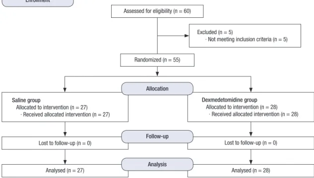
Fig. 1. COMSORT flow diagram to illustrate the study design.
Table 1. Demographic data and perioperative outcomes Demographic parameters Saline group
(n = 27)
Dexmedetomidine group (n = 28) P value
Age, yr 62.0 ± 13.9 60.7 ± 12.7 0.89
Gender (M/F) 15/12 16/12 0.36
Height, cm 163.1 ± 8.0 163.9 ± 9.0 0.73
Weight, kg 63.2 ± 9.1 67.7 ± 10.9 0.31
Hypertension (yes/no) 13/14 15/13 0.42
Type of surgery, No. (%) Recto-sigmoid cancer Prostate cancer Gynecological cancer
18 (66.7) 3 (11.1) 6 (22.2)
19 (67.9) 4 (14.3) 5 (17.9)
0.56
Baseline MAP, mmHg 91.8 ± 10.1 96.2 ± 9.5 0.21
Blood loss, mL 209.6 ± 28.5 182.5 ± 23.1 0.34
Crystalloid administered, mL 2,175.6 ± 167.6 1,933.9 ± 160.2 0.37 Colloid administered, mL 316.7 ± 34.6 230.4 ± 25.2 0.29
Urine output, mL 708.9 ± 41.7 634.3 ± 41.0 0.51
Duration of surgery, min 308.9 ± 38.7 318.2 ± 24.4 0.79 Duration of position, min 207.6 ± 33.1 251.6 ± 27.9 0.28 Duration of anesthesia, min 360.6 ± 51.4 375.2 ± 29.5 0.70 Remifentanil administered, μg 1,448.2 ± 174.3 1,235.7 ± 167.7 0.42 Results are presented as mean (± 1 SD) or number (percentage).
MAP, mean arterial pressure.
Fig. 3. Comparisons of intraocular pressure between normotensive and hypertensive patients in saline (A) and dexmedetomidine (B) groups. Time values indicated by T1 through T16 are as in Fig. 2.
IOP, intraocular pressure.
T1 T2 T3 T4 T5 T6 T7 T8 T9 T10 T11 T12 T13 T14 T15 T16
IOP (mmHg)
Time 40
35 30 25 20 15 10 5 0
Normotensive Hypertensive
A T1 T2 T3 T4 T5 T6 T7 T8 T9 T10 T11 T12 T13 T14 T15 T16
IOP (mmHg)
Time 35
30 25 20 15 10 5 0
Normotensive Hypertensive
B
RESULTS
In total, 60 patients were recruited for the study; five were ex- cluded based on the exclusion criteria (Fig. 1). Demographic data and perioperative outcomes are shown in Table 1. No pa- tient complained of any vision change or eye discomfort in the recovery room.
There was no significant difference in the preoperative base- line IOP between the saline and dexmedetomidine groups. The IOP increased sharply after adopting the steep Trendelenburg position, and an increased IOP was maintained during the sus- tained Trendelenburg position. The IOP at T14 was about 11.3 mmHg higher than that at T1 in the saline group, but only about 4.2 mmHg higher than at T1 in the dexmedetomidine group (Fig. 2A).
The OPP was reduced during the steep Trendelenburg posi-
tion in both the saline and dexmedetomidine groups. The de- gree of decrease in the OPP during the steep Trendelenburg position compared with the baseline OPP was less in the dex- medetomidine group than in the saline group; however, the difference was not statistically significant (18.3 vs. 20.6 mmHg at T14 in the dexmedetomidine and saline groups, respectively;
Fig. 2B).
The number of patients with hypertension in the saline group was 13 and that in the dexmedetomidine group was 15. Demo- graphic data between normotensive and hypertensive patients in each group did not show any statistical differences. Patients with underlying hypertension showed slightly higher IOP during the entire period of surgery than normotensive patients in both groups. The difference was not statistically significant (Fig. 3).
The PIP values correlated with the IOP during the surgery (r = 0.881 and 0.739 for the saline and dexmedetomidine groups,
Fig. 2. Comparison of intraocular pressure (A) and ocular perfusion pressure (B) between groups. T1 = before anesthetic induction; T2 = before administration of the study drug; T3 = after administration of anesthetic induction agents; T4 = immediately after tracheal intubation; T5-T8 = 1, 3, 5, and 10 minutes after tracheal intubation; T9 = im- mediately after intraperitoneal CO2 insufflation; T10 = immediately after steep Trendelenburg position; T11-T13 = 1, 2, and 4 hours after onset of steep Trendelenburg position;
T14 = just before supine position; T15, T16 = 10 and 30 minutes after supine position.
IOP, intraocular pressure; OPP, ocular perfusion pressure.
*Bonferroni correction for multiple comparisons, adjusted P value for significance P < 0.003.
IOP (mmHg)
Time
T1 T2 T3 T4 T5 T6 T7 T8 T9 T10 T11 T12 T13 T14 T15 T16 40
35 30 25 20 15 10 5 0
Saline group Dexmedetomidine group
* * *
* * * *
*
* * * *
T1 T2 T3 T4 T5 T6 T7 T8 T9 T10 T11 T12 T13 T14 T15 T16 Time
OPP (mmHg)
120
100
80
60
40
20
0
Saline group Dexmedetomidine group
* *
A B
Fig 4. (A and B) Correlations between peak inspiratory pressure and intraocular pressure. r = 0.881 and 0.739, respectively, for saline (A) and dexmedetomidine groups (B);
P < 0.001. (C and D) Correlations between mean inspiratory pressure and intraocular pressure. r = 0.812 and 0.739, respectively, for saline (C) and dexmedetomidine (D) groups;
P < 0.001.
IOP (mmHg)
PIP (cmH2O)
0 5 10 15 20 25 30 35 40
45 40 35 30 25 20 15 10 5 0
r = 0.881 P < 0.001
A
IOP (mmHg)
Pmean (cmH2O)
0 5 10 15 20 25 45
40 35 30 25 20 15 10 5 0
r = 0.812 P < 0.001
C
IOP (mmHg)
Pmean (cmH2O)
0 2 4 6 8 10 12 14 16 18 20 50
45 40 35 30 25 20 15 10 5 0
r = 0.739 P < 0.001
D
IOP (mmHg)
PIP (cmH2O)
0 5 10 15 20 25 30 35
r = 0.793 P < 0.001
B 50
45 40 35 30 25 20 15 10 5 0
respectively; P < 0.001). The Pmean values showed a similar pattern (r = 0.812 and 0.739, respectively; P < 0.001; Fig. 4).
DISCUSSION
During laparoscopic lower abdominal procedures in today’s surgical environment, the surgeon’s visualization is assisted by displacing the bowel cephalad away from the surgical field. This can be done by placing the patient in a steep Trendelenburg position. However, an increase in IOP is inevitable in the steep Trendelenburg position. The increased IOP is caused by the in- creased episcleral venous pressure due to gravity (2,10).
This study showed that the IOP increased immediately after entering the steep Trendelenburg position, and was not re- duced during the sustained position. This differs from the re- port of Hayreh, who postulated that the IOP would eventually be normalized because of increased drainage of the aqueous humor and decreased episcleral venous pressure that would occur as a result of autoregulatory mechanisms during the steep Trendelenburg position (11). The results of the present study show that it is essential to devise a means to actively attenuate IOP to prevent ophthalmologic complications when it increases significantly during a sustained steep Trendelenburg position.
The study results demonstrated attenuation of the increase in IOP with continuous infusion of dexmedetomidine in patients
receiving surgery under a steep Trendelenburg position of more than 30°. This effect persisted during the sustained steep Tren- delenburg position, indicating that dexmedetomidine is effec- tive in attenuating the increase of IOP associated with this sur- gical position.
Among various agents known to mitigate IOP, dexmedetomi- dine, an α-2 agonist, is theoretically considered to be the most appropriate agent for attenuation of the increase in IOP during the steep Trendelenburg position. This is because an α-2 ago- nist decreases the production of aqueous humor by provoking direct vasoconstriction of afferent vessels in the ciliary body and facilitates the drainage of aqueous humor by decreasing the sympathetically mediated vasomotor tone in the ocular drainage system; the mechanism of the increase in IOP in the steep Trendelenburg position is increased aqueous humor pro- duction and episcleral congestion (12,13). Additionally, sys- temically administered α-2 agonists demonstrate a neuropro- tective effect on retinal ganglion cell against the increase in IOP.
Thus, an α-2 agonist is an appropriate agent for attenuating the increase in IOP and protecting vision (14).
We chose to administer dexmedetomidine continuously rather than as a bolus. Dexmedetomidine has terminal half-life of 2 hours and a relatively shorter context-sensitive half-life, compared with the long surgery duration. Considering this, it should be effective to administer dexmedetomidine continu-
ously during a sustained steep Trendelenburg position. Con- tinuously infused dexmedetomidine in this study contributed to attenuating the increase in IOP during the sustained Tren- delenburg position, regardless of the duration of surgery.
Various perioperative factors must be considered that influ- ence IOP in the steep Trendelenburg position. Hemodynamic maintenance, ventilation strategy, and fluid management are factors that are manageable by the anesthesiologist. However, underlying systemic hypertension, body position, CO2 insuffla- tion, and duration of surgery in the steep Trendelenburg posi- tion are largely non-adjustable. In this study, we attempted to maintain an intraoperative MAP between ± 20% of the baseline value by modulating the depth of anesthesia, and keep the EtCO2
level between 30 and 35 mmHg by regulating the respiratory rate. Intraoperative fluid was administered according to strict guidelines. By strictly adjusting factors that are manageable by the anesthesiologist, we tried to minimize the influence of those factors on the IOP. As a result, it was possible to evaluate the un- adulterated effect of body position, study drugs, and systemic hypertension on the IOP.
Previous studies have not revealed a clear consensus about the relationship between systemic hypertension and the incre- ase in IOP (4,15-17). We hypothesized that the IOP would be related to the presence of hypertension because ocular perfu- sion is influenced by vascular dysregulation and abnormal blood pressure (18). However, this study showed no relationship be- tween the IOP and systemic hypertension, consistent with the study of Czarnik et al. (19). Mitchell et al. reported that poorly controlled hypertension increased the risk of open-angle glau- coma, whereas controlled hypertension was not a risk factor for glaucoma (16). In light of this, we attribute our findings to the fact that all patients with underlying hypertension involved in this study were already managed with antihypertensive agents.
Furthermore, intravenous agents used for anesthetic induction and continuously administered inhalational anesthetics for an- esthetic maintenance played a role in decreasing the IOP, which counteracted the increase in IOP caused by hypertension (20).
Postoperative ophthalmological complications are intimately related to the surgical position (21). Against general expecta- tions, most of these episodes do not appear to be related to di- rect pressure on the periorbital area or the globe itself, but rath- er to alterations in the blood flow to the eyeball or the optic nerve, by decreased perfusion or embolism (22). OPP is a commonly used variable to predict the perfusion to the eyeball or optic nerve, which is defined as the difference between the MAP and the IOP. Because the increase in IOP can lower the OPP despite maintenance of a normal MAP, it is important to understand how the IOP and OPP change in anesthetized patients in a steep Trendelenburg position. This study showed that the OPP de- creased during the steep Trendelenburg position in both the dexmedetomidine and saline groups, consistent with the re-
sults of Molloy (2). However, there was no significant difference in the OPP between the dexmedetomidine and saline groups.
This result conflicts with our hypothesis that dexmedetomidine would significantly decrease the MAP and result in a negative influence on the OPP by its sympatholytic effect as an α-2 ago- nist. The reason why the OPP was maintained despite adminis- tration of dexmedetomidine is interpreted in terms of the MAP being maintained in a constant range, especially in hyperten- sive patients, by regulating the depth of anesthesia diligently with sevoflurane and remifentanil. Patients in present study, especially hypertensive patients, had blood pressure in well- controlled manner. However, there are patients with poorly con- trolled blood pressure in daily life, and blood pressure tends to be poorly controlled during anesthesia and surgery in those patients. For those patients, maintaining IOP is important to maintain OPP. The anesthesiologist should keep in mind that maintaining a proper MAP by regulating fluid management and the depth of anesthesia and attenuating the increase in the IOP during the steep Trendelenburg position is important for maintaining the OPP and consequently preventing postopera- tive ophthalmological complications.
We also evaluated the relationships between the PIP or Pmean and the IOP, and found that both the PIP and Pmean were cor- related significantly with the IOP. This might be a result of a de- creased outflow of aqueous humor through the episcleral vein as a consequence of the increased intrathoracic pressure due to the increase in the PIP. The increased CVP during the steep Tren- delenburg position in our study supports this explanation.
We used a hand-held Tono-Pen AVIA tonometer to measure intraoperative IOP. The mechanism of the tonometer is as fol- lows. The tonometer operates on the principle of the Imbert-Fick law: P = F/A, where P = intraocular pressure, F = the amount of force exerted by the tonometer to flatten a specific area of the eye, and A = the area flattened. The tonometer contains a strain gauge that converts IOP measurements to an electrical signal (23). The tonometer was selected as our instrument for mea- surements because of its speed, ability for use on multiple pa- tients because of its disposable latex tip covers, ease of use, ac- curacy in a variety of positions, and reliability (24). Setogawa and Kawai (25) measured and compared IOP with an intraocu- lar needle transducer and with a hand-held tonometer in rab- bits, and showed a good correlation. Thus, the IOP measured with the Tono-Pen AVIA in this study is trustworthy.
This study has a couple of limitations. Although dexmedeto- midine alleviated the increase in IOP in the steep Trendelenburg position, we could not find any objective evidence for decreased incidence or severity of ophthalmological complications due to the use of dexmedetomidine. In fact, no patient complained of postoperative ophthalmological complications despite the in- creased IOP observed during the steep Trendelenburg position in our study. This might be a result of the relative rarity of oph-
thalmological complications. According to a literature review, ophthalmological complications, such as visual loss, may occur as a result of sustained increase in IOP during surgery (26,27).
Thus, we should not overlook the potential harm of a sustained increase in IOP due to the steep Trendelenburg position. Sec- ond, we included several kinds of surgeries that may demand different degrees of Trendelenburg position unlike the study of Kim et al. (28) which included the patients undergoing robot- assisted laparoscopic radical prostatectomy. However, general, gynecological, and urological surgeons in our institution demand similar degrees of steep Trendelenburg position (30°-45°) for laparoscopic or robot-assisted surgery due to recto-sigmoid co- lon cancer, prostate cancer, or gynecological cancer. Thus, the effect of different degree of Trendelenburg position might have been minimized.
In conclusion, steep Trendelenburg position during resulted in a considerable increase in IOP and a decrease in OPP during surgery. Continuous infusion of dexmedetomidine is a valuable means for attenuating the increase in IOP without triggering any additional decrease in OPP during surgery, even in sustained steep Trendelenburg position. The IOP in patients with under- lying hypertension did not show any significant difference from that in normotensive patients.
ACKNOWLEDGMENT
We express our gratitude to Professor Yong-gyu Park (Depart- ment of Statistics, College of Medicine, Seoul St. Mary’s Hospi- tal, The Catholic University of Korea) for his support with statis- tical analysis.
DISCLOSURE
The authors have no potential conflicts of interest to disclose.
AUTHOR CONTRIBUTION
Conception and coordination of the study: Lee J. Design of ethi- cal issues: Joo J, Koh H, Lee K, Lee J. Acquisition of data: Lee J, Joo J. Data review: Lee J, Joo J, Koh H, Lee K. Statistical analysis:
Lee J, Joo J. Manuscript preparation: Lee J, Joo J. Manuscript ap- proval: all authors.
ORCID
Jin Joo http://orcid.org/0000-0002-4260-9397 Hyunjung Koh http://orcid.org/0000-0002-9634-5120 Kusang Lee http://orcid.org/0000-0003-0172-4054 Jaemin Lee http://orcid.org/0000-0002-0224-7141
REFERENCES
1. Awad H, Santilli S, Ohr M, Roth A, Yan W, Fernandez S, Roth S, Patel V.
The effects of steep trendelenburg positioning on intraocular pressure during robotic radical prostatectomy. Anesth Analg 2009; 109: 473-8.
2. Molloy BL. Implications for postoperative visual loss: steep trendelen- burg position and effects on intraocular pressure. AANA J 2011; 79: 115- 21.
3. Weber ED, Colyer MH, Lesser RL, Subramanian PS. Posterior ischemic optic neuropathy after minimally invasive prostatectomy. J Neurooph- thalmol 2007; 27: 285-7.
4. Vaajanen A, Mervaala E, Oksala O, Vapaatalo H. Is there a relationship between blood pressure and intraocular pressure? An experimental study in hypertensive rats. Curr Eye Res 2008; 33: 325-32.
5. Farag E, Sessler DI, Kovaci B, Wang L, Mascha EJ, Bell G, Kalfas I, Rock- wood E, Kurz A. Effects of crystalloid versus colloid and the alpha-2 ago- nist brimonidine versus placebo on intraocular pressure during prone spine surgery: a factorial randomized trial. Anesthesiology 2012; 116: 807- 15.
6. Kaya FN, Yavascaoglu B, Baykara M, Altun GT, Gülhan N, Ata F. Effect of oral gabapentin on the intraocular pressure and haemodynamic respons- es induced by tracheal intubation. Acta Anaesthesiol Scand 2008; 52: 1076- 80.
7. Pal CK, Ray M, Sen A, Hajra B, Mukherjee D, Ghanta AK. Changes in in- traocular pressure following administration of suxamethonium and en- dotracheal intubation: influence of dexmedetomidine premedication.
Indian J Anaesth 2011; 55: 573-7.
8. Lam AK, Douthwaite WA. Does the change of anterior chamber depth or/and episcleral venous pressure cause intraocular pressure change in postural variation? Optom Vis Sci 1997; 74: 664-7.
9. Cantor LB. The evolving pharmacotherapeutic profile of brimonidine, an alpha 2-adrenergic agonist, after four years of continuous use. Expert Opin Pharmacother 2000; 1: 815-34.
10. Friberg TR, Weinreb RN. Ocular manifestations of gravity inversion. JAMA 1985; 253: 1755-7.
11. Hayreh SS. Ischemic optic neuropathy. Prog Retin Eye Res 2009; 28: 34-62.
12. Macri FJ, Cevario SJ. Clonidine. Arch Ophthalmol 1978; 96: 2111-3.
13. Vartiainen J, MacDonald E, Urtti A, Rouhiainen H, Virtanen R. Dexme- detomidine-induced ocular hypotension in rabbits with normal or ele- vated intraocular pressures. Invest Ophthalmol Vis Sci 1992; 33: 2019-23.
14. Donello JE, Padillo EU, Webster ML, Wheeler LA, Gil DW. alpha(2)-adre- noceptor agonists inhibit vitreal glutamate and aspartate accumulation and preserve retinal function after transient ischemia. J Pharmacol Exp Ther 2001; 296: 216-23.
15. Klein BE, Klein R, Knudtson MD. Intraocular pressure and systemic blood pressure: longitudinal perspective: the Beaver Dam Eye Study. Br J Oph- thalmol 2005; 89: 284-7.
16. Mitchell P, Lee AJ, Rochtchina E, Wang JJ. Open-angle glaucoma and sys- temic hypertension: the blue mountains eye study. J Glaucoma 2004; 13:
319-26.
17. Xu L, Wang H, Wang Y, Jonas JB. Intraocular pressure correlated with ar- terial blood pressure: the beijing eye study. Am J Ophthalmol 2007; 144:
461-2.
18. Gherghel D, Hosking SL, Orgül S. Autonomic nervous system, circadian
rhythms, and primary open-angle glaucoma. Surv Ophthalmol 2004; 49:
491-508.
19. Czarnik T, Gawda R, Kolodziej W, Latka D, Sznajd-Weron K, Weron R. As- sociations between intracranial pressure, intraocular pressure and mean arterial pressure in patients with traumatic and non-traumatic brain in- juries. Injury 2009; 40: 33-9.
20. Ding C, Wang P, Tian N. Effect of general anesthetics on IOP in elevated IOP mouse model. Exp Eye Res 2011; 92: 512-20.
21. Benumof JL, Mazzei W, Roth S, Barach P, Lam AM, Lee LA. Multifactorial etiology of postoperative vision loss. Anesthesiology 2002; 96: 1531-2.
22. Stambough JL, Dolan D, Werner R, Godfrey E. Ophthalmologic compli- cations associated with prone positioning in spine surgery. J Am Acad Orthop Surg 2007; 15: 156-65.
23. Cheng MA, Todorov A, Tempelhoff R, McHugh T, Crowder CM, Laurys- sen C. The effect of prone positioning on intraocular pressure in anesthe- tized patients. Anesthesiology 2001; 95: 1351-5.
24. Beneyto P, Barajas MA, Garcia-de-Blas F, Del Cura I, Sanz T, Vello R, Sal-
vador C. Predictive value of tonometry with Tono-pen XL in primary care.
Br J Gen Pract 2007; 57: 653-4.
25. Setogawa A, Kawai. Measurement of intraocular pressure by both inva- sive and noninvasive techniques in rabbits exposed to head-down tilt.
Jpn J Physiol 1998; 48: 25-31.
26. Pinkney TD, King AJ, Walter C, Wilson TR, Maxwell-Armstrong C, Ache- son AG. Raised intraocular pressure (IOP) and perioperative visual loss in laparoscopic colorectal surgery: a catastrophe waiting to happen? A systematic review of evidence from other surgical specialities. Tech Colo- proctol 2012; 16: 331-5.
27. Nickels TJ, Manlapaz MR, Farag E. Perioperative visual loss after spine surgery. World J Orthop 2014; 5: 100-6.
28. Kim NY, Yoo YC, Park H, Choi YD, Kim CY, Bai SJ. The effect of dexme- detomidine on intraocular pressure increase in patients during robot-as- sisted laparoscopic radical prostatectomy in the steep trendelenburg po- sition. J Endourol 2015; 29: 310-6.