Original Article
고지방 식이 유도 고지혈증에 대한
ChondroT의 혈액 내 지질대사에 미치는 영향
윤찬석*⋅김도형*⋅나창수†⋅정지원‡⋅김지훈*⋅김선길*⋅최지민*⋅김선종*
동신대학교 한의과대학 한방재활의학과교실*, 경혈학교실†, 전라남도 영암군보건소‡
Observational Study of ChondroT's Improvement of Blood Metabolites in High-fat Diet-induced Hyperlipidemia
Chan Suk Yoon, K.M.D.*, Do Hyeong Kim, K.M.D.*, Chang Su Na, K.M.D.†, Ji Won Jeong, K.M.D.‡, Ji Hoon Kim, K.M.D.*, Sun Gil Kim, K.M.D.*, Ji Min Choi, K.M.D.*, Seon Jong Kim, K.M.D.*
Departments of Korean Medicine Rehabilitation*, Meridian and Acupoint†, College of Korean Medicine, Dongshin University, Yeongam Public Health Center‡
RECEIVED September 9, 2020 ACCEPTED September 21, 2020 CORRESPONDING TO
Seon Jong Kim, Department of Korean Medicine Rehabilitation, Mokpo Oriental Hospital of Dongshin University, 313 Baengnyeon-daero, Mokpo 58665, Korea
TEL (061) 280-7905 FAX (061) 280-7788 E-mail [email protected] Copyright © 2021 The Society of Korean Medicine Rehabilitation
Objectives The objective of the study was to investigate effects of ChondroT by im- provement of blood metabolites in high-fat diet (HFD)-induced hyperlipidemia rat model.
Methods Sprague-Dawley rats were randomly assigned to intact, control, simvasta- tin, and CT100, CT200 and CT400 (each n=6). For observing cholesterol change, ani- mals were first fed high fat diet for 5 weeks and then high fat diet and drugs for 3 weeks. At the end of the experiment, total cholesterol, triglyceride, high density lip- oprotein-cholesterol (HDL-C) and low density lipoprotein-cholesterol (LDL-C) were analyzed by obtained blood collection. Further, amplified leptin, peroxisome pro- liferator activated receptor (PPAR) and adiponectin DNA were observed by reverse transcription polymerase chain reaction analysis.
Results Observing the effect of ChondroT on the change of lipid metabolism in hyper- lipidemia-induced rats, triglyceride and total cholesterol were significantly decreased in SV100 group, HDL-C was significantly increased in SV100, CT100 and CT200 groups, and LDL-C was significantly decreased in SV100, CT100, CT200 and CT400 groups, compared to the control group. Leptin level in hyperlipidemia-induced rats was significantly decreased in CT100 and CT200 groups, compared to the control group. The effect of ChondroT on adiponectin level in hyperlipidemia-induced rats was significantly increased in SV100, CT100 and CT200 groups. PPAR level in hyper- lipidemia-induced rats was significantly decreased in SV100, CT200 and CT400 groups. Platelete activating factor level in hyperlipidemia-induced rats was significantly decreased in CT100 and CT200 groups.
Conclusions Based on these results, it could be suggested that ChondroT has certain effects of improving blood metabolites in HFD-induced hyperlipidemia. (J Korean Med Rehabil 2021;31(1):81-93)
Key words Osteoarthritis, ChondroT, High-fat diet, Hyperlipidemias
서론»»»
2000년대에 들어와 비만과 골관절염(osteoarthritis, OA)의 관계에 대한 연구들이 활발히 진행되고 있다1,2). 이 러한 연구들을 통해 비만은 OA와 대사증후군(metabolic syndrome, MetS)의 연관성에 있어서 주요한 기여요소 로 주목되고 있으며, 연구결과 체중감소에 따른 OA 증 상의 경감이 확인되었다3). 그러나 OA는 비만환자의 체 중부하가 없는 관절에서도 흔하게 발생하여 체중부하 에 따른 기계적인 현상이라기보다 인체대사체계에 따 른 현상임을 암시하고 있다1).
이러한 추세에 따라 최근에는 OA를 단순한 생물 기 계학적인 관절부하에 의한 질환으로서의 인식에서 벗 어나 새로운 아형 OA로서의 대사성 골관절염(metabolic OA, MOA)의 개념이 대두되고 있다4).
ChondroT5)는 현재 관절질환 치료 목적으로 개발 중 인 신약으로 Osterici Radix, Lonicerae Folium, Angelicae Gigantis Radix, Clematidis Radix, Phellodendri Cortex 등 5가지 한약재로 구성된 물 추출물이다. ChondroT는 세포 배양을 통한 in vitro 연구에서 SW1353 연골세포의 성장을 촉진하고 interleukin (IL)-1β 유도 matrix metalloproteinase-1 발현을 유의하게 억제하고, lipopolysaccharide 유도 RAW264.7 세포에서 nuclear factor-κB의 활성화 및 IL-1β, IL-6, prostglandin E2 및 산화 질소(nitric oxide)와 같은 염증 매개체의 생성을 감소시켰으며6), 쥐 실험을 통한 혈청학적 inflammatory cytokine인 tumor necrosis fac- tor-α, IL-1β, IL-6의 유의한 감소, aminotransferase인 as- partate aminotransferase의 유의한 감소, albumin, white blood cell 및 lymphocytes의 유의한 감소와 plantar with- drawal response의 reaction time의 증가, apoptosis 촉진 신호인 Bax의 유의한 감소, 억제신호인 Bcl-2의 유의한 증가, BAX/Bcl-2 ratio의 유의한 감소 및 interferon-γ의 유의한 감소가 확인되었다5,7).
또한 Kim 등8)의 선행연구에서 높은 항응집 효과를 나타내는 등 기존의 전통적인 OA의 염증개선 및 진통 효과 이외의 효과에 대한 가능성을 확인하였다.
최근의 메타분석에서는 식물성 sterols와 stanols (2 g/day) 의 섭취가 low density lipoprotein-cholesterol (LDL-C)을 각각 8.2% 및 9.3% 낮춘다는 연구결과가 도출되는 등 식물성 원료의 효능이 주목받고 있어9), 식물성 한약재
로 구성된 ChondroT의 OA에 대한 전통적인 연골보호 및 진통효과 외에 지질대사산물에 대한 개선효과를 검 증하여 비만과 MetS에 의한 혈액 대사산물로 유발되는 OA 위험인자 억제효과를 확인하고자 하였다.
본 연구를 통해 고지방 식이(high-fat diet, HFD)로 유 도된 hyperlipidemia 동물 모델에서 ChondroT의 혈액 내 지질 대사산물 개선 효과를 실험적으로 관찰하고자 triglyceride (TG), total cholesterol (TCHO), high density lipoprotein-cholesterol (HDL-C), LDL-C, adipokines 중 leptin과 adiponectin, peroxisome proliferator activated receptor (PPAR), platelete activating factor (PAF), throm- boxane B2 (TXB2) 등의 변화를 관찰하여 유의한 지견 을 얻었기에 보고하는 바이다.
재료 및 방법»»»
1. 재료
1) 동물
체중 약 160~180 g인 Sprague Dawley계의 수컷 6주 령 흰쥐 36마리를 ㈜샘타코(오산, 한국)에서 공급받아 모든 개체의 건강상태에 대한 외관검사를 실시한 뒤 고 형사료와 물을 충분히 공급하면서 1주일 동안 사육실 환경(실내온도 24±2℃, 습도 50±5%, 12시간 dark/light) 에 적응시킨 후 체중변화 및 일반적인 건강상태를 관찰 한 후 건강한 개체를 선별하여 30마리를 시험에 투여하 였다(동물실험계획 승인번호: 2018-01-01).
2) 고지방 사료
고지방 식이 사료(Research Diets, Inc., New Brunswick, NJ, USA)의 구성은 protein (20%), fat (60%), carbohy- drate (20%)로 칼로리 구성은 casein 800 kcal/g, L-cystine 12 kcal/g, maltodextrin 500 kcal/g, sucrose 275.2 kcal/g, soybean oil 225 kcal/g, lard 2205 kcal/g, vitamin mix 40 kcal/g로 총 4.057 kcal/g이었다. 고지방 식이 사료를 5 주간 공급한 후 3주간 고지방 식이 사료와 약물을 동시 에 공급하였으며 실험은 총 8주동안 시행하였다.
3) 약재
사용한 약재는 강활(Osterici Radix), 금은화(Lonicerae Folium), 당귀(Angelicae Gigantis Radix), 위령선(Clematidis Radix), 황백(Phellodendri Cortex)으로 (주)옴니허브(대 구, 한국)에서 구입하여 사용하였다(Table Ⅰ).
지표 성분을 확인하기 위하여 high-performance liquid chromatography를 통해 금은화 중 chlorogenic acid, 당 귀 중 총 decursin, decursinol angelate, 황백 중 berberine chloride의 3종 성분을 확인하였다7,10).
4) 양성 대조 약물
양성 대조군으로는 simvastatin (Sigma-Aldrich, St.
Louis, MO, USA)을 구입하여 100 mg/kg 농도로 음용 투여하였다.
2. 실험 방법
1) 군 분리
실험군은 일반 사료를 공급한 정상군(Intact, n=6), 고지 방 식이 사료를 공급하고 무처치한 대조군(Control, n=6), 고지방 식이 사료를 공급하며 ChondroT를 100 mg/kg,
200 mg/kg, 400 mg/kg 농도별로 공급한 실험군(100 mg/kg, 200 mg/kg, 400 mg/kg, 각 n=6)으로 분리하였다. 양성 대조군(Positive Control, n=6)은 고지방 식이 사료를 공 급하고 simvastatin를 100 mg/kg 농도로 음용 투여하였 다(Table Ⅱ).
2) 시료 추출
강활, 금은화, 당귀, 위령선, 황백을 배합한 복합 시료 ChondroT (강활 : 당귀 : 금은화 : 위령선 : 황백 = 6 : 4 : 4 : 4 : 3)를 준비하였다. 복합 시료 ChondroT 총량 을 각각 105 g으로 정하였고, 비율에 따라 강활 30 g, 당귀 20 g, 금은화 20 g, 위령선 20 g, 황백 15 g을 정제 수 1,000 mL에 넣고 3시간 동안 열수추출법을 통해 추 출하였다. ChondroT 시료는 50 mL가 되도록 농축하여 실험에 적용하였다. 50 mL로 농축된 시료를 동결 건조하 여 분말을 얻었으며, 얻어진 분말을 기준으로 100 mg/kg, 200 mg/kg, 400 mg/kg으로 희석하여 음용 투여하였다.
3) 체중 측정
체중은 전자 저울(㈜카스, 양주, 한국)로 측정하였으며, 실험기간인 8주 동안 매 7일에 1회씩 측정하여 주(week) 간 체중 변화를 관찰하였다.
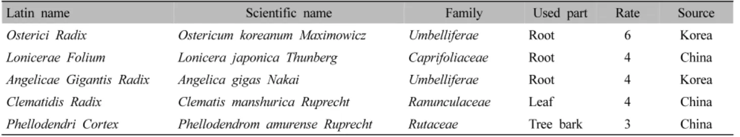
Latin name Scientific name Family Used part Rate Source
Osterici Radix Ostericum koreanum Maximowicz Umbelliferae Root 6 Korea
Lonicerae Folium Lonicera japonica Thunberg Caprifoliaceae Root 4 China
Angelicae Gigantis Radix Angelica gigas Nakai Umbelliferae Root 4 Korea
Clematidis Radix Clematis manshurica Ruprecht Ranunculaceae Leaf 4 China
Phellodendri Cortex Phellodendrom amurense Ruprecht Rutaceae Tree bark 3 China Table I. Composition of ChondroT and the Used Parts of 5 Herbs
Group HFD feeding ChondroT Simvastatin n Remark
Intact X X X 6
Control O X X 6
SV100 O X O 6 100 mg/kg
CT100 O O X 6 100 mg/kg
CT200 O O X 6 200 mg/kg
CT400 O O X 6 400 mg/kg
HFD: high-fat diet.
Table II. The Experimental Design and Assignment of the Study
4) 혈청 분리
채혈에 의하여 얻어진 혈액은 VS 6000CFI (Vision Scientific Co., Ltd., Daejeon, Korea)를 사용하여 3,000 rpm 으로 20분간 시행하여 혈청을 분리하였다.
5) 지질대사 측정
분리된 혈청으로 지질대사 측정 항목인 TCHO은 TCHO-PⅢ Slide (Fujifilm Corporation, Tokyo, Japan) 를, HDLC은 HDL-C-PⅢ Slide (Fujifilm Corporation)를, TG는 TG-PⅢ Slide (Fujifilm Corporation)를 사용하여 Fuji Dri-Chem Clinical Chemistry Analyzer (DRI-Chem 4000ie; Fujifilm Corporation)로 측정하였으며, LDL-C은 TCHO 수치에서 TG/5 수치를 빼고 HDLC 수치를 뺀 값 으로 계산하였다.
6) Total RNA 분리 및 reverse transcription poly- merase chain reaction (RT-PCR)
(1) Total RNA 분리
Total RNA의 분리는 liver 부위의 조직(50 mg)을 800 uL Trizol Reagent (Life Technologies, Carlsbad, CA, USA) 를 넣고 precellys 24 (Bertin Technologies, Montigny-le- Bretonneux, France)에서 균질화하고, 균질액에 200 μL 의 chloroform (Sigma-Aldrich)을 가하여 15초 동안 잘 흔들어 혼합한 후 실온상태에서 5분 방치하고 난 다음 세 포 유잔물을 제거하기 위하여 4℃, 14,000 rpm에서 5분 동안 원심분리(Centrifuge 5415 R; Eppendorf, Hamburg, Germany)하였다. 원심분리로 얻어진 상층액에 500 μL 의 isopropanol Sigma-Aldrich)을 첨가하여 실온상태에 서 5분동안 방치한 후 RNA pellet을 얻기 위하여 4℃, 14,000 rpm에서 8분간 원심분리하고, 원심분리로 생긴 pellet에 냉장 보관된 70% ethanol과 함께 diethyl pyrocar- bonate (DEPC)를 넣고 4℃, 7,500 rpm에서 5분간 원심 분리 후 pellet만 남기고 모두 제거하고, 남은 ethanol은 실온에서 5분간 방치시켜 건조시킨 다음 DEPC-treated water에 녹여 spectrophotometer (Biophotometer; Eppendorf) 에서 OD260 값을 읽어 RNA의 순도 및 농도를 정량하 였다.
(2) RT-PCR
분리된 total RNA 5 μg과 2.5 μL Oligo (dT), DEPC-
treated water를 RT premix (Bioneer, Daejeon, Korea)에 넣고 Mastercycler gradient (Eppendorf)를 이용, 50 μL cDNA를 합성하여 PCR 증폭을 위한 template으로 사용 하였다. 또한 해당 실험에 housekeeping 유전자인 glyc- eraldehyde-3-phosphate dehydrogenase (sense primer:
5'-ATCCCATCACCATCTTCCAG-3', antisense primer:
5'-CCTGCTTTCACCACCTCCTT-3')를 internal control 로서 사용하였다. Reverse transcription temperature cy- cle은 42℃에서 1시간 동안 cDNA synthesis, 94℃에서 5분동안 denature 그리고 4℃에서 5분동안 cooling시키 는 단계를 거쳤다. Leptin, PPAR, adiponectin 유전자에 대한 백서의 특이 primer는 PCR-premix kit (Bioneer)를 사용하였다. PCR은 cDNA, sense primer, antisense pri- mer, DEPC-treated water를 PCR premix (Bioneer)에 넣 었다. PCR temperature cycle은 cDNA의 증폭을 위하여 95℃에서 300초동안 pre-denaturation, 94℃에서 40초동 안 melting, leptin은 59℃, PPAR과 adiponectin은 58℃
에서 40초동안 annealing, 72℃에서 90초동안 extension하 는 과정을 40회 반복 수행하고 마지막 cycle에서 72℃에 서 600초동안 extension 단계를 거쳐 leptin primer (sense primer: 5'-CCTGTGGCTTTGGTCCTATCTG-3', antisense primer: 5'-AGGCAAGCTGGTGAGGATCTG-3), PPAR primer (sense primer: 5'-CTCCTGTTGACCCAGAGCAT-3", antisense primer: 5'-CAACCATTGGGTCAGCTCTT-3), adiponectin primer (sense primer: 5'-GGAACTTGTGCA GGTTGGAT-3', antisense primer: 5'-GCTTCTCCAGGC TCTCCTTT-3')을 이용하여 유전자증폭을 Mastercycler gradient에서 시행하였다.
이렇게 증폭된 leptin, PPAR, adiponectin의 DNA를 Greenview nucleic acid gel stain (1:10,000; IO Rodeo Inc., Pasadena, CA, USA)를 포함한 1.5% agarose gel상 에서 0.5×TBE buffer (80 mM Tris-HCL, 80 mM boric acid, 2 mM EDTA, pH 8.3)로 100 V에서 전기 영동하여 관찰한 후 Image Station (Samsung, 서울, Korea)을 이용 하여 촬영하였으며, Alphaease FC StandAlone Software (Alpha Innotech, San Leandro, CA, USA)를 이용하여 측정하였다.
3. 통계처리
실험 성적은 평균값과 표준오차(mean±standard error) 를 통해 표시하였다. 각 실험군 간의 통계학적 분석은 Window용 SPSS (Version 10.05; IBM Co., Armonk, NY, USA)를 이용하였으며, 비모수적 방법 중 n<30의 경우 에 사용하는 Mann-Whitney U test를 시행하였다. 전체 실험결과 중 신뢰구간 p<0.05와 p<0.01에서 통계적 유 의성을 부여하였다.
결과»»»
1. 지질대사 변화
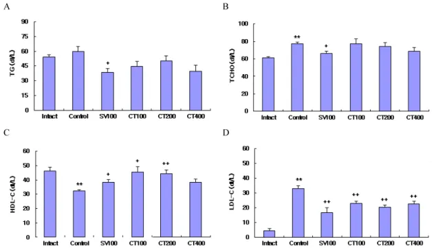
ChondroT가 고지혈증이 유발된 흰쥐의 지질대사 함 량 변화에 미치는 영향을 관찰한 결과, TG는 대조군에 비하여 SV100군은 유의한 감소를 나타내었으며, TCHO 는 대조군이 정상군에 비하여 유의한 증가를 나타내었 고, 대조군에 비하여 SV100군은 유의한 감소를 나타내
었으며, HDL-C는 대조군이 정상군에 비하여 유의한 감 소를 나타내었고, 대조군에 비하여 SV100, CT100군, CT200군은 유의한 증가를 나타내었으며, LDL-C는 대 조군이 정상군에 비하여 유의한 증가를 나타내었고, 대 조군에 비하여 SV100, CT100군, CT200군, CT400군은 유의한 감소를 나타내었다(Fig. 1, Table Ⅲ).
2. 아디포카인 활성
1) Leptin
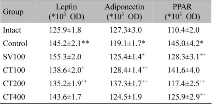
ChondroT가 고지혈증이 유발된 흰쥐의 leptin 함량 변화에 미치는 영향을 관찰한 결과, 정상군은 125.9±1.8 (*103 OD), 대조군은 145.2±2.1 (*103 OD)를 나타내어 대조군이 정상군에 비하여 유의한 증가를 나타내었으며, SV100군은 155.3±2.0 (*103 OD), CT100군은 138.6±2.0 (*103 OD), CT200군은 135.2±1.9 (*103 OD), CT400군 은 143.6±1.7 (*103 OD)를 나타내어 대조군에 비하여 CT100군, CT200군은 유의한 감소를 나타내었다(Fig. 2, Table Ⅳ).
A B
C D
Fig. 1. Effect of ChondroT treatment on TG (A), TCHO (B), HDL-C (C) and LDL-C (D) in hyperlipidemia rats induced HFD.
Intact: no HFD and no treatment, Control: HFD, no treatment, SV100: HFD, simvastatin 100 mg/kg treatment, CT100, CT200, CT400: HFD, ChondroT treatment, 100 mg/kg, 200 mg/kg, 400 mg/kg. TG: triglyceride, TCHO: total-cholesterol, HDL-C: high density lipoprotein cholesterol, LDL-C: low density lipoprotein-cholesterol, HFD: high-fat diet. Values are expressed mean±standard error. **p<0.01 compared with intact, +p<0.05, ++p<0.01 compared with control.
2) Adiponectin
ChondroT가 고지혈증이 유발된 흰쥐의 adiponectin 함 량 변화에 미치는 영향을 관찰한 결과, 정상군은 127.3±3.0 (*103 OD), 대조군은 119.1±1.7 (*103 OD)을 나타내어 대조군이 정상군에 비하여 유의한 감소를 나타내었으며, SV100군은 125.4±1.4 (*103 OD), CT100군은 128.4±1.4 (*103 OD), CT200군은 137.3±1.7 (*103 OD), CT400군 은 124.5±1.9 (*103 OD)를 나타내어 대조군에 비하여 SV100군, CT100군, CT200군은 유의한 증가를 나타내
었다(Fig. 3, Table Ⅳ).
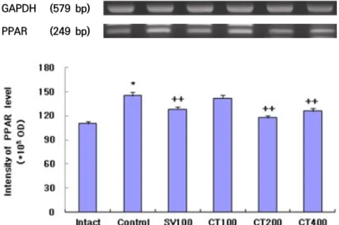
3) PPAR
ChondroT가 고지혈증이 유발된 흰쥐의 PPAR 함량 변화에 미치는 영향을 관찰한 결과, 정상군은 110.4±2.0 (*103 OD), 대조군은 145.0±4.2 (*103 OD)를 나타내어 대조군이 정상군에 비하여 유의한 증가를 나타내었으며, SV100군은 128.3±3.1 (*103 OD), CT100군은 141.6±4.0 (*103 OD), CT200군은 117.4±2.5 (*103 OD), CT400군 은 125.9±2.9 (*103 OD)를 나타내어 대조군에 비하여
Group TG (dL/L) TCHO (dL/L) HDL-C (dL/L) LDL-C (dL/L)
Intact 54.0±2.3 61.0±1.5 46.0±2.8 4.2±1.7
Control 59.5±5.2 77.3±2.1** 32.5±0.6** 32.9±1.9**
SV100 38.6±3.9+ 66.0±2.4+ 38.4±1.8+ 16.6±3.3++
CT100 44.5±4.9 77.2±5.4 45.3±3.8+ 22.9±1.3++
CT200 50.2±5.2 74.3±4.3 44.2±2.9++ 20.1±1.7++
CT400 39.8±5.7 68.8±4.4 38.3±2.3 22.5±1.9++
Values are expressed mean±standard error.
Intact: no HFD and no treatment, Control: HFD, no treatment, SV100: HFD, simvastatin 100 mg/kg treatment, CT100, CT200, CT400: HFD, ChondroT treatment, 100 mg/kg, 200 mg/kg, 400 mg/kg. TG: triglyceride, TCHO: total-cholesterol, HDL-C: high density lipoprotein cholesterol, LDL-C: low density lipoprotein-cholesterol, HFD: high-fat diet.
**p<0.01 compared with intact, +p<0.05, ++p<0.01 compared with control.
Table III. Changes on the Ser um TCHO, TG, HDL-C and LDL-C Contents after ChondroT Treatment in Hyperlipidemia Rats Induced HFD
GAPDH (579 bp) Leptin (250 bp)
Fig. 2. Effect of ChondroT treatment on leptin in hyperlipidemia rats induced HFD. Intact: no HFD and no treatment, Control:
HFD, no treatment, SV100: HFD, simvastatin 100 mg/kg treatment, CT100, CT200, CT400: HFD, ChondroT treatment, 100 mg/kg, 200 mg/kg, 400 mg/kg. GAPDH: glyceraldehyde-3-phosphate dehydrogenase, HFD: high-fat diet. Values are expressed mean±standard error. **p<0.01 compared with intact, +p<0.05,
++p<0.01 compared with control.
Group Leptin
(*103 OD) Adiponectin
(*103 OD) PPAR (*103 OD)
Intact 125.9±1.8 127.3±3.0 110.4±2.0
Control 145.2±2.1** 119.1±1.7* 145.0±4.2*
SV100 155.3±2.0 125.4±1.4+ 128.3±3.1++
CT100 138.6±2.0+ 128.4±1.4++ 141.6±4.0 CT200 135.2±1.9++ 137.3±1.7++ 117.4±2.5++
CT400 143.6±1.7 124.5±1.9 125.9±2.9++
Values are expressed mean±standard error.
Intact: no HFD and no treatment, Control: HFD, no treatment, SV100: HFD, simvastatin 100 mg/kg treatment, CT100, CT200, CT400: HFD, ChondroT treatment, 100 mg/kg, 200 mg/kg, 400 mg/kg. GAPDH: glyceraldehyde-3-phosphate dehydrogenase, PPAR: peroxisome proliferator activated receptor, HFD: high-fat diet.
*p<0.05, **p<0.01 compared with intact, +p<0.05, ++p<0.01 compared with control.
Table Ⅳ. Changes on the Serum Leptin, Adiponectin and PPAR Contents after ChondroT Treatment in Hyperlipidemia Rats Induced HFD
SV100군, CT200군, CT400군은 유의한 감소를 나타내 었다(Fig. 4, Table Ⅳ).
3. PAF 변화
ChondroT가 고지혈증이 유발된 흰쥐의 PAF 함량 변화 에 미치는 영향을 관찰한 결과, 정상군은 10.9±1.1 pg/mL, 대조군은 18.9±2.3 pg/mL를 나타내어 대조군이 정상군 에 비하여 유의한 증가를 나타내었으며, SV100군은 12.7±2.7 pg/mL, CT100군은 11.9±0.9 pg/mL, CT200군 은 13.1±1.1 pg/mL, CT400군은 13.1±1.4 pg/mL를 나타 내어 대조군에 비하여 CT100군, CT200군은 유의한 감 소를 나타내었다(Fig. 5, Table Ⅴ).
GAPDH (579 bp) Adiponectin (221 bp)
Fig. 3. Effect of ChondroT treatment on adiponectin in hyperlipidemia rats induced HFD. Intact: no HFD and no treatment, Control: HFD, no treatment, SV100: HFD, simvastatin 100 mg/kg treatment, CT100, CT200, CT400: HFD, ChondroT treatment, 100 mg/kg, 200 mg/kg, 400 mg/kg. GAPDH:
glyceraldehyde-3-phosphate dehydrogenase, HFD: high-fat diet.
Values are expressed mean±standard error. *p<0.05 compared with intact, +p<0.05, ++p<0.01 compared with control.
GAPDH (579 bp) PPAR (249 bp)
Fig. 4. Effect of ChondroT treatment on PPAR in hyperlipidemia rats induced HFD. Intact: no HFD and no treatment, Control:
HFD, no treatment, SV100: HFD, simvastatin 100 mg/kg treatment, CT100, CT200, CT400: HFD, ChondroT treatment, 100 mg/kg, 200 mg/kg, 400 mg/kg. GAPDH: glyceraldehyde- 3-phosphate dehydrogenase, PPAR: peroxisome proliferator activated receptor, HFD: high-fat diet. Values are expressed mean±standard error.
*p<0.05 compared with intact, ++p<0.01 compared with control.
Fig. 5. Effect of ChondroT treatment on PAF in hyperlipidemia rats induced HFD. Intact: no HFD and no treatment, Control:
HFD, no treatment, SV100: HFD, simvastatin 100 mg/kg treatment, CT100, CT200, CT400: HFD, ChondroT treatment, 100 mg/kg, 200 mg/kg, 400 mg/kg. PAF: platelete activating factor, HFD:
high-fat diet. Values are expressed mean±standard error. *p<0.05 compared with intact, +p<0.05 compared with control.
Group PAF (pg/mL) TXB2 (pg/mL)
Intact 10.9±1.1 64.6±5.5
Control 18.9±2.3* 68.5±3.5
SV100 12.7±2.7 71.2±4.6
CT100 11.9±0.9+ 74.3±7.5
CT200 13.1±1.1+ 61.7±2.2
CT400 13.1±1.4 62.0±8.6
Values are expressed Mean±standard error.
Intact: no HFD and no treatment, Control: HFD, no treatment, SV100: HFD, simvastatin 100 mg/kg treatment, CT100, CT200, CT400: HFD, ChondroT treatment, 100 mg/kg, 200 mg/kg, 400 mg/kg. PAF: platelete activating factor, TXB2: thromboxane B2, HFD: high-fat diet.
*p<0.05 compared with intact, +p<0.05 compared with control.
Table Ⅴ. Changes on the PAF and TXB2 Contents after ChondroT Treatment in Hyperlipidemia Rats Induced HFD
4. TXB2 변화
ChondroT가 고지혈증이 유발된 흰쥐의 TXB2 함량 변화에 미치는 영향을 관찰한 결과, 정상군은 64.6±5.5 pg/mL, 대조군은 68.5±3.5 pg/mL를 나타내었고, SV100 군은 71.2±4.6 pg/mL, CT100군은 74.3±7.5 pg/mL, CT200 군은 61.7±2.2 pg/mL, CT400군은 62.0±8.6 pg/mL를 나 타내어 대조군에 비하여 실험군은 유의한 차이를 나타 내지 않았다(Fig. 6, Table Ⅴ).
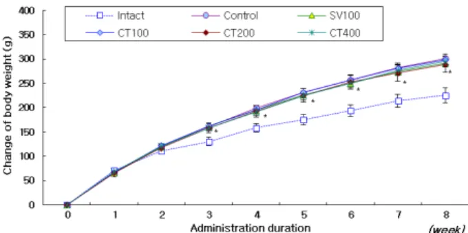
5. 체중 변화
ChondroT가 고지혈증이 유발된 흰쥐의 체중변화를 관찰한 결과, 3주째부터 대조군이 정상군에 비하여 유 의한 증가를 나타내었고, 대조군에 비하여 실험군들은 유의한 차이를 나타내지 않았다(Fig. 7, Table Ⅵ).
고찰»»»
현대의 풍부한 에너지, 지방 및 당이 많이 함유된 음 식과 음료의 소비, 서구화된 식이는 HFD 섭취의 증가 로 이어진다11). HFD는 체중 증가, 공복혈당 및 인슐린 수준의 변화를 초래하고, metabolite profile에 뚜렷하고 장기적인 변화를 나타낸다12).
대사증후군(MetS)은 내당능 장애, 고혈압, 고콜레스 테롤혈증, 고중성지방혈증 및 비만을 포함한 신체증후 군을 지칭하는 말로서13), 과도한 당과 포화지방의 만성 적 섭취는 MetS 뿐만 아니라 2형 당뇨(type 2 diabetes mellitus, T2DM)로의 이환에도 영향을 준다는 연구결과 와 이러한 식이섭취로 인한 혈액의 대사산물의 수준에 따라 발생한 세포 및 인체 구성의 변화가 OA의 병원임 을 암시하는 연구도 진행되었다11,14).
TG는 공복상태에서 LDL-C, 식후엔 chylomicron에 Fig. 6. Effect of ChondroT treatment on TXB2 in hyperlipidemia
rats induced HFD. Intact: no HFD and no treatment, Control:
HFD, no treatment, SV100: HFD, simvastatin 100 mg/kg treatment, CT100, CT200, CT400: HFD, ChondroT treatment, 100 mg/kg, 200 mg/kg, 400 mg/kg. TXB2: thromboxane B2, HFD: high-fat diet.
Group 0 week 1 week 2 week 3 week 4 week 5 week 6 week 7 week 8 week
Intact Control SV100 CT100 CT200 CT400
0.00±0.00 0.00±0.00 0.00±0.00 0.00±0.00 0.00±0.00 0.00±0.00
70.50±2.48 64.88±3.08 66.17±2.70 67.00±2.14 65.50±2.20 68.50±3.62
110.83±4.83 119.88±6.69 118.83±5.26 121.33±2.72 117.00±6.02 120.17±5.02
129.83±8.52 159.75±9.90*
161.17±6.37 161.50±5.29 158.17±9.87 158.00±5.23
158.50±8.90 199.63±13.23*
194.67±8.50 195.50±7.68 192.83±12.20 191.50±7.14
175.00±10.16 230.75±15.97*
225.67±10.06 231.83±7.57 225.00±13.99 224.50±8.77
194.00±11.82 256.75±18.06*
249.67±11.82 257.17±8.72 252.50±14.38 250.17±9.10
213.50±13.21 281.63±20.00*
274.00±13.01 281.33±10.32 271.17±17.10 276.50±10.43
225.50±15.09 300.63±20.55*
291.50±14.02 298.17±11.35 289.00±15.72 294.83±11.06 Values are expressed mean±standard error.
Intact: no HFD and no treatment, Control: HFD, no treatment, SV100: HFD, simvastatin 100 mg/kg treatment, CT100, CT200, CT400: HFD, ChondroT treatment, 100 mg/kg, 200 mg/kg, 400 mg/kg. HFD: high-fat diet.
*p<0.05 compared with Intact.
Table Ⅵ. Changes of Body Weight after ChondroT Treatment in Hyperlipidemia Rats Induced HFD
Fig. 7. Effect of ChondroT treatment on change of body weight in hyperlipidemia rats induced HFD. Intact: no HFD and no treatment, Control: HFD, no treatment, SV100: HFD, simvastatin 100 mg/kg treatment, CT100, CT200, CT400: HFD, ChondroT treatment, 100 mg/kg, 200 mg/kg, 400 mg/kg. HFD: high-fat diet. Values are expressed mean±standard error. *p<0.05 compared with intact.
의해 운반된다15). 식후 증가된 TG 및 TG 함유 지단백 은 동맥경화 관련 질환, 인슐린 저항성 및 T2DM, 가족 력에 의한 고콜레스테롤혈증 및 고지질혈증의 병리적 변화와 밀접한 관계가 있다16). TG, TG 함유 지단백, 유 리 지방산 및 산화 콜레스테롤 등이 풍부한 HFD는 혈 관내피세포의 기능이상과 동맥경화의 병리적 변화에 핵심적 역할을 한다17,18).
여러 다른 실험연구들에서 고콜레스테롤혈증은 연골 악화 요인보다 골극 생성과의 연관성이 높은 것으로 보 고되고 있으며19,20), 활액막(synovial membrane, SM)에 서 대식세포, 내피세포 및 섬유모세포는 지방 조직과 함께 활액기질을 구성하는데, 이들은 체내 지질 수준에 따라 민감하게 변화한다19,21).
지방세포에서 분비하는 adipokines는 중추 및 말초 모두에서 혈압조절, 에너지 소비, 음식 섭취, 지혈, 세포 대사 및 염증 등과 관련된 다발적 기능을 나타낸다.
Adipokines는 OA 이환 시 SM, 연골, 관절내 지방조직에서 도 합성되는데 OA 병리과정 중 전염증(proinflammation), 동화 및 이화과정에 관여하는 것으로 밝혀졌다22-24).
대표적인 adipokines인 leptin은 OA 발병 기전에서 핵 심적 역할을 한다. Dumond 등의 연구에서 OA 환자로 부터 얻은 활액(synovial fluid) 내 leptin의 존재와 leptin 수준과 신체질량지수(body mass index) 사이의 유의한 상관관계를 확인하였으며, leptin의 관절 내 주사는 메 신저 RNA (mRNA)와 단백 동화 작용을 발휘할 수 있 는 단백질 수준과 관련하여 인슐린유사성장인자-1 (IGF-1)의 합성과 형질전환증식인자β(TGF-β)를 강력하 게 자극하는 것으로 나타났다25).
또 하나의 대표적인 adipokines인 adiponectin은 비만 과 T2DM에서 그 수준이 감소하며, 감소된 adiponectin은 심혈관질환, T2DM 및 MetS에 광범위한 영향을 미칠 수 있다는 증거가 속속 나타나고 있다26-28). 그러나 비만인 사람 중 정상군에 비해 류마티스 관절염(rheumatoid ar- thritis, RA) 환자군에서 혈장 내 수준이 높고 OA 환자 군의 혈장 내 수준은 OA 관절의 활액 내 수준의 100배 에 달하는 등 논쟁의 여지가 있다29,30). SM과 무릎 아래 지방체(infrapatellar fat pad)는 OA 관절에서 adiponectin 의 주 공급원이며, 이곳에서의 adiponectin 조절장애가 관절염과의 연관성이 있음을 알 수 있다22,23). 그런데 Unger의 토끼 실험에서 SM에서의 leptin 및 adiponectin
유전자 발현은 대조군과 비교하여 OA 및 OA-HFD 그 룹에서 감소하였으나, 혈장내 순환 leptin은 OA-HFD군 에서 현저히 증가했다31). 이러한 연구들을 통해 지질대 사이상에 의한 SM의 adipokines의 조절장애가 RA 및 OA의 위험요인이 될 수 있음을 추론할 수 있다.
HFD는 외과적 방법으로 무릎관절의 OA를 유도한 토끼 실험에서 SM의 염증을 악화시킨다. 토끼의 SM에 는 대식세포의 침윤 증가와 더불어 지질대사로 remod- eling된 지방조직과 염증전구물질(proinflammatory cy- tokines)의 증가가 나타났다. HFD는 지방세포의 이상지 질혈증과 대식세포의 침윤증가에 의한 지질독성을 초 래하고, 이는 OA와 MetS를 가진 환자의 관절기능 악화 에 결정적인 역할을 하는 것으로 추정된다32).
또한 쥐의 전방십자인대(anterior cruciate ligament)에 대한 외과적 처치로 불안정성을 유도한 실험에서 자당 (sucrose)과 HFD로 유발된 비만은 불안정성을 유도한 쥐의 무릎의 OA 발현에 독립적인 위험요인으로 나타났 다. 체지방 비율은 식이유도비만(diet induced obesity)에 의한 염증성 변화를 촉진하여 MOA에 핵심적인 역할을 한다33).
PPAR은 핵 수용체 superfamily의 하나로 말초 인슐 린 감수성, 지방 생성 및 포도당 항상성에 관여하는 유 전자의 발현을 조절한다. PPAR의 강제 발현은 중성 지 질의 축적 및 지방 세포-특이적 유전자 패턴의 발현에 의해 성숙한 백색 지방세포로의 분화를 촉진하여 지방 축적을 초래한다34,35). 특히 PPARγ 활성화로 인한 지방 세포 전구세포의 분화를 고려하면 PPARγ를 활성화시 키는 화합물은 암세포의 분화를 유도할 수 있다. 강력 한 PPARγ 작동제인 TZD 유도체 efatutazone은 역형성 갑상선 암종, 비소세포 폐암 및 췌장암 등 여러 종류의 암의 분화를 유도하는 것으로 보고되었다36-38). 따라서 PPAR 수의 증가 및 과도한 활성화는 지방축적 및 이상 분화를 통한 신생물 분화의 위험요인이 될 수 있다.
또한 과도한 당과 포화지방의 만성적 섭취는 MetS뿐 아니라 T2DM으로의 이환에도 영향을 준다는 연구결과 뿐만 아니라 이러한 식이섭취로 인한 혈액의 대사산물 의 수준에 따라 발생한 세포 및 인체 구성의 변화가 OA의 병원임을 암시하는 연구에서 T2DM 환자도 OA 위험인자에 노출되어 있음을 추론할 수 있다14).
T2DM 환자는 비정상적인 혈장지질 프로파일을 보
인다. 당뇨병성 이상지질혈증은 고중성지방혈증, 저밀 도의 HDL-C 및 고아포지단백B (hyperapolipoprotein B) 의 정량적 변화와 더불어 역콜레스테롤 수송 및 항산화 특성 등 HDL-C 및 LDL-C의 질적 변화도 발생된다39,40).
이상에서 살펴본 바와 같이 HFD는 MetS뿐 아니라 OA에 대한 위험인자로 대두되고 있으며14), SM의 adi- pokines의 조절장애를 유발하며22,23,32), 지방세포 분화 등에 영향을 미쳐 PPAR 등의 변화로 OA 및 암 등의 위 험인자로도 지목되고 있다34-38).
본 관찰실험에서도 HFD 투여로 대조군에서 체중 및 혈청 내 TG, TCHO, LDL-C, leptin, PPAR, PAF의 유의 한 증가와 HDL-C 및 adiponectin의 유의한 감소가 나타 났으나 TXB2는 유의한 변화가 없었다.
ChondroT 용량별 투여로 TG는 대조군에 비하여 감 소하였으나 통계적으로 유의한 수준으로 감소하지 않 았으며, TCHO도 대조군에 비하여 유의한 변화는 없었 다. 그러나 HDL-C, LDL-C에 미치는 영향을 관찰한 결 과, HDL-C의 경우 대조군에 비하여 CT100 및 CT200군 에서 유의하게 증가하였고, LDL-C의 경우 CT100/200/400 모든 군에서 통계적으로 유의하게 감소하였다(Fig. 1, Table Ⅲ).
Adipokines에 미치는 영향을 관찰한 결과, leptin은 대조군에 비해 CT100/200군에서 통계적으로 유의하게 감소하였으며(Fig. 2, Table Ⅳ), adiponectin은 대조군에 비하여 CT100/200군에서 통계적으로 유의하게 증가하였 다(Fig. 3, Table Ⅳ). PPAR은 대조군에 비해 CT200/400 군에서 유의하게 감소하였다(Fig. 4, Table Ⅳ).
그런데 지질대사가 개선되어 수준이 증가하는 것이 좋 은 HDL-C 및 adiponectin과 감소하는 것이 좋은 LDL-C, leptin 및 PPAR의 변화를 살펴보면 ChondroT의 투여용 량이 100 mg/kg에서 200 mg/kg으로 증가할 때는 HDL-C 를 제외하고 모두 개선되나 400 mg/kg으로 투여된 CT400 군에서는 HDL-C를 포함하여 모두 CT200군에 비해 악 화되는 결과를 보여 용량에 따른 독성실험 등 향후 추 가적인 연구가 필요할 것으로 보인다.
한편 simvastatin을 투여한 양성대조군 SV100군과의 비교결과, 통계적으로 유의한 수준의 변화를 보인 결과 중, HDL-C는 CT100군에서 SV100군보다 많은 증가를 보였고, LDL-C는 CT200 및 CT400에서 SV100군과 비 슷한 수준의 감소를 보였다. Leptin과 PPAR은 CT200군
에서, PAF는 CT100군에서 SV100군보다 많은 감소를 보였으며, adiponectin은 CT200군에서 SV100군보다 많 은 증가를 보였다(Figs. 1~5, Tables Ⅲ~Ⅴ).
스타틴은 콜레스테롤의 생체합성 저해제로서 HMG-CoA 환원효소 억제제로도 알려져 있으며, 콜레스테롤 및 그 전구체의 세포 내 수준을 낮추고 간의 LDL-C 수용체의 상향조절을 통해 LDL-C의 이화작용을 강화하여 장 내 지단백의 생성을 감소시킨다41).
그러나 스타틴은 용량의존적으로 근육병을 유발할 수 있다. 스타틴의 용량 증가와 인체 대사 노출 증가는 크레아틴 분해효소(creatine kinase)의 증가와 근육독성 을 증가시킨다. 또한 스타틴은 인슐린 분비장애 및 저 하된 인슐린 감수성으로 인한 혈당 항상성 변화로 T2DM을 유발한다는 보고들이 잇따르고 있다42,43). 최 신 연구에서는 용량의존적으로 골감소증(osteoporosis) 의 유발 가능성에 대한 보고도 있다44).
PAF는 급성 염증 과정의 강력한 자극제인 알레르기 반응의 인지질 매개체로 면역 또는 비면역 자극에 의 한 활성화 시 다양한 염증 세포에 의해 생성되어 혈관투 과성 및 혈액응고에 관여한다45). 실험결과 CT100군 및 CT200군에서 대조군에 비해 유의한 감소를 나타냈다 (Fig. 5, Table Ⅴ).
TXB2는 최근 연구에서 노령 환자와 심방세동 등 심 혈관질환 기왕력이 있는 사람에게서 높게 나타나 죽상 경화증 등 혈관염증과의 상관관계가 있는 것으로 추정 되고 있다46). TXA2는 혈소판 응집, 혈관 수축 및 증식 을 촉진하는 급성 및 만성 효과로 많은 심혈관 질환에 의 병인으로 지목되고 있다. 그러나 TXA2는 약 30초 정도의 매우 짧은 반감기를 가지며 TXB2로 추가로 대 사되고 소변으로 배설되므로 TBX2로 혈관염증의 정도 를 추정한다47,48). 실험결과 군간 통계적 유의성 있는 변 화는 나타나지 않았으나 CT200/400군에서 낮아지는 경 향을 보였다(Fig. 6, Table Ⅴ). 따라서 ChondroT 투여가 혈관염증에 영향을 주는 인자를 자극하지 않는다는 것을 추론할 수 있다. 이러한 결과가 PAF의 유의적 감소와 상 관관계가 있는지 현재로서 알 수 없으나, CT200/400군 에서 통계적으로 유의하지 않으나 농도가 낮아지는 경 향이 있어 향후 연구를 통해 혈관염증을 개선할 가능성 을 확인할 필요가 있다고 보여진다.
체중변화는 HFD 투여 3주부터 정상군에 비해 대조
군의 유의한 증가가 나타나기 시작했으나 약물의 종류 및 용량에 따른 유의한 변화는 관찰되지 않았다(Fig. 7, Table Ⅵ).
결론»»»
ChondroT 용량별 투여가 HFD로 유발된 hyperlipidemia 에 미치는 영향을 지질대사, 아이포카인 활성, PAF, TBX2 변화를 통해 관찰한 결과 다음과 같은 결론을 얻었다.
1. ChondroT가 고지혈증이 유발된 흰쥐의 지질대사 함량 변화에 미치는 영향을 관찰한 결과, 대조군 에 비하여 TG와 TCHO의 경우 SV100군에서 유 의한 감소를 나타내었고, HDL-C의 경우 SV100, CT100군, CT200군에서 유의한 증가를 나타내었고, LDL-C의 경우 SV100, CT100군, CT200군, CT400 군에서 유의한 감소를 나타내었다.
2. ChondroT가 고지혈증이 유발된 흰쥐의 leptin 함량 변화에 미치는 영향을 관찰한 결과, 대조군에 비 하여 CT100군, CT200군에서 유의한 감소를 나타 내었다.
3. ChondroT가 고지혈증이 유발된 흰쥐의 adiponectin 함량 변화에 미치는 영향을 관찰한 결과, 대조군 에 비하여 SV100군, CT100군, CT200군에서 유의 한 증가를 나타내었다.
4. ChondroT가 고지혈증이 유발된 흰쥐의 PPAR 함 량 변화에 미치는 영향을 관찰한 결과, 대조군에 비하여 SV100군, CT200군, CT400군에서 유의한 감소를 나타내었다.
5. ChondroT가 고지혈증이 유발된 흰쥐의 PAF 함량 변화에 미치는 영향을 관찰한 결과, 대조군에 비 하여 CT100군, CT200군에서 유의한 감소를 나타 내었다.
이상과 같이 ChondroT의 용량별 투여 실험결과, ChondroT는 HFD로 유발된 이상지질대사에 의해 혈청 내에 증가된 LDL-C, leptin, PPAR, PAF의 유의한 감소 효과와 HDL-C 및 adiponectin의 유의한 증가를 통해 체 내 이상지질대사산물의 수준을 개선하고, 염증위험인 자들을 감소시키는 효과가 관찰되었다.
따라서 기존에 확인된 OA 치료효과 뿐만 아니라 지
질대사산물 개선에 따른 고지혈증 및 MetS 개선에도 효과가 있음을 추론할 수 있었다. 더불어 스타틴을 투 여한 SV100군과 유사한 수준의 혈중지질 개선효과 및 혈관염증개선 가능성도 확인되었다.
특히 CT200군에서 HDL-C, LDL-C, leptin, adiponectin, PPAR 및 PAF의 통계적으로 유의한 수준의 개선이 이 루어진 반면 ChondroT 투여에 따른 TXB2의 유의한 변 화가 없어 현재 개발 중인 OA 치료제로서의 효능뿐 아 니라 HFD로 유발될 수 있는 hyperlipidemia 및 MetS로 발생할 수 있는 MOA 예방과 함께 심혈관질환에서 혈 관염증을 개선할 수 있는 가능성도 향후 임상시험 등 더 진행된 연구를 통해 확인하는 것이 필요할 것으로 생각한다.
References»»»
1. Aspden RM. Obesity punches above its weight in osteoarthritis. Nat Rev Rheumatol. 2011;7(1):65-8.
2. Pottie P, Presle N, Terlain B, Netter P, Mainard D, Berenbaum F. Obesity and osteoarthritis: more complex than predicted! Ann Rheum Dis. 2006;65(11):1403-5.
3. Niu J, Clancy M, Aliabadi P, Vasan R, Felson DT.
Metabolic syndrome, its components, and knee osteo- arthritis: the framingham osteoarthritis study. Arthritis Rheumatol Hoboken NJ. 2017;69(6):1194-203.
4. Zhuo Q, Yang W, Chen J, Wang Y. Metabolic syndrome meets osteoarthritis. Nat Rev Rheumatol. 2012;8(12):
729-37.
5. Won JY, Jeong JW, Na CS, Kim SJ. Analgesic effects of ChondroT in collagenase-induced osteoarthritis rat model. J Korean Med Rehabil. 2016;26(3):17-30.
6. Park JU, Kim SJ, Na CS, Choi CH, Seo CS, Son JK, Kang BY, Kim YR. Chondroprotective and anti-in- flammatory effects of ChondroT, a new complex herbal medication. BMC Complement Altern Med. 2016;
16(1):213.
7. Jeong JW, Bae KJ, Kim SG, Kwak DW, Moon YJ, Choi CH, Kim YR, Na CS, Kim SJ. Anti-osteoarthritic ef- fects of ChondroT in a rat model of collagenase-induced osteoarthritis. BMC Complement Altern Med. 2018;
18(1):131.
8. Kim SG, Jeong JW, Lim YH, Kim JH, Na CS, Kim SJ. A study on the anti-condensing effect of ChondroT components. J Korean Med Rehabil. 2018;28(2):47-60.
9. Musa-Veloso K, Poon TH, Elliot JA, Chung C. A com-
parison of the LDL-cholesterol lowering efficacy of plant stanols and plant sterols over a continuous dose range: results of a meta-analysis of randomized, place- bo-controlled trials. Prostaglandins Leukot Essent Fatty Acids. 2011;85(1):9-28.
10. Bae KJ, Jeong JW, Choi CH, Won JY, Kim TG, Kim YR, Na CS, Kim SJ. Antiosteoarthritic effects of ChondroT in a rat model of monosodium iodoacetate-in- duced osteoarthritis. Evid Based Complement Alternat Med. 2018;2018:1-11.
11. Nolan CJ, Damm P, Prentki M. Type 2 diabetes across generations: from pathophysiology to prevention and management. Lancet Lond Engl. 2011;378(9786):169-81.
12. Datta P, Zhang Y, Parousis A, Sharma A, Rossomacha E, Endisha H, Wu B, Kacprzak I, Mahomed NN, Gandhi R, Rockel JS, Kapoor M. High-fat diet-induced acceleration of osteoarthritis is associated with a distinct and sustained plasma metabolite signature. Sci Rep.
2017;7(1):8205.
13. Grundy SM, Cleeman JI, Daniels SR, Donato KA, Eckel RH, Franklin BA, Gordon DJ, Krauss RM, Savage PJ, SmithJr SC, Spertus JA, Costa F. Diagnosis and management of the metabolic syndrome: an American Heart Association/National Heart, Lung, and Blood Institute Scientific Statement. Circulation.
2005;112(17):2735-52.
14. van Dam RM, Willett WC, Rimm EB, Stampfer MJ, Hu FB. Dietary fat and meat intake in relation to risk of type 2 diabetes in men. Diabetes Care. 2002;25(3):
417-24.
15. Alipour A, Elte JWF, van Zaanen HCT, Rietveld AP, Cabezas MC. Postprandial inflammation and endothelial dysfuction. Biochem Soc Trans. 2007;35(Pt 3):466-9.
16. Wallace JP, Johnson B, Padilla J, Mather K. Postprandial lipaemia, oxidative stress and endothelial function: a review. Int J Clin Pract. 2010;64(3):389-403.
17. Lambert JE, Parks EJ. Postprandial metabolism of meal triglyceride in humans. Biochim Biophys Acta.
2012;1821(5):721-6.
18. Ghosh A, Gao L, Thakur A, Siu PM, Lai CWK. Role of free fatty acids in endothelial dysfunction. J Biomed Sci. 2017;24(1):50.
19. de Munter W, Blom AB, Helsen MM, Walgreen B, van der Kraan PM, Joosten LAB, van den Berg WB, van Lent PLEM. Cholesterol accumulation caused by low density lipoprotein receptor deficiency or a cholester- ol-rich diet results in ectopic bone formation during ex- perimental osteoarthritis. Arthritis Res Ther. 2013;15(6):
R178.
20. de Munter W, van der Kraan PM, van den Berg WB, van Lent PLEM. High systemic levels of low-density
lipoprotein cholesterol: fuel to the flames in inflammatory osteoarthritis? Rheumatol Oxf Engl. 2016;55(1):16-24.
21. Chung S, Parks JS. Dietary cholesterol effects on adi- pose tissue inflammation. Curr Opin Lipidol. 2016;
27(1):19-25.
22. Hu P, Bao J, Wu L. The emerging role of adipokines in osteoarthritis: a narrative review. Mol Biol Rep.
2011;38(2):873-8.
23. Presle N, Pottie P, Dumond H, Guillaume C, Lapicque F, Pallu S, Mainard D, Netter P, Terlain B. Differential distribution of adipokines between serum and synovial fluid in patients with osteoarthritis. Contribution of joint tissues to their articular production. Osteoarthritis Cartilage. 2006;14(7):690-5.
24. de Boer TN, van Spil WE, Huisman AM, Polak AA, Bijlsma JWJ, Lafeber FPJG, Mastbergen SC. Serum adipokines in osteoarthritis; comparison with controls and relationship with local parameters of synovial in- flammation and cartilage damage. Osteoarthritis Cartilage.
2012;20(8):846-53.
25. Dumond H, Presle N, Terlain B, Mainard D, Loeuille D, Netter P, Pottie P. Evidence for a key role of leptin in osteoarthritis. Arthritis Rheum. 2003;48(11):3118-29.
26. Yamauchi T, Kamon J, Ito Y, Tsuchida A, Yokomizo T, Kita S, Sugiyama T, Miyagishi M, Hara K, Tsunoda M, Murakami K, Ohteki T, Uchida S, Takekawa S, Waki H, Tsuno NH, Shibata Y, Terauchi Y, Froguel P, Tobe K, Koyasu S, Taira K, Kitamura T, Shimizu T, Nagai R, Kadowaki T. Cloning of adiponectin receptors that mediate antidiabetic metabolic effects. Nature. 2003;
423(6941):762-9.
27. Weyer C, Funahashi T, Tanaka S, Hotta K, Matsuzawa Y, Pratley RE, Tataranni PA. Hypoadiponectinemia in obesity and type 2 diabetes: close association with in- sulin resistance and hyperinsulinemia. J Clin Endocrinol Metab. 2001;86(5):1930-5.
28. Gilardini L, McTernan PG, Girola A, da Silva NF, Alberti L, Kumar S, Invitti C. Adiponectin is a candi- date marker of metabolic syndrome in obese children and adolescents. Atherosclerosis. 2006;189(2):401-7.
29. Otero M, Lago R, Gomez R, Lago F, Dieguez C, Gomez-Reino JJ, Gualillo O. Changes in plasma levels of fat-derived hormones adiponectin, leptin, resistin and visfatin in patients with rheumatoid arthritis. Ann Rheum Dis. 2006;65(9):1198-201.
30. Chen TH, Chen L, Hsieh MS, Chang CP, Chou DT, Tsai SH. Evidence for a protective role for adiponectin in osteoarthritis. Biochim Biophys Acta. 2006;1762(8):
711-8.
31. Unger RH. Longevity, lipotoxicity and leptin: the adipo- cyte defense against feasting and famine. Biochimie.
2005;87(1):57-64.
32. Larralaga-Vera A, Lamuedra A, Prez-Baos S, Prieto-Potin I, Pena L, Herrero-Beaumont G, Largo R. Increased synovial lipodystrophy induced by high fat diet ag- gravates synovitis in experimental osteoarthritis.
Arthritis Res Ther. 2017;19(1):264.
33. Collins KH, Reimer RA, Seerattan RA, Leonard TR, Herzog W. Using diet-induced obesity to understand a metabolic subtype of osteoarthritis in rats. Osteoarthritis Cartilage. 2015;23(6):957-65.
34. Willson TM, Lambert MH, Kliewer SA. Peroxisome proliferator-activated receptor gamma and metabolic disease. Annu Rev Biochem. 2001;70:341-67.
35. Kim HI, Ahn YH. Role of peroxisome proliferator-acti- vated receptor-gamma in the glucose-sensing apparatus of liver and beta-cells. Diabetes. 2004;53 Suppl 1:S60-5.
36. Shimazaki N, Togashi N, Hanai M, Isoyama T, Wada K, Fujita T, Fujiwara K, Kurakata S. Anti-tumour activ- ity of CS-7017, a selective peroxisome proliferator-acti- vated receptor gamma agonist of thiazolidinedione class, in human tumour xenografts and a syngeneic tu- mour implant model. Eur J Cancer Oxf Engl 1990.
2008;44(12):1734-43.
37. Smallridge RC, Copland JA, Brose MS, Wadsworth T, Houvras Y, Menefee ME, Bible KC, Shah MH, Gramza AW, Klopper JP, Marlow LA, Heckman MG, von Roemeling R. Efatutazone, an oral PPAR-γ agonist, in combination with paclitaxel in anaplastic thyroid can- cer: results of a multicenter phase 1 trial. J Clin Endocrinol Metab. 2013;98(6):2392-400.
38. Serizawa M, Murakami H, Watanabe M, Takahashi T, Yamamoto N, Koh Y. Peroxisome proliferator-activated receptor γ agonist efatutazone impairs transforming growth factor β2-induced motility of epidermal growth factor receptor tyrosine kinase inhibitor-resistant lung cancer cells. Cancer Sci. 2014;105(6):683-9.
39. Toth PP, Simko RJ, Palli SR, Koselleck D, Quimbo RA, Cziraky MJ. The impact of serum lipids on risk for mi-
croangiopathy in patients with type 2 diabetes mellitus.
Cardiovasc Diabetol. 2012;11:109.
40. Kontush A, Chapman MJ. Antiatherogenic function of HDL particle subpopulations: focus on antioxidative activities. Curr Opin Lipidol. 2010;21(4):312-8.
41. Gylling H, Plat J, Turley S, Ginsberg HN, Ellegard L, Jessup W, Jones PJ, Lutjohann D, Maerz W, Masana L, Silbernagel G, Staels B, Boren J, Catapano AL, de Backer G, Deanfield J, Descamps OS, Kovanen PT, Riccardi G, Tokgozoglu L, Chapman MJ. Plant sterols and plant stanols in the management of dyslipidaemia and prevention of cardiovascular disease. Atherosclerosis.
2014;232(2):346-60.
42. Jacobson TA. Toward “pain-free” statin prescribing:
clinical algorithm for diagnosis and management of myalgia. Mayo Clin Proc. 2008;83(6):687-700.
43. Aiman U, Najmi A, Khan RA. Statin induced diabetes and its clinical implications. J Pharmacol Pharmacother.
2014;5(3):181-5.
44. Leutner M, Matzhold C, Bellach L, Deischinger C, Harreiter J, Thurner S, Klimek P, Kautzky-Willer A.
Diagnosis of osteoporosis in statin-treated patients is dose-dependent. Annals of the Rheumatic Diseases.
2019;78:1706-11.
45. Lee W. Medical dictionary. Department of Pharmacology, Yonsei University. 5th ed. Paju:Gunja Publishing. 2012.
46. Pastori D, Pignatelli P, Farcomeni A, Nocella C, Bartimoccia S, Carnevale R, Violi F. Age-related in- crease of thromboxane B 2 and risk of cardiovascular disease in atrial fibrillation. Oncotarget. 2016;7(26):
39143-7.
47. Smyth EM. Thromboxane and the thromboxane re- ceptor in cardiovascular disease. Clin Lipidol. 2010;
5(2):209-19.
48. Fontana P, Zufferey A, Daali Y, Reny JL. Antiplatelet therapy: targeting the TxA2 pathway. J Cardiovasc Transl Res. 2014;7(1):29-38.