Fortified Antioxidative Potential by Chrysoeriol through the Regulation of the Nrf2/MAPK-mediated HO-1 Signaling Pathway in RAW 264.7 Cells
Chung Mu Park*
Department of Clinical Laboratory Science, Dong-Eui University, Busan 47340, Korea
Received October 17, 2017 /Revised December 26, 2017 /Accepted December 27, 2017
Chrysoeriol is a widespread flavone, and it is usually found in alfalfa, which has been used as a tradi- tional medicine to treat dyspepsia, asthma, and urinary system disorders. Recently, analysis has been conducted on the anti-inflammatory activity of chrysoeriol, but information on its antioxidative ca- pacity is limited. In this study, the antioxidative potential of chrysoeriol against oxidative damage and its molecular mechanisms were evaluated by analysis of the cell viability, reactive oxygen species (ROS) formation, and Western blots in the RAW 264.7 cell line. Chrysoeriol significantly scavenged lipopolysaccharide (LPS)-induced intracellular ROS formation in a dose-dependent manner, without any cytotoxicity. Heme oxygenase-1 (HO-1), a phase II enzyme that exerts antioxidative activity, was also potently induced by chrysoeriol treatment, which corresponded to the translocation of nuclear factor-erythroid 2 p45-related factor 2 (Nrf2) into the nucleus. Moreover, mitogen-activated protein kinase (MAPK) and phosphoinositide 3-kinase (PI3K) were analyzed due to their important role in maintaining cellular redox homeostasis against oxidative stress. As a result, chrysoeriol-induced HO-1 upregulation was mediated by extracellular signal–regulated kinase (ERK), c-Jun NH2-terminal kinase (JNK), and p38 phosphorylation. To identify the antioxidative potential exerted by HO-1, tert-butyl hy- droperoxide (t-BHP)-induced oxidative damage was applied and mitigated by chrysoeriol treatment, which was confirmed by the HO-1 selective inhibitor and inducer, respectively. Consequently, chrys- oeriol strongly strengthened the HO-1-mediated antioxidative potential through the regulation of the Nrf2/MAPK signaling pathways.
Key words : Chrysoeriol, heme oxygenase-1, mitogen activated protein kinase, nuclear factor-eryth- roid 2 p45-related factor 2
*Corresponding author
*Tel : +82-890-2685, Fax : +82-505-182-6877
*E-mail : [email protected]
This is an Open-Access article distributed under the terms of the Creative Commons Attribution Non-Commercial License (http://creativecommons.org/licenses/by-nc/3.0) which permits unrestricted non-commercial use, distribution, and reproduction in any medium, provided the original work is properly cited.
Journal of Life Science 2018 Vol. 28. No. 1. 43~49 DOI : https://doi.org/10.5352/JLS.2018.28.1.43
Introduction
Excessively produced reactive oxygen species (ROS) can damage various cellular components, which contributes to the pathogenesis of various disease including cancer, aging and atherosclerosis [19]. In order to survive under the cir- cumstance of oxidative stress, cells should have evolved their own antioxidative mechanisms to eliminate the ROS production through the induction of intracellular phase II enzymes [15]. Among these phase II enzymes, heme oxygen- ase (HO)-1 has exhibited a critical role in maintaining a cel- lular redox homeostasis against the oxidative stress [18, 24].
In advance of the HO-1 protein induction, transcription is
regulated by an inducible transcription factor, nuclear fac- tor-erythroid 2 p45-related factor 2 (Nrf2). Nrf2 is activated in response to various extracellular stimuli, including the ox- idative stress [11, 15]. Activated Nrf2 translocates into the nucleus and binds the antioxidant response element (ARE), which induces the HO-1 overexpression. In addition, the nu- clear translocation of Nrf2 requires the activation of the up- stream signaling molecules, such as the mitogen activated protein kinases (MAPKs) and phosphoinositide 3-kinase (PI3K) [7]. MAPKs are made up of three subfamilies, includ- ing extracellular signal-regulated protein kinase (ERK), c-Jun NH2-terminal kinase (JNK) and p38 MAPK [9]. Upon the oxidative stress, the phosphorylated signaling kinases medi- ate the Nrf2 activation, and the antioxidative/cytoprotective cascades are initiated [26]. MAPKs and PI3K are activated by the extracellular oxidative responses, whose signaling cascade is manifested by a transcription factor, Nrf2 [16, 26].
Therefore, Nrf2 is a pivotal transcriptional regulator for the antioxidative genes induction and MAPKs; PI3K are key molecules for the Nrf2-mediated signaling transduction [9].
So far, a lot of natural resources which have radical scav-
enging capacity have been identified, and they are proposed as therapeutic agents to attenuate the progress of the oxida- taive stress induced various disorders [13, 23]. Chrysoeriol (4', 5, 7-Trihydroxy-3'-methoxyflavone) is abundant in the aerial parts of alfalfa (Medicago sativa L.) and has been used in folk medicine to treat dyspepsia, asthma and urinary sys- tem disorders [21]. Chrysoeriol also has been reported to exert inhibitory activities against inflammation obesity and oxidation [4]. Among them, several reports about anti- oxidative activity of chrysoeriol were usually focused on radical scavenging activity. Therefore, the present study aimed to analyze both the antioxidative potential of chrys- oeriol against the oxidative damage and its underlying mo- lecular mechanism in RAW 264.7 cells.
Materials and Methods
Reagents
Chrysoeriol was obtained from Chromadex (Irvine, CA, USA). Dimethyl sulfoxide (DMSO), U0126, SP600125, SB 202190, LY294002, DCFH-DA (2’, 7’-dichlorofluorescin diac- etate) and sodium dodecyl sulfate (SDS) were purchased from the Sigma Chemical Co. (St. Louis, MO, USA). Tin pro- toporphyrin (SnPP) and cobalt protoporphyrin (CoPP) were obtained from Frontier scientific (Logan, UT, USA). Antibod- ies against HO-1, phospho-extracellular signal-regulated kin- ase (ERK), ERK, phospho-c-Jun NH2-terminal kinase (JNK), JNK, phospho-p38, p38, Nrf2, β-actin and poly (ADP-ribose) polymerase (PARP) as well as the horseradish peroxidase (HRP)-conjugated anti-rabbit IgG were purchased from Cell Signaling Technology (Boston, MA, USA).
Cell culture and treatment
The RAW 264.7 murine macrophage cell line was ob- tained from American Type Culture Collection (TIB-71;
Rockville, MD, USA) and cultured in Dulbecco’s modified Eagle medium (DMEM), supplemented with 10% fetal bo- vine serum (FBS) and 2 mM L-glutamine.
Cell viability
The cell viability was determined using 3-(4,5-dimethyl- thiazol-2-yl)–5-(3-carboxymethoxyphenyl)-2-(4-sulfo- phenyl)-2H-tetrazolium, inner salt (MTS) assay purchased from Promega (Madison, WI, USA), which measures the mi- tochondrial activity in living cells as previously described [17]. The cells were seeded in 96-well plates and attached
for 24 hr. Then the cells were treated with chrysoeriol and selective inhibitors at an indicated concentration and time.
The cells were then treated with MTS for 1 hr at 37℃. The absorbance of the formazan product was measured at 490 nm with a multi-detection reader (Bio-Tek Instruments Inc., Winooski, VT, USA).
Intracellular reactive oxygen species formation assay The ROS scavenging activity was measured by the cell permeable fluorescent dye that is based on the ROS-depend- ent oxidation of DCFH-DA to DCF described previously [25, 27]. RAW 264.7 cells were stained with 50 μM of DCFH-DA for 2 hr. The cells were then pre-incubated with chrysoeriol for 2 hr and subsequently incubated with t-BHP (0.5 mM) for 30 min to induce the ROS generation. The dye fluo- rescence was measured in a multi-detection reader (Bio-Tek Instruments Inc.) at an excitation and emission wavelength of 485 nm and 530 nm, respectively.
Isolation of cytoplasmic and nucleic protein The nuclear and the cytoplasmic extracts were prepared using an NE-PER Nuclear and Cytoplasmic Extraction Reagents kit (Thermo Fisher Scientific, Waltham, MA, USA), according to the manufacturer’s instructions.
Western blot analysis
The cells (5×10⁶ cells/dish) in 100-mm dishes were in- cubated with or without indicated concentrations for an in- dicated duration of time. The cells were washed twice with PBS, scraped into 0.5 ml of ice-cold protein extraction sol- ution (M-PER, Thermo Fisher Scientific) for 10 min on the ice. The lysis buffer containing the disrupted cells was centri- fuged at 13,000× g for 5 min. The protein samples (50 μg) from each lysate were separated on a 10% SDS-polyacryla- mide gel and electrotransferred to polyvinylidene fluoride (PVDF) membrane (Bio-rad, Hercules, CA, USA). The mem- branes were blocked for 1 hr at room temperature with 5%
nonfat dry milk in TBST solution. The reactions were then incubated at 4℃ overnight with a 1:1,000 dilution of each primary antibody. After the overnight incubation, the mem- branes were washed and then further incubated with a 1:1,000 dilution of horseradish peroxidase-conjugated an- ti-rabbit IgG for 2 hr at room temperature. The blots were developed with an ECL developing solution (Santa Cruz Biotechnology), and the data were quantified using the Gel Doc EQ System (Bio-Rad).
Fig. 1. Chrysoeriol scavenged LPS-induced ROS generation in RAW 264.7 cells. The cells were stained with 50 μM of DCFH-DA for 2 hr. Then, the cells were pre-incubated with either chrysoeriol for 2 hr and subsequently in- cubated with LPS (1 μg/ml) for 30 min to induce the ROS generation. Data represent the mean ± standard de- viation of triplicate experiments. Values sharing the same superscript are not significantly different at p<0.05 by Duncan’s multiple range test.
Statistical analysis
Oneway ANOVA with Duncan’s multiple range test was performed using the SPSS program (version.10.0, SPSS Inc., Chicago, IL, USA). All data were expressed as mean ± the standard deviation of three independent experiments. p<0.05 was considered as statistically significant.
Results and Discussion
Chrysoeriol induced HO-1 expression in accordance with the nuclear Nrf2 accumulation in RAW 264.7 cells
This study aimed to investigate the antioxidative potential of chrysoeriol through the HO-1 induction and its molecular mechanism in RAW 264.7 cells.
Radical scavenging assay was conducted to identify whether chrysoeriol has the antioxidative activity or not. Fig.
1 indicates that chrysoeriol significantly attenuated the ROS production in a dose dependent manner in RAW 264.7 cells, which suggests chrysoeriol could be a promising candidate for an antioxidant. HO-1 is the rate limiting the enzyme which catalyzes heme to biliverdin, carbon monoxide and free iron. As the by-products of HO-1 catabolism, CO and biliverdin/bilirubin, have shown to provide protection against the oxidative and the inflammatory stimuli [7]. CO, a gaseous by-product of heme metabolism, contributes to the attenuation of proinflammatory processes. Bilirubin po-
tently scavenged peroxyl radicals in vitro as strong as α- tocopherol and protected cells from the hydrogen per- oxide-induced oxidative damage [2, 22]. In addition, the fer- ritin induction provides an outstanding antioxidative ca- pacity by a rapid segregation of free cytosolic iron, an im- portant catalyst of oxygen-oriented radical production [6].
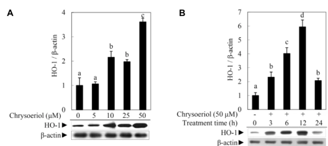
Fig. 2A and Fig. 2B indicate that chrysoeriol significantly induced the HO-1 expression at 50 μM for 12 hr treatment without any cytotoxicity (data not shown).
Nrf2, one of transcription factors, has known to play an important role in the induction of the antioxidant response element (ARE)-dependent genes expression, including the phase II enzymes [8]. Nrf2 is localized as an inactive form in the cytoplasm, which is anchored by the cytoskele- ton-associated protein, Kelch-like ECH-associated protein 1 (Keap1). Extracellular stimuli (i.e., ROS and electrophiles) provide signals for the dissociation of Nrf2-Keap1 complex leading to a nuclear translocation of Nrf2 and the induction ARE-related genes, including HO-1 [14]. To investigate whether Nrf2 translocates into the nucleus, a nuclear protein was extracted and determined by Western blot analysis. Fig.
3 shows that the nuclear translocation of Nrf2 was initiated when RAW 264.7 cells were exposed against 50 μM of chrys- oeriol and the Nrf2 activation was constantly increased by an uninterrupted exposure, which was in accordance with the accelerated HO-1 expression.
ERK, JNK and p38 MAPKs regulated HO-1 expre- ssion in RAW 264.7 cells
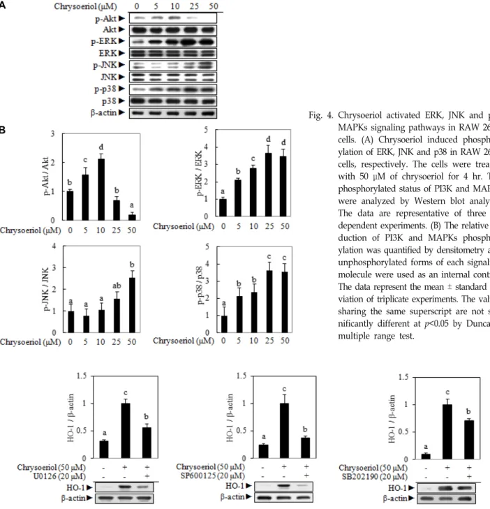
MAPKs or PI3K/Akt could be a candidate among various upstream signaling pathways for Nrf2 related HO-1 regu- lation in RAW 264.7 cells [12]. Chrysoeriol phosphorylates ERK, JNK and p38 MAPKs, while PI3K/Akt signaling mole- cule did not give any visible effect (Fig. 4). It has been re- ported that the modulation of intracellular kinase cascades was involved in the regulation of the HO-1 expression.
Quercetin was an effective inducer of HO-1 gene through an activation of ERK but not JNK and p38 in macrophages [5]. In addition, the bark extract from Lannea coromandelica induced HO-1 expression through upregulation of Nrf2- mediated pathway via the phosphorylation of p38 and JNK [1, 10]. This study further investigated whether the activa- tion of the p38 and JNK MAPKs signaling pathways was an inevitable event for the HO-1 expression through a phar- macological study applied to three well-known selective in- hibitors, including U0126 (for ERK), SP600125 (for JNK) and
A B
Fig. 2. Chrysoeriol induced HO-1 protein expression in RAW 264.7 cells. (A) Chrysoeriol induced HO-1 protein expression as a function of concentration. RAW 264.7 cells were treated for 4 hr with chrysoeriol at the indicated concentrations (0, 5, 10, 25 or 50 μM). The HO-1 protein expression was analyzed by Western blot analysis. The data are representative of three independent experiments. (B) Chrysodriol induced HO-1 protein expression as a function of time. RAW 264.7 cells were treated with 50 μM of chrysoeriol for indicated durations (0, 3, 6, 12 or 24 hr). The HO-1 protein expression was analyzed by Western blot analysis. The data are representative of three independent experiments. The relative induction of the HO-1 protein expression was quantified by densitometry and β-actin was used as an internal control. The data represent the mean
± standard deviation of triplicate experiments. The values sharing the same superscript are not significantly different at p<0.05 by Duncan’s multiple range test.
Fig. 3. Chrysoeriol induced Nrf2 activation, the nuclear translocation of Nrf2, in RAW 264.7 cells. The cells were treated for 4 hr with chrysoeriol at the indicated concentrations (0, 5, 10, 25 or 50 μM). The Nrf2 nuclear trans- location was determined from a nuclear extract by Western blot analysis.
The data are representative of three independent experiments. The relative induction of Nrf2 translocation was quantified by densitometry and PARP was used as an internal control. The data represent the mean ± standard deviation of triplicate experiments. The values sharing the same super- script are not significantly different at p<0.05 by Duncan’s multiple range test. Nrf2, nuclear factor-erythroid 2 p45-related factor 2; PARP, poly (ADP-ribose) polymerase.
SB202190 (for p38). Fig. 5 shows that the addition of each selective inhibitor significantly abolished the HO-1 protein expression induced by chrysoeriol. These results suggest that the chrysoeriol induced HO-1 expression is regulated by the ERK, JNK and p38 signaling molecules in RAW 264.7 cells.
Pretreatment of chrysoeriol protected RAW 264.7 cells against oxidative stress induced cell death
The exposure to ROS can be led cell death, as a result of severe damage to cellular lipid, protein and DNA. In this study, t-BHP, one of the organic hydroperoxides, was ap- plied to induce an oxidative damage in RAW 264.7 cells.
The t-BHP is metabolized by cytochrome P450, which leads
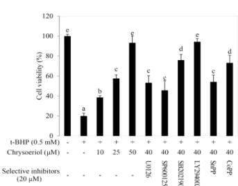
the formation of toxic peroxyl and alkoxyl radicals and ini- tiates a lipid peroxidation leading to cell death. The con- dition for cytotoxic damage issued from t-BHP on RAW 264.7 cells was described in previous report [20]. Cells were pre-incubated with 40 μM of chrysoeriol for 12 hr in order to induce the HO-1 expression in the presence or the absence of each selective inhibitor or inducer. The selective in- hibitors, as well as SnPP and CoPP (HO-1 inhibitor and in- ducer, respectively), were treated as 20 μM [3]. The untreated and the chrysoeriol treated cells with or without the in- hibitors or the inducers were exposed to 0.5 mM of t-BHP for 3 hr in order to induce a cytotoxic damage. A sharp in- crease in cytotoxicity was shown in Fig. 6 as a result of the
A
B
Fig. 4. Chrysoeriol activated ERK, JNK and p38 MAPKs signaling pathways in RAW 264.7 cells. (A) Chrysoeriol induced phosphor- ylation of ERK, JNK and p38 in RAW 264.7 cells, respectively. The cells were treated with 50 μM of chrysoeriol for 4 hr. The phosphorylated status of PI3K and MAPKs were analyzed by Western blot analysis.
The data are representative of three in- dependent experiments. (B) The relative in- duction of PI3K and MAPKs phosphor- ylation was quantified by densitometry and unphosphorylated forms of each signaling molecule were used as an internal control.
The data represent the mean ± standard de- viation of triplicate experiments. The values sharing the same superscript are not sig- nificantly different at p<0.05 by Duncan’s multiple range test.
Fig. 5. Chrysoeriol induced HO-1 expression depends on the ERK, JNK and p38 MAPK signaling pathways. Chrysoeriol was co-treated with selective inhibitors of each MAPK signaling molecule in RAW 264.7 cells. Fifty microliter of chrysoeriol was co-treated with 20 μM of selective inhibitors for ERK, JNK and p38. The HO-1 protein expression was analyzed by Western blot analysis.
The data are representative of three independent experiments. The relative induction of HO-1 protein expression was quantified by densitometry and β-actin was used as an internal control. The data represent the mean ± standard deviation of triplicate experiments. The values sharing the same superscript are not significantly different at p<0.05 by Duncan’s multiple range test.
lipid peroxidation, which was significantly attenuated by chrysoeriol treatment. On the other hand, the cytoprotective potential against the oxidative stress could not be exerted on U0126, SP600125, SB202190 and SnPP (ERK, JNK, p38 in- hibitors and HO-1 inhibitor, respectively) treated cells since
the abolished HO-1 expression. Consequently, these results suggest that the Nrf2 mediated HO-1 expression by chrys- oeriol strengthens the antioxidative potential against the t-BHP-induced oxidative damage through the modulation of ERK, JNK and p38 signaling pathways in RAW 264.7 cells.
Fig. 6. The antioxidative potential of crysoeriol against the t-BHP- induced oxidative damage in RAW 264.7 cells. The cells were treated with indicated concentrations of chrysoeriol (0, 10, 25 or 50 μM) for 12 hr in the presence or absence of each selective inhibitor or inducer. The untreated and the chrysoeriol treated cells with or without inhibitor or inducer were exposed to 0.5 mM t-BHP for 3 hr. The data represent the mean ± standard deviation of tripli- cate experiments. The values sharing the same super- script are not significantly different at p<0.05 by Dun- can’s multiple range test.
Acknowledgements
This work was supported by Dong-Eui University Grant (201702540001) and Blue-Bio Industry Regional Innovation Center (RIC08-06-07) at Dong-Eui University as a RIC pro- gram under Ministry of Trade, Industry and Energy (MOTIE) and Busan city.
References
1. Alam, M. B., Kwon, K. R. and Lee, S. H. 2017. Lannea coro- mandelica (Houtt.) Merr. induces heme oxygenase 1 (HO-1) expression and reduces oxidative stress via the p38/c-Jun N-terminal kinase-nuclear factor erythroid 2-related factor 2 (p38/JNK-NRF2)-mediated antioxidant pathway. Int. J.
Mol. Sci. 18, 226.
2. Baranano, D. E., Rao, M., Ferris, C. D. and Snyder, S. H. 2002.
Biliverdin reductase: a major physiologic cytoprotectant.
Proc. Natl. Acad. Sci. USA. 99, 16093-16098.
3. Bussolati, B., Ahmed, A., Pemberton, H., Landis, R. C., Di Carlo, F., Haskard, D. O. and Mason, J. C. 2004. Bifunctional role for VEGF-induced heme oxygenase-1 in vivo: induction of angiogenesis and inhibition of leukocytic infiltration.
Blood 103, 761-766.
4. Cha, B. Y., Shi, W. L., Yonezawa, T., Teruya, T., Nagai, K.
and Woo, J. T. 2009. An inhibitory effect of chrysoeriol on
platelet-derived growth factor (PDGF)-induced proliferation and PDGF receptor signaling in human aortic smooth mus- cle cells. J. Pharmacol. Sci. 110, 105-110.
5. Chow, J. M., Shen, S. C., Huan, S. K., Lin, H. Y. and Chen, Y. C. 2005. Quercetin, but not rutin and quercitrin, pre- vention of H2O2-induced apoptosis via anti-oxidant activity and heme oxygenase 1 gene expression in macrophages.
Biochem. Pharmacol. 69, 1839-1851.
6. Eisenstein, R. S., Garcia-Mayol, D., Pettingell, W. and Munro, H. N. 1991. Regulation of ferritin and heme oxygenase syn- thesis in rat fibroblasts by different forms of iron. Proc. Natl.
Acad. Sci. USA. 88, 688-692.
7. Farombi, E. O. and Surh, Y. J. 2006. Heme oxygenase-1 as a potential therapeutic target for hepatoprotection. J. Bio- chem. Mol. Biol. 39, 479-491.
8. Itoh, K., Chiba, T., Takahashi, S., Ishii, T., Igarashi, K., Katoh, Y., Oyake, T., Hayashi, N., Satoh, K., Hatayama, I., Yamamoto, M. and Nabeshima, Y. 1997. An Nrf2/small Maf heterodimer mediates the induction of phase II detoxifying enzyme genes through antioxidant response elements.
Biochem. Biophys. Res. Commun. 236, 313-322.
9. Johnson, G. L. and Lapadat, R. 2002. Mitogen-activated pro- tein kinase pathways mediated by ERK, JNK, and p38 pro- tein kinases. Science 298, 1911-1912.
10. Jeong, Y. H., Park, J. S., Kim, D. H. and Kim, H. S. 2016.
Lonchocarpine increases Nrf2/ARE-mediated antioxidant enzyme expression by modulating AMPK and MAPK sig- naling in brain astrocytes. Biomol. Ther. 24, 581-588.
11. Keum, Y. S. 2012. Regulation of Nrf2-mediated phase II de- toxification and anti-oxidant genes. Biomol. Ther. 20, 144-151.
12. Kim, J., Cha, Y. N. and Surh, Y. J. 2010. A protective role of nuclear factor-erythroid 2-related factor-2 (Nrf2) in in- flammatory disorders. Mutat. Res. 690, 12-23.
13. Kwon, E. J., Park, H. J., Nam, H., Lee, S. G., Hong, S., Kim, M. M., Lee, K. R., Hong, I., Lee, D. G. and Oh, Y. 2014.
Whitening and antioxidant effects of a mixture of Poria cocas, Glycyrrhiza uralensis, and Ulmus macrocarpa extracts. J. Life Sci. 24, 1063-1069.
14. Lee, J. S. and Surh, Y. J. 2005. Nrf2 as a novel molecular target for chemoprevention. Cancer Lett. 224, 171-184.
15. Ma, Q. 2013. Role of nrf2 in oxidative stress and toxicity.
Annu. Rev. Pharmacol. Toxicol. 53, 401-426.
16. Martin, D., Rojo, A. I., Salinas, M., Diaz, R., Gallardo, G., Alam, J., De Galarreta, C. M. and Cuadrado, A. 2004. Regu- lation of heme oxygenase-1 expression through the phos- phatidylinositol 3-kinase/Akt pathway and the Nrf2 tran- scription factor in response to the antioxidant phytochem- ical carnosol. J. Biol. Chem. 279, 8919-8929.
17. Pradhan, P., Giri, J., Rieken, F., Koch, C., Mykhaylyk, O., Doblinger, M., Banerjee, R., Bahadur, D. and Plank, C. 2010.
Targeted temperature sensitive magnetic liposomes for ther- mo-chemotherapy. J. Control. Release 142, 108-121.
18. Shi, X. and Zhou, B. 2010. The role of Nrf2 and MAPK path- ways in PFOS-induced oxidative stress in zebrafish embryos. Toxicol. Sci. 115, 391-400.
19. Sies, H. 1997. Oxidative stress: oxidants and antioxidants.
초록:생쥐 대식세포에서 HO-1 발현 유도를 통한 chrysoeriol의 항산화 효과
박충무*
(동의대학교 임상병리학과)
Chrysoeriol은 alfalfa에서 주로 발견되는, 식물계에 많이 분포하고 있는 flavone으로 전통의학에서 소화불량, 천식, 비뇨기계 이상의 치료에 사용되어 왔다. 최근의 연구에서는 항염증 효과가 있는 것으로 밝혀졌으나 항산화 효과에 대한 분석은 없었다. 본 연구에서는 chrysoeriol의 항산화 효과와 그 분자적 기전을 RAW 264.7 cell에서 세포생존율, reactive oxygen species (ROS)와 Western blot분석을 통해 알아보고자 하였다. Chrysoeriol은 lip- opolysaccharide (LPS)에 의해 발생한 ROS를 세포독성없이 농도의존적으로 제거하였다. 그리고 항산화효과를 보 이는 2상 효소 중 하나인 heme oxygenase (HO)-1의 발현을 강하게 유도하였고, 그와 동시에 전사인자인 Nrf2의 핵내 이동도 촉진하는 것으로 밝혀졌다. 특히, 산화스트레스에 대한 세포내 산화환원항상성 유지에 중요한 역할을 하고 있는 것으로 알려진 mitogen activated protein kinase (MAPK)와 phosphoinositide 3-kinase (PI3K)의 분석 결과, chrysoeriol은 extracellular signal regulated kinase (ERK), c-Jun NH2-terminal kinase (JNK)와 p38의 인산 화를 통해 HO-1의 발현을 유도하는 것으로 나타났다. HO-1에 의한 항산화 효과를 확인하기 위하여 chrysoeriol을 전처리한 후 t-BHP에 의한 산화 스트레스에 세포를 노출시킨 결과, chrysoeriol 처리에 의해 세포사멸이 줄어드는 것을 확인하였고, HO-1의 유도제와 억제제의 처리에 따라 세포생존율 또한 조절되는 것을 확인할 수 있었다. 따 라서, chrysoeriol은 HO-1의 발현을 유도하여 항산화 효과를 높이고 이것은 Nrf2/MAPK 신호전달 체계에 의한다 는 것을 알 수 있었다.
Exp. Physiol. 82, 291-295.
20. Song, Y. S. and Park, C. M. 2014. Luteolin and luteo- lin-7-O-glucoside strengthen antioxidative potential through the modulation of Nrf2/MAPK mediated HO-1 signaling cascade in RAW 264.7 cells. Food Chem. Toxicol. 65, 70-75.
21. Stochmal, A., Simonet, A. M., Macias, F. A. and Oleszek, W. 2001. Alfalfa (Medicago sativa L.) flavonoids. 2. Tricin and chrysoeriol glycosides from aerial parts. J. Agric. Food Chem.
49, 5310-5314.
22. Stocker, R., Yamamoto, Y., McDonagh, A. F., Glazer, A. N.
and Ames, B. N. 1987. Bilirubin is an antioxidant of possible physiological importance. Science 235, 1043-1046.
23. Vitaglione, P., Morisco, F., Caporaso, N. and Fogliano, V.
2004. Dietary antioxidant compounds and liver health. Crit.
Rev. Food Sci. Nutr. 44, 575-586.
24. Yoo, O. K., Lee, Y. G., Do, K. H. and Keum, Y. S. 2017.
Ethanol extracts of Rheum undulatum and Inula japonica pro- tect against oxidative damages on human keratinocyte HaCaT cells through the induction of ARE/NRF2-depend- ent phase II cytoprotective enzymes. J. Life Sci. 27, 310-317.
25. Yoon, J. W., Kim, S. J. and Park, D. S. 2016. Anti-oxidative and anti-inflammatory effects of Cheongajihwang-Tang ex- tract on RAW264.7 cells. J. Kor. Med. Rehabil. 26, 51-58.
26. Yu, R., Chen, C., Mo, Y. Y., Hebbar, V., Owuor, E. D., Tan, T. H. and Kong, A. N. 2000. Activation of mitogen-activated protein kinase pathways induces antioxidant response ele- ment-mediated gene expression via a Nrf2-dependent mechanism. J. Biol. Chem. 275, 39907-39913.
27. Wang, H. and Joseph, J. A. 1999. Quantifying cellular oxida- tive stress by dichlorofluorescein assay using microplate reader. Free Radic. Biol. Med. 27, 612-616.