169
Copyright © 2017 The Korean Society of Fisheries and Aquatic Science pISSN:0374-8111, eISSN:2287-8815
서 론
넙치
(Paralichtys olivaceus)
는가자미목넙치과에속하는해 산어종으로서완전양식기술이확립되어있으며,
식용으로서 의기호도가높아우리나라의대표적인양식어종으로서자리 매김하고있다. 2015
년넙치류의양식생산량은45,759
톤(
약5,040
억원)
으로전체어류양식생산량의약53%
를차지하고 있다(KOSIS, 2016).
하지만고밀도사육,
병원체의출현,
어장 의노후화등으로인해다양한감염성질병이발생하여경제적 으로피해를입고있다.
항체검출
enzyme-linked lmmunosorbent assay (ELISA)
는 혈중에존재하는특이항체를검출하는방법으로저렴한비용 으로다수의샘플처리가가능하고,
민감도와특이도가높아육 상동물에서는병원체의감염이력의파악,
백신의효능검증,
건강상태모니터링등의목적으로널리사용되고있다
(Crowther, 1995; OIE, 2016).
포유류의항체는immunoglobulin A (IgA), IgD, IgE, IgG
및IgM
으로구성되어있으며,
일반적으로체 내에 항원이 유입되게 되면초기에는IgM
이 생성되며 다음 으로IgG
가 생성되어 항원에 더 효과적으로 반응하게 된다(Crowther, 1995; Peter, 2014).
경골어류의항체는주로IgM, IgD
및IgT/IgZ
로구성되어있고,
이중IgM
은경골어류의혈 청과점액에서가장많이존재하며감염성병원체의중화,
세포 성세포독성매개,
보체경로의활성등의기능을하나포유류의 항체(IgG)
에비해친화성과다양성이낮다(Wilson and Warr, 1992; Kaattari and Piganelli, 1996; Pilström and Bengtén, 1996; Tort et al., 2003; Ye et al., 2013).
하지만어류도항원에 노출되면항원에대한특이적인항체가생성되며(Yoshimizu et al., 1992; LaPatra, 1996; Kim et al., 2009; Kim et al., 2011),
넙치(Paralichthys olivaceus)의 immunoglobulin M에 대한 단클론 항체 생산
김위식·김기홍·김춘섭 1 ·오명주*
전남대학교 수산생명의학과, 1㈜엔바이로젠
Production of Monoclonal Antibodies Against the Immunoglobulin M of Olive Flounder Paralichthys Olivaceus
Wi-Sik Kim, Ki-Hong Kim, Choon-sup Kim
1
and Myung-Joo Oh*Department of Aqualife Medicine, Chonnam National University, Yeosu 59626, Korea
1EnbioGene, Yeosu 59771, Korea
Immunoglobulin M (IgM) was purified from olive flounder Paralichthys olivaceus sera using mannan-binding protein (MBP) and protein L affinity columns (designated as MBPIgM and ProLIgM, respectively). A monoclonal antibody (MAb) against olive flounder IgM was produced. The MBPIgM and ProLIgM had apparent molecular weights of 77, 73, and 28 kDa in SDS-PAGE. Nine hybridomas secreting MAbs against olive flounder IgM were established: five MAbs for MBPIgM (1, 2, 3, 4, and 5) and four for ProLIgM (6, 7, 8, and 9). Western blotting indicated that seven MAbs recognized heavy (H; MAbs 1, 2, 3, 4, 5, 6, and 7) chains and one recognized light (L; MAb 9) chains of IgM, while MAb 8 did not recognize IgM. The results of enzyme-linked immunosorbent assay (ELISA) with bovine serum albumin (BSA, antigen) and the nine MAbs revealed that the optical density (OD) values of sera differed significantly between BSA- and non-immunized fish, despite some sera from non-immunized fish with slight high OD values.
These results suggest that the MAbs produced in this study reacted specifically with the IgM from olive flounder.
Key words: Immunoglobulin M, Olive flounder, Monoclonal antibody, Mannan-binding protein affinity column, Protein L affinity column
This is an Open Access article distributed under the terms of the Creative Commons Attribution Non-Commercial Licens (http://creativecommons.org/licenses/by-nc/3.0/) which permits unrestricted non-commercial use, distribution, and reproduction in any medium, provided the original work is properly cited.
http://dx.doi.org/10.5657/KFAS.2017.0169 Korean J Fish Aquat Sci 50(2) 169-174, April 2017
Received 10 February 2017; Revised 20 March 2017; Accepted 23 March 2017
*Corresponding author: Tel: +82. 61. 659. 7173 Fax: +82. 61. 659. 7170
E-mail address: [email protected]
김위식
ㆍ
김기홍ㆍ
김춘섭ㆍ
오명주170
혈액중에존재하는특이항체검사를통해병원체에감염되지 않은친어선별
,
양식장에서의병원체에대한감염이력등의조 사가가능한것으로보고되어있다(LaPatra, 1996; Watanabe et al., 1998; 2000; Kibenge et al., 2002).
어류는사육환경에따라 면역반응이달라지므로어류를대상으로한항체검출ELISA
를실시하기위해서는사육환경에따른특이항체반응에대한 정보가필요하며,
또한실험을실시하는데있어서는2
차항체 로서어류IgM
을인식하는항체가필요하다.
본연구에서는넙치를대상으로항체검출
ELISA
를실시하기위한기초연구로서넙치
IgM
에대한단클론항체(monoclonal antibody, MAb)
를생산하고자하였다.
넙치의IgM
은mannan binding protein (MBP) affinity column
과protein L affinity column
을사용하 여정제한후항원으로사용하였다.
재료 및 방법
넙치 혈청 분리
넙치의혈청은전남대학교수산과학연구소에서사육중인건 강한넙치
(
체중: 1.0-1.5 kg)
의미부정맥에서채혈하여분리하 였다.
채혈한혈액은상온에서30
분간방치한후6,000 rpm
에 서20
분간원심분리하여혈청을분리하였다.
분리한혈청은실 험에사용하기전까지-80℃
에보관하였다.
항 bovine serum albumin (BSA) 넙치 혈청 생산
항BSA
넙치혈청은건강한넙치(
체중: 1.0-1.5 kg) 2
마리에BSA
를1 mg/200 µL
씩복강에주사한후17-20℃
에사육하 면서0, 7, 14, 21
일째미부정맥에서채혈하여원심분리(6,000 rpm, 4℃, 20 min)
하여얻었으며,
대조구의혈청은2
마리의넙 치에phosphate-buffered saline (PBS, pH 7.5)
를200 µL
씩복 강에주사한후위와동일한방법으로분리하였다.
넙치 IgM 정제
1) MBP affinity column
Shin et al. (2007)
이언급한방법에따라ImmunoPure IgM purification kit (Pierce, USA)
를사용하여넙치IgM
을정제하 였다. ImmunoPure MBP column
을preparation buffer 5 mL
로수세한후binding buffer 20 mL
로column
을equilibration
하였다.
넙치혈청과binding buffer
를1:1
로혼합한후column
에넣고4℃
에서30
분간반응시킨후, binding buffer 42 mL
로 수세하였다. Elution buffer 3 mL
를column
에넣고1
시간동안 실온에서반응시킨후1 mL
씩수집하였다.
정제된IgM (MB- PIgM)
은실험에사용하기전까지-80℃
에보관하였다. 2) Protein L affinity column
Protein L resin (GenScript, USA)
을column
에넣고멸균된 증류수와binding buffer (20 mM Na
2HPO
4, 0.15 M NaCl, pH 8)
로수세한후,
넙치혈청과binding buffer
를1:1
로혼합하여column
에넣어주었다. 4℃
에서15
분간반응시킨후binding buffer
로수세하였고, elution buffer (0.1 M glycine, pH 2.5)
로 수집한후1M Tris-HCl (pH 8.5)
로중화시켰다.
정제된IgM (ProLIgM)
은실험에사용하기전까지-80℃
에보관하였다. MAb 생산
정제된 넙치
IgM (MBPIgM
과ProLIgM)
과complete Freund’s adjuvant (Gibco, USA)
를동량으로 섞어100 μL
씩2
마리의BALB/c
마우스의복강에1
차접종하였고, 2
주후 에정제된넙치IgM
으로2
차접종하였다.
다시1
주후에정제 된넙치IgM
으로3
차접종한후,
마우스로부터비장을분리한 후polyethylene glycol (Roche, Germany)
을사용하여SP2/0 myeloma
세포와융합시킨후fetal bovine serum
이10%
첨가 된HAT
배지(0.1 mM hypoxanthine, 4×10
-4mM aminopro- tein, 1.6×10
-2mM thymidine in Dulbecco’s modified eagle medium)
로suspension
시킨후96 well plate
에분주하여37℃
로설정된
CO
2배양기에서배양하였다.
양성hybridoma
는정 제된넙치IgM
을사용하여ELISA
법으로선별하였고3
회이상 제한희석법으로클로닝하였다.
선별된MAb
의isotyping
은마 우스Ig isotyping ELISA kit (BD Biosciences, USA)
를사용하 여결정하였다.
M 1 2
kDa
116
66
53
38
30 25
77 73
28
kDa 100 75
50 37
25 20
H
MBPIgM ProLIgM
1 2 3 4 5 1 2 3 4 5
M
6 7 8 9 6 7 8 9
kDa
100 75 50
37
25 20
H
L
MBPIgM ProLIgM
M
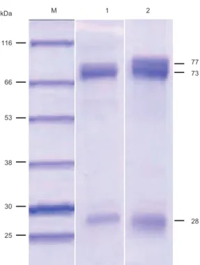
Fig. 1. SDS-PAGE analysis of olive flounder Paralichthys olivaceus IgM purified with mannan-binding protein (MBP) and protein L affinity columns. Lanes are M, molecular marker; 1, IgM purified with MBP affinity column; 2, IgM purified with protein L affinity column.
넙치 IgM에 대한 단클론 항체 생산
171
Sodium dodecyl sulfate-polyacrylamide gel electrophoresis (SDS-PAGE)와 western blotting
Laemmli (1970)
의 방법에 준해SDS-PAGE
를 실시하였 다.
넙치IgM
의인식부위를확인하기위해,
정제된넙치IgM (MBPIgM
와ProLIgM)
을12% polyacrylamide gel
에loading
한후100 V
에서전기영동을하였다.
전기영동종료후gel
을coomassie brilliant blue
를사용하여염색하였다. Western blot- ting
은Towbin et al. (1979)
의방법에따라실시하였다.
전기 영동한넙치IgM
을nitrocellulose membrane (Bio-Rad, USA)
에옮긴후,
본연구에서제작한MAb (1
차항체)
와alkaline phosphatase (AP)
가붙어있는goat anti-mouse IgG (Sigma, USA, 2
차항체)
로반응시키고AP
가붙어있는substrate kit (Bio-Rad, USA)
를사용하여발색하였다.
ELISA
넙치
IgM
에대한MAb
를생산하는hybridoma
의선별과제작 된MAb
가넙치IgM
을인식하는지를조사하기위하여ELISA
를실시하였다. Hybridoma
선별은Kim et al. (2008)
의ELISA
방법을변형하여실시하였다.
정제된넙치IgM (MBPIgM
와ProLIgM)
을coating buffer (0.5M carbonate-bicarbonate, pH 9.6)
로희석한후ELISA plate (Nunc, Denmark)
에50 µL (250 ng/well)
씩분주한후, 4℃
에서overnight
하여항원을coating
하였다
. Tween-20
이0.05%
포함된PBS (T-PBS)
로3
번세척 하였고5% skim milk
를380 µL
씩넣어25℃
에서1
시간동안blocking
시켰다. 1
차항체로는본연구에서제작한hybridoma
의배양액을사용하여50 µL
씩분주하였고, 2
차항체로서5%
skim milk
로1,000
배 희석된peroxidase conjugated
항마우 스IgG
염소혈청(Younginfrontier, Korea)
을50 µL
씩분주하 였다.
각각의항체는25℃
에서1
시간동안반응하였다. T-PBS
로5
번수세하였고ELISA
발색액(100 mM Na
2HPO
4, 50 mM citric acid, 1 mg/mL o-phenylenediamine, 0.03% H
2O
2)
을각well
에50 µL
씩분주한후25℃
에서15
분동안발색하였다.
각well
에2 N H
2SO
4를50 µL
씩넣어발색반응을중지시킨후microplate photometer (Multiskan, USA)
로492 nm
에서흡광 도(optical density, OD)
값을측정하였다.
제작된MAb
가넙치IgM
을인식하는지를조사하기위하여BSA
에대한특이항체 를검출하는ELISA
를Kim et al. (2007, 2011)
의방법에준해 실시하였다. ELISA plate
에5 µg BSA/50 µL
를각well
에분주 한후, 37℃
에서overnight
하여항원을coating
하였다. 1
차항체로는넙치혈청
(BSA
면역및비면역넙치로부터분리한혈청
)
을5% skim milk
로희석(40
배)
하여1
시간반응시킨후50 µL
씩분주하였고, 2
차항체로는본연구에서제작한MAb
를50 µL
씩분주하였으며, 3
차항체는peroxidase conjugated
항M 1 2
kDa
116
66
53
38
30 25
77 73
28
kDa 100 75 50 37
25 20
H
MBPIgM ProLIgM
1 2 3 4 5 1 2 3 4 5
M
6 7 8 9 6 7 8 9
kDa
100 75 50
37
25 20
H
L
MBPIgM ProLIgM
M
Fig. 2. Western blotting of purified olive flounder Paralichthys oli- vaceus IgMs (MBPIgM and ProLIgM) immunostained with 5 anti- MBPIgM monoclonal antibodies (MAbs). M, molecular marker;
1-5, MAbs against MBPIgM; MBPIgM, olive flounder IgM puri- fied with mannan-binding protein affinity column; ProLIgM, olive flounder IgM purified with protein L affinity column.
66
53
38
30 25
77 73
28
kDa 100 75 50 37
25 20
H
MBPIgM ProLIgM
1 2 3 4 5 1 2 3 4 5
M
6 7 8 9 6 7 8 9
kDa
100 75 50
37
25 20
H
L
MBPIgM ProLIgM
M
Fig. 3. Western blotting of purified olive flounder Paralichthys oli- vaceus IgMs (MBPIgM and ProLIgM) immunostained with 4 anti- ProLIgM monoclonal antibodies (MAbs). M, molecular marker;
6-9, MAbs against ProLIgM; MBPIgM, olive flounder IgM puri- fied with mannan-binding protein affinity column; ProLIgM, olive flounder IgM purified with protein L affinity column.
김위식
ㆍ
김기홍ㆍ
김춘섭ㆍ
오명주172
Fig. 4. Antibody detection enzyme-linked lmmunosorbent assay with bovine serum albumin (antigen) and 10 monoclonal antibodies [1-9, commercial antibody (CA)]. BSA1 and BSA2, sera from BSA immunization of olive flounder Paralichthys olivaceus; Cont1 and cont2, sera from non-immunization of olive flounder.
0.0 0.5 1.0 1.5 2.0 2.5 3.0 3.5 4.0
0 7 14 21
ELISA absorbance (OD 492 nm)
Days after infection BSA1
BSA2 Cont1 Cont2
0.0 0.5 1.0 1.5 2.0 2.5 3.0 3.5 4.0
0 7 14 21
ELISA absorbance (OD 492 nm) Days after infection
BSA1 BSA2 Cont1 Cont2
0.0 0.5 1.0 1.5 2.0 2.5 3.0 3.5 4.0
0 7 14 21
ELISA absorbance (OD 492 nm)
Days after infection BSA1
BSA2 Cont1 Cont2
0.0 0.5 1.0 1.5 2.0 2.5 3.0 3.5 4.0
0 7 14 21
ELISA absorbance (OD 492 nm)
Days after infection BSA1
BSA2 Cont1 Cont2 0.0
0.5 1.0 1.5 2.0 2.5 3.0 3.5 4.0
0 7 14 21
ELISA absorbance (OD 492 nm)
Days after infection BSA1
BSA2 CON1 CON2
0.0 0.5 1.0 1.5 2.0 2.5 3.0 3.5 4.0
0 7 14 21
ELISA absorbance (OD 492 nm)
Days after infection BSA1
BSA2 CON1 CON2
0.0 0.5 1.0 1.5 2.0 2.5 3.0 3.5 4.0
0 7 14 21
ELISA absorbance (OD 492 nm)
Days after infection BSA1
BSA2 Cont1 Cont2
0.0 0.5 1.0 1.5 2.0 2.5 3.0 3.5 4.0
0 7 14 21
ELISA absorbance (OD 492 nm)
Days after infection BSA1
BSA2 Cont1 Cont2
0.0 0.5 1.0 1.5 2.0 2.5 3.0 3.5 4.0
0 7 14 21
ELISA absorbance (OD 492 nm)
Days after infection BSA1
BSA2 Cont1 Cont2
0.0 0.5 1.0 1.5 2.0 2.5 3.0 3.5 4.0
0 7 14 21
ELISA absorbance (OD 492 nm)
Days after infection BSA1
BSA2 Cont1 Cont2
6
8 9
7 2
3 4
5 CA
1
마우스
IgG
염소혈청을50 µL
씩분주하였다.
항체반응과발 색조건은위와동일하게실시하였다.
양성대조구로는시판중 인항넙치IgM
에대한MAb (Diagnostics Ltd., Scotland)
를사 용하여위와동일하게ELISA
를실시하였다.
결과 및 고찰
본연구에서는넙치를대상으로한항체검출
ELISA
를실시 하는데,
반드시필요한넙치IgM
에대한항체를생산하기위 하여MBP
와protein L affinity column
을사용하여넙치IgM
를정제한후,
정제된IgM (MBPIgM
와ProLIgM)
을사용하여MAb
를제작하였다. MBP
와protein L affinity column
을사 용하여넙치Ig
를정제한후SDS-PAGE
를실시한결과, MB- PIgM
과ProLIgM
모두에서약77, 73, 28 kDa
의분자량이관 찰되었다(Fig. 1). ProLIgM
에서는넙치혈청로트에따라서약77, 73, 28 kDa
의분자량이외에도약57 kDa
의분자량이관찰 되기도하였다(data not shown).
넙치IgM
의분자량은SDS- PAGE
상에서heavy (H) chain
과light (L) chain
이각각75 kDa
과23 kDa (Nishida et al., 1998), 77, 72 kDa
과28, 26 kDa (Jang et al., 2004), 74 kDa
과24 kDa (Li et al., 2007), 68, 66 kDa
과27, 25 kDa (Shin et al., 2007)
로보고되어있다.
본연구 의정제한Ig
은기존에보고된넙치IgM
의분자량과유사하므 로정제된Ig
는IgM
으로사료된다. Shin et al. (2007)
은MBP affinity column
을사용하여넙치Ig
를정제한결과68 kDa (H chain)
과25, 27 kDa (L chain)
의분자량을얻을수있었는데본 연구에서는동일한MBP affinity column
을사용했음에도불 구하고분자량에차이를보였다.
이는전기영동에사용한항원 의양또는분자량측정시에발생하는오차로인한것으로추정 된다. Protein L
은IgM, IgG, IgA, IgD
및IgE
의L chain
과특 이적으로결합하는 단백질로보고되어 있는데(Björck, 1988;
Nilson et al., 1993),
본연구의결과(
넙치IgM
정제결과)
넙치IgM
도protein L
과특이적으로결합한것으로사료된다.
MBPIgM
과ProLIgM
을마우스에면역시킨후마우스의비 장조직과SP2/0 myeloma
세포를융합시켜hybridoma
를제 작하였다. Hybridoma
로부터 생성되는 항체를ELISA
법으 로선별한후,
제한희석법으로클로닝하여최종적으로9
개의MAb
를선별하였다. MBPIgM
에대한5
개의MAb (MBPIgM- MAb:1, 2, 3, 4, 5)
와ProLIgM
에대한4
개의MAb (ProLIgM- MAb:6, 7, 8, 9). 9
개의MAb
에대한isotyping
결과, H chain
은IgG1 (1, 2, 3, 4, 5, 9), IgG2a (7), IgG2b (6, 8)
로나타났으 며, L chain
은모두kappa
로확인되었다(data not shown). 9
개 의MAb
와정제된넙치IgM
을사용하여western blotting
을실 시한결과, MBPIgM-MAb 5
개는MBPIgM
과ProLIgM
의H chain
을인식하였다(Fig. 2). ProLIgM-MAb
의경우, 6
과7
은MBPIgM
과ProLIgM
모두H chain
을강하게인식하였고, 9
는L chain
에반응하였다(Fig. 3). 8
은H chain
과L chain
모두 에반응하지않았다.
본연구에서제작된
MAb
가항체검출ELISA
에사용가능한 지를평가하기위해,
넙치에BSA
를면역시킨후0, 7, 14, 21
일 째혈청을분리하여BSA
에대한항체가를측정하였다(Fig. 4).
ProLIgM-MAb 4
개(6, 7, 8, 9)
는BSA
면역넙치혈청에대해 시간이경과됨에따라항체가가증가되는것으로나타났으며,
대조구의혈청(BSA
비면역혈청)
에서는뚜렷한항체가의증가 가관찰되지않았다. Kim et al. (2011)
의연구에따르면BSA
면 역후(
사육수온:15℃) 14
일째부터BSA
에대한특이항체가검 출되는개체가관찰되며, 21
일째모든개체에서BSA
에대한특 이항체가검출되었다고보고하였다.
본연구의결과는Kim et al. (2011)
의연구결과와유사하기때문에높은OD
값은BSA
에대한특이넙치항체를인식한것으로사료된다.
또한,
양성 대조구(
시판되고있는항넙치IgM
에대한MAb
사용)
에서도 유사한OD
값을보였기때문에,
본연구에서제작된MAb
는넙 치IgM
을특이적으로인식하는것으로사료된다.
그러나MAb 6
을제외한7, 8, 9
및시판용항넙치IgM
에대한MAb
에서는BSA
비면역넙치혈청과BSA
를면역시키기전에취한혈청에 서도0.1-1.1
의OD
값이관찰되었다(Fig. 4). MBPIgM-MAb 5
개(1, 2, 3, 4, 5)
는3
개의ProLIgM-MAb (7, 8, 9)
와유사한결과를보여
BSA
면역혈청과대조구의혈청이뚜렷이구분되었다
.
하지만BSA
비면역넙치혈청과BSA
를면역시키기전에 취한혈청에서도OD
값(0.08-0.8)
이관찰되었다(Fig. 4).
본연구를통해서는
BSA
비면역혈청에서보이는OD
값의원인에대해서알수는없으나항체검출
ELISA
방법에서넙치혈청을
5% skim milk
로전처리과정을경유하여실시했기때문에(Kim et al., 2007)
넙치IgM
이블로킹제에비특이적으로흡착 하여발생하는높은백그라운드와는관계가없을것으로사료 된다.
향후위의원인을규명하는연구가수행되어야할것이다.
넙치의
Ig
을정제하는 방법으로는sephacryl S-300 gel fil- tration
을사용하는 방법(Bang et al., 1996), salting-out, ion- exchange chromatography
및gel filtration
을 이용한 방법(Nishida et al., 1998), immune affinity column
을사용하는방 법(Jang et al., 2004; Shin et al., 2007), MBP affinity column
을사용하는방법(Shin et al., 2007)
이보고되어있다.
본연구에 서는처음으로protein L affinity column
을사용하여넙치IgM
을정제하였다. Protein L affinity column
은기존의방법들에 비해(salting-out, ion-exchange chromatography
및gel filtra- tion
을이용한방법, immune affinity column
을사용하는방법)
간편하면서도빠른시간내에 넙치IgM
을정제할수있는장 점을지니고있다.
또한ProLIgM
을사용하여제작한MAb
는MBPIgM-MAb
와마찬가지로넙치IgM
을특이적으로인식하 는것으로확인되었다.
사 사
본연구는
2015
년해양수산부재원으로한국해양과학기술진흥원의지원을받아수행되었습니다
(
수산동물바이러스전염김위식
ㆍ
김기홍ㆍ
김춘섭ㆍ
오명주174
병진단용항체생산
).
References
Bang JD, Kim JW, Lee SD, Park SI, Chun SG, Jeong CS and Park JW. 1996. Humoral immune response of flounder to
Edwardsiella tarda: the presence of various sizes of immu-
noglobulins in flounder. Dis Aquat Org 26, 197-203.Björck L. 1988. Protein L. A novel bacterial cell wall protein with affinity for Ig L chains. J Immunol 140, 1194-1197.
Crowther JR. 1995. ELISA. Theory and practice. Humana Press Inc., New Jersey, U.S.A.
Jang HN, JK Woo, YH Cho, SB Kyong and SH Choi. 2004.
Characterization of monoclonal antibodies against heavy and light chains of flounder (Paralichthys olivaceus) immu- noglobulin. J Biochem Mol Biol 37, 314-319.
Kaattari SL and Piganelli JD. 1996. The specific immune sys- tem: humoral defense. In the fish immune system: organism, pathogen and environment. Iwama G and Nakanishi T, eds.
Academic Press, San Diego, U.S.A., 207-254.
Kibenge MT, Opazo B, Rojas AH and Kibenge FSB. 2002.
Serological evidence of infectious salmon anaemia virus (ISAV) infection in farmed fishes, using an indirect enzyme- linked immunosorbent assay (ELISA). Dis Aquat Org 51, 1-11.
Kim CS, Jang MS, Kim WS, Kim JO, Kim DW, Kim DH, Han HJ, Jung SJ and Oh MJ. 2009. Evaluation of the stability of IgM and specific antibody response of sevenband grouper
Epinephelus septemfasciatus for application of antibody-
detection ELISA. J Fish Pathol 22, 335-342.Kim WS, Mochizuki M, Nishizawa T and Yoshimizu M. 2008.
Detection of specific antibodies against infectious hemato- poietic necrosis virus from rainbow trout sera by ELISA us- ing two novirhabdoviruses. Fish Pathol 43, 112-116.
Kim WS, Jang MS, Jung SJ, Kim SR, Park MA, Lee JH, My- eong JI and Oh MJ. 2011. Specific antibody response of ol- ive flounder Paralichthys olivaceus by water temperature. J Fish Pathol 24, 39-45.
Kim WS, Nishizawa T and Yoshimizu M. 2007. Non-specific adsorption of fish immunoglobulin M (IgM) to blocking re- agents on ELISA plate wells. Dis Aquat Org 78, 55-59.
KOSIS (Korean statistical information service ). 2016. Result of fish farm trends survey in 2015[Internet]. Retrieved from http://kosis.kr/eng/statisticsList/statisticsList_01List.
jsp?vwcd=MT_ETITLE&parmTabId=M_01_01 on March 2016.
Laemmli UK. 1970. Cleavage of structural proteins during the assembly of the head of bacteriophage T4. Nature 227, 680- LaPatra SE. 1996. The use of serological techniques for virus 685.
surveillance and certification of finfish. Annu Rev Fish Dis 6, 15-28.
Li Q, Zhan W, Xing J and Sheng X. 2007. Production, charac- terization and applicability of monoclonal antibodies to im- munoglobulin of Japanese flounder (Paralichthys olivaceus).
Fish Shellfish Immun 23, 982-990.
Nilson BH, Lögdberg L, Kastern W, Björck L and Akerström B. 1993. Purification of antibodies using protein L-binding framework structures in the light chain variable domain. J Immunol Methods 164, 33-40.
Nishida H, Enokida T, Hiramatsu N, Hara A and Yoshimizu M.
1998. Purification of Immunoglobulin M (IgM) in serum of Japanese flounder (Paralichthys olivaceus). Bull Faculty Fish Hokkaido Univ 49, 157-164.
Peter P. 2014. The immune system. Garland Science, Connecti- cut, U.S.A.
Pilström L and Bengtén E. 1996. Immunoglobulin in fish-genes, expression and structure. Fish Shellfish Immun 6, 243-262.
Shin GW, Kim YR, Shin YS, Lee EG, Oh MJ, Yoshida T and Jung TS. 2007. Purification of two different immunoglobu- lins (Igs) from olive flounder Paralichthys olivaceus and analysis of Lactococcus garvieae antigens by the Igs. Fish Pathol 42, 19-28.
Tort L, Balasch JC and Mackenzie S. 2003. Fish immune sys- tem. A crossroads between innate and adaptive responses.
Num 22, 277-286.
Towbin H, Staehelin T and Gordon J. 1979. Electrophoretic transfer of proteins from polyacrylamide gels to nitrocellu- lose sheets; procedure and some applications. Proc Natl Acad Sci USA 76, 4350-4354.
Watanabe K, Nishizawa T and Yoshimizu M. 2000. Selection of brood stock candidates of barfin flounder using an ELISA system with recombinant protein of barfin flounder nervous necrosis virus. Dis Aquat Org 41, 219-223.
Watanabe K, Suzuki S, Nishizawa T, Suzuki K, Yoshimizu M and Ezura Y. 1998. Control strategy for viral nervous necro- sis of barfin flounder. Fish Pathol 33, 445-446.
Wilson MR and Warr GW. 1992. Fish immunoglobulins and the genes that encode them. Annu Rev Fish Dis 2, 201-221.
World organization for animal health (OIE). 2016. Manual of di- agnostic tests and vaccines for terrestrial animals[Internet].
Retrieved from http://www.oie.int/manual-of-diagnostic- tests-and-vaccines-for-terrestrial-animals/ on May 2016.
Ye J, Kaattari IM, Ma C and Kaattari S. 2013. The teleost humor- al immune response. Fish Shellfish Immunol 35, 1719-1728.
http://dx.doi.org/10.1016/j.fsi.2013.10.015.
Yoshimizu M, Direkbusarakom S, Nomura T, Ezura Y and Kimura T. 1992. Detection of antibody against Aeromonas

![Fig. 4. Antibody detection enzyme-linked lmmunosorbent assay with bovine serum albumin (antigen) and 10 monoclonal antibodies [1-9, commercial antibody (CA)]](https://thumb-ap.123doks.com/thumbv2/123dokinfo/5547529.469285/4.892.165.703.143.1010/antibody-detection-lmmunosorbent-albumin-monoclonal-antibodies-commercial-antibody.webp)
