Arthroscopic rotator cuff repair: Double row technique
울산대 고 상 훈
Rotator cuff tear
1. Most common cause of chronic shoulder pain 2. Rotator cuff disease
초기: 건염과 활액 낭염으로 시작하여 통증을 일으키고 진행: 힘줄이 약해져서 실밥이 풀어지듯이 회전근 개 파열 발생
Rotator cuff disease
Stage 2 impingement, Tendonitis, fibrosis, Partial-thickness tears, Full-thickness tears : complete cuff tears, Irreparable tears Cuff arthropathy
Arthroscopic repair
Advantage of arthroscopic repair
Smaller incisions: cosmisis, Access to the G-H joint for inspection
Treatment of intra-articular lesions, No need for detachment of the deltoid, Less soft tissue dissection, Less pain, More rapid rehabilitation
Consideration at arthroscopic repair
Patient age, Patient activity level,Concomittent lesion,Range of motion, Symptom, Tear location
Advantage of single row repair
Relative simple technique than double row technique, not technical difficulty of performing double row
No time consumption than double row
No high cost than double row: additional anchor not needed No potential expense of vascularity
Disadvantage of single row repair
Warner, Iannotti, Flatow Complex and Revision Problems in Shoulder Surgery 2nd edition High rate of re-tear, Anchor failure, Suture loosening, Less superior compression of torn cuff after rotator cuff repair, Not re-establishing normal footprint, Tendon failure after
Integrity after arthroscopic repair
Galatz, Yamaguchi et al. JBJS-A 2004Outcome and repair integrity, Complete arthroscopic repair, Large and massive tear, Arthroscopic repair : high re-tear, Above 90%
Mode of failure for rotator cuff repair
Mode of failure for repair with suture anchors identified at revision surgery prospectively followed 342 consecutive torn rotator cuff
re-tear rate: 25%~90%
most common: tendon pulling through sutures
than tear in new location & anchor pulling out of bone
weak link in rotator cuff repairs with suture anchors: tendon-suture interface
Technique for arthroscopic cuff repair
Simple arthroscopic repair, Double row foot print repair Apreleva & Warner et al. 2002
Waltrip & Andrews et al. 2003 Lo & Burkhart 2003
De Beer et al 2002
Transosseous equivalent repair
Millett et al 2004: Mattress double anchor, footprint repair
ElAttrache, Tibone et al. J Shoulder Elbow Surgery 2007: Suture bridge using push lock Reinforced cuff repair
Modified Mason-Allen stitch, Arthroscopic Mac stitch, MacGillivary and Ma 2004, Ma et al. JBJS-A 2006
Anatomical footprint reconstruction - Biomechanical study -
Apreleva & Warner et al. 2002 Arthroscopy
Single row with SAS(suture anchor simple)--> covered 67% original SS insertion More lateral anchor placement -->increase repair-site area
Large contact area btw tendon and bone -->improve biological healing Waltrip & Andrews et al. 2003 AJSM
Initial fixation strength of double layer repair : exceed single layer repairs Either suture anchor or trans-osseous tunnel
Double row rotator cuff repair
Lo & Burkhart 2003 ArthroscopyMedial row : just lateral to articular surface of head Lateral row : just medial to drop-off of GT
May potentially improve strength and healing of rotator cuff repair constructs
Double row rotator cuff repair
De Beer et al 200258 cases by foorprint reconstruction 90% excellent and good results 89% intact cuff shown by ultrasound
Our Double row rotator cuff repair
Double row repairDouble row repair
Mazzocca, Millett et al. AJSM 2005
Single row similar to double-row in load to failure, cyclic displacement, gap formation Double-row larger footprint than single-row
Fealy, Warren et al. Arthroscopy 2006
No differences in strength at 2-anchor and 4-anchor repair Park JY et al. KJSES 2006
No difference on small & medium tear Significant abduction strength on large tear
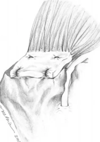
Fig. 1. Double row suture anchor repair
Technique of double row repair
Place medial row suture anchor, Place suture 15mm from cuff margin, Place lateral row anchors, Pass lateral row suture 10 mm from cuff margin, Lateral suture as simple suture loop, Sucure suture limbs of medial anchors, completion of medial row repair, Tie and sucure medial row suture
Transosseous tunnel cuff repair
Apreleva, Warner et al. Arthroscopy 20023-dimensional (3-D) reconstruction: area of original supraspinatus insertion, repair-site area after 4 recon method
TOS (Transosseous simple): 20% larger repair-site area than TOM (transosseous mattress suture)
SAS (suture-anchor simple suture) SAM (suture-anchor mattress suture) TOS repair :
better potential for healing
ultimately, greater strength of repair Maxwell Park, Louis U. Bigliani et al. AJSM 2005
transosseous tunnel repair : more contact and greater pressure distribution for suture anchor
Mattress double anchor footprint repair
Similar to transosseous techniques: Novel arthroscopic cuff repair use of 2 suture anchors independently -->connected by suture loop Suture linked between 2 bio anchors
distributes the stress across 2 anchors
restores a large surface area for healing between the rotator cuff and the tuberosity
Mattress double anchor
Alternative suture configuration with interlocking of the sutures, To prevent cutout from the tendon Sutures criss-cross from 4 separate anchors, Interlocked, Maximize tendon compression, Maximize repair surface area
Suture bridges using push lock
Transosseous equivalent rotator cuff repair 6 fresh-frozen human shoulders
4 suture bridges:
medial 2 anchors
lateral interference screw Suture limb bridge over tendon Results
Improve contact area : nearly twice more than double-row (77.6% vs 39.6%) Improve pressure area : 30% more than double-row repair
Transosseous-equivalent repair
4 suture bridges and 2 Bio-Tenodesis screws
medial & lateral fixation points : same as traditional open transosseous technique medial row suture limbs : not cut after tying
Lateral row :
two 4.5-mm holes : 1 cm distal to the lateral edge of the footprint in line with the medial suture anchors
distanced 12.5 mm apart in the anterior-posterior direction cannulated bio-Tenodesis 5.5-mm screw
suture limb from each medial suture anchor : minimum 4 kg of tension
bio-tenodesis screw : 45�angle screw countersunk 1 mm
each suture limb tied to a limb from the loop previously passed 2 knots tied over bio-Tenodesis screw
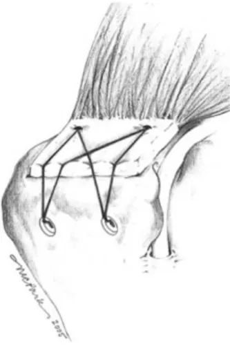
Transosseous-equivalent repair
Fig. 2. Transosseous-equivalent repair
except only 1 Bio-Tenodesis screw, 1 cm distal to the lateral edge of the footprint each medial anchor : 1 remaining suture limb
Biomechanics of transosseous-equivalent
Mean ultimate load to failure : greater to double-row Gap formation : not different to double-rowStiffness : not statistically different Advantage transosseous-equivalent repair Diagrams of Reinforced Technique
Arthroscopic Mason-Allen stitch
Scheibel, Habermeyer Arthroscopy 2003
Arthroscopic Mason-Allen stitch reported
MacGillivray et al. Arthroscopy 2004Tendon-suture interface : weak link in arthroscopic rotator cuff repair Simply arthroscopic modification of modified Mason-Allen stitch
Seperation & Combination at the site of repair as a horizontal mattress loop
vertical simple loop
Massive Cuff Stitch
Ma et al. JBJS-A 2004Biomechanical study using sheep infraspinatus, MCS : increase the strength of tendon- suture interface, Horizontal mattress loop act as checkrein for vertical simple loop, To prevent suture pull-out through tendon, Ultimate tensile strength, Three times than
Fig. 3.
simple suture repair, Similar than open modified Mason-Allen stitch MCS combined with Two row, Biomechanical study
Massive Cuff Stitch, Similar cyclic loading & load to failure than double row fixation
New reinforced stitch
Castagna et al. Chir Organi Mov 2005 New reinforced stitch simple stitch with horizontal stitch Resistance to loading similar to modified Mason-Allen
greater than simple and mattress stitch Baleani et al. Clin Biomech 2006 No difference in two ‘reinforced’stitches simple stitch closed over horizontal loop
modified Mason-Allen
maximum grasping power : only using with high-strength suture material Simple stitch closed over horizontal loop
attractive alternative for arthroscopic repair than to modified Mason-Allen for open repair