Introduction
Bicondylar tibial plateau fractures (TPFs) usually accompany severe comminuted fractures and soft tissue injuries because of high energy trauma. Based on the complexity of the fracture that involves both medial and lateral condyles, extensive dissection of soft tissue can aggravate soft tissue injury because soft tissue of
proximal tibia is very thin13). As a result, complications, such as skin necrosis, superficial or deep infection, and nonunion, can occur.
Treatment of bicondylar TPFs is still a controversial issue and is generally difficult because patients can suffer from postopera
tive arthritis and functional disability of the knee joint47). Many authors have reported that conventional open reduction and internal fixation (ORIF) in bicondylar fractures can cause soft tissue injuries, leading to complications such as nonunion, knee joint stiffness, and metal failure810). Several fixation methods can be employed to solve soft tissue problems including the use of a hybrid external fixator11,12) and staged treatment using a tempo
rary external fixator1315). Some authors have reported favorable clinical outcomes with staged treatment using a temporary exter
nal fixator9,16). The benefits of temporary external fixation include immediate osseous stabilization, prevention of further articular damage, access to wounds, increased patient comfort, ease of subsequent reduction, and potential for decreased narcotic re
Staged Treatment of Bicondylar Tibial Plateau Fracture (Schatzker Type V or VI) Using Temporary External Fixator: Correlation between Clinical and Radiological Outcomes
Seung Min Ryu, MD, PhD, Han Seok Yang, MD, and Oog Jin Shon, MD
Department of Orthopedic Surgery, Yeungnam University Medical Center, Daegu, Korea
Purpose: This study is to investigate clinical and radiological results of staged treatment using a temporary external fixator in bicondylar tibial plateau fractures (TPFs) and to evaluate correlation between prognostic factors and postoperative clinical outcomes.
Materials and Methods: Twentyfour bicondylar TPF patients were selected. All patients were operated by a temporary external fixator first and then open reduction and internal fixation with dual plating. Clinical and radiological outcomes were evaluated.
Results: The mean American Knee Society score (AKSS) was 85.3. The mean Western Ontario and McMaster Universities Osteoarthritis index was 11.2. The mean range of motion (ROM) was 123.4°. The mean medial tibial plateau angle (mTPA) was 88.3°, and the mean proximal posterior tibial angle (PPTA) was 8.4°. Compared with the uninjured limb, the mean difference of mTPA was 1.5° and that of PPTA was 4.0°. The difference of PPTA and the AKSS demonstrated negative correlation (p=0.007). Patients with normal mTPA showed better ROM than those with abnormal mTPA (p=0.041).
Conclusions: Staged treatment using a temporary external fixator in bicondylar TPFs showed good clinical and radiological outcomes. Surgeons should evaluate the reduction status intraoperatively by fluoroscopy and also refer to the uninjured limb radiologically.
Keywords: Tibia, Plateau, Bicondylar, Fracture, External fixator pISSN 2234-0726 · eISSN 2234-2451
Knee Surgery & Related Research
Received February 7, 2017; Revised May 7, 2017;
Accepted June 19, 2017
Correspondence to: Oog Jin Shon, MD
Department of Orthopedic Surgery, Yeungnam University Medical Center, 170 Hyeonchungro, Namgu, Daegu 42415, Korea Tel: +82536203640, Fax: +82536284020
Email: maestro[email protected]
261
This is an Open Access article distributed under the terms of the Creative Commons Attribution NonCommercial License (http://creativecommons.org/licenses/bync/4.0/) which permits unrestricted noncommercial use, distribution, and reproduction in any medium, provided the original work is properly cited.
Copyright © 2018 KOREAN KNEE SOCIETY www.jksrr.org
quirements14).
In the present study, authors have investigated clinical and ra
diological results of staged treatment using a temporary external fixator in bicondylar TPFs and evaluated correlation between prognostic factors and postoperative clinical outcomes.
Materials and Methods
1. Patients
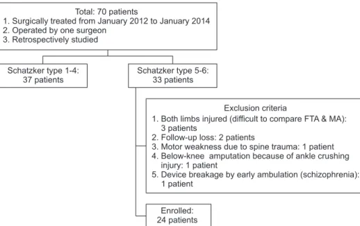
This study was approved by our hospital’s Institutional Review Board. Of the 70 patients who underwent operation for TPFs in our hospital between January 2012 and January 2014, 33 patients who presented with a Schatzker type V or VI fracture were se
lected. Patients were excluded if they had injuries in both limbs or an injury to the spinal cord with motor weakness, underwent aboveknee amputation or early ambulation with metal failure, or were lost to followup. Ultimately, 24 patients were selected and retrospectively investigated (Fig. 1). The mean followup was 24.2 months (range, 15 to 32 months). Clinical details of the patients are described in Table 1.
2. Surgical Technique and Rehabilitation
Patients were positioned supine on the radiolucent operat
ing table before receiving general or spinal anesthesia. In cases where patients were diagnosed with compartment syndrome, the operator immediately made dual incisions (anterolateral and posteromedial incisions) on the calf followed by fasciotomy to all compartments. Compartment syndrome was diagnosed clinically by using 5P physical signs and symptoms (pain, pallor, pulseless
ness, paresthesia, and paralysis). In case of an open fracture, mas
sive irrigation and wound debridement was done first and then a temporary external fixator was applied. However, in case of a closed fracture, an external fixator was applied immediately.
Pins of temporary external fixators were carefully applied con
sidering the position of medial and lateral plating. Subsequently, on a daily basis, authors carefully observed soft tissue of patients and planned appropriate time for final internal fixation with dual plating. In secondary plate fixation, under supine position, sepa
rate skin incisions which were anterolateral and posteromedial for dual plating (tubular plate or proximal medial plate [Synthes, Oberdorf, Switzerland], proximal lateral locking plate [Zimmer, Warsaw, IN, USA]) were done and the distance between skin in
cisions was kept to be more than 8 cm (Fig. 2).
Basically, antibiotics were used for approximately 5 to 7 days after the second operation. However, antibiotics were used longer if the patient had other complications due to systemic trauma or open fractures upon confirming the wound status.
We did not apply any splint or cast immobilization to prevent iatrogenic paralysis. The stitches were removed about 2 weeks af
ter surgery. Range of motion (ROM) exercises using a continuous passive motion machine were started about 1 week after surgery and were increased gradually thereafter. Weight bearing was at
tempted at 6 to 8 weeks after surgery, and the degree of bony union was periodically evaluated.
3. Evaluation Methods
Authors evaluated the clinical and radiological outcomes as well as complications. Clinically, the American Knee Society score (AKSS), the Western Ontario and McMaster Universities Osteoarthritis index (WOMAC), the ROM, and bone union time
Total: 70 patients
1. Surgically treated from January 2012 to January 2014 2. Operated by one surgeon
3. Retrospectively studied
Schatzker type 1-4:
37 patients
Schatzker type 5-6:
33 patients
Enrolled:
24 patients
Exclusion criteria
1. Both limbs injured (difficult to compare FTA & MA):
3 patients
2. Follow-up loss: 2 patients
3. Motor weakness due to spine trauma: 1 patient 4. Below-knee amputation because of ankle crushing
1 patient
5. Device breakage by early ambulation (schizophrenia):
1 patient injury:
Fig. 1. Patient enrollment flow chart. FTA:
femoral tibial angle, MA: mechanical axis.
Table 1. Clinical Details of the Patients CaseSexAge (yr)VectorAOTypePreop OAAssociated injuryDays from injury to internal fixationAntibiotics durationGA classificationCompartment syndromeComplication 1F70Pedestrian TAC1VOAIpsilateral tibia shaft fracture1325 2F74Motorcycle TAC1VIOAIpsilateral distal tibia open fracture724IIIA 3F65Pedestrian TAC1VIpsilateral fibular head fracture914 4F64Motorcycle TAC1VI714Compartment 5M48Pedestrian TAC2VIpsilateral fibular avulsion fracture & LCL rupture511 6F49Motorcycle TAC2V1418Compartment 7M37F/DC2V1436Compartment 8F48In car TAC2VIOA813II 9F54Motorcycle TAC2V915 10M71Bicycle TAC2VOA2142IIIBCompartmentRevision due to metal failure 11F76F/DC2VI1419Compartment 12M57Motorcycle TAC3VIIpsilateral popliteal artery intima injury3044CompartmentTKA due to postoperative OA 13F60In car TAC3VIOA631 14M57In car TAC3VIOAIpsilateral femur shaft fracture1425IIIB 15M79Motorcycle TAC3VIOA1823IICompartment 16M36F/DC3V736 17M61In car TAC3VI1334IIIA 18M60Pedestrian TAC3V628IIIA 19M61F/DC3VIOAContralateral calcaneus fracture1419Compartment 20F59Bicycle TAC3VI915 21F65Pedestrian TAC3VIOAIpsilateral pelvic ring fracture, lateral malleolar fracture1431Compartment 22M57In car TAC3VIOAIpsilateral MCL & LCL rupture, fibular shaft fracture1065 23M58F/DC3VI1327Compartment 24M51F/DC3VI2243IIIA AO: the AO Foundation and Orthopaedic Trauma Association classification, Preop: preoperative, OA: osteoarthritis, GA: Gustilo and Anderson classification of open fracture, TA: traffic accident, LCL: lateral collateral ligament, F/D: fall down, TKA: total knee arthroplasty, MCL: medial collateral ligament.
were investigated after surgery. Bone union was considered ob
tained when formation of callus on the fracture site was clinically evident on the anteroposterior and lateral radiographs and when patients do not feel pain on the fracture site on weight bearing.
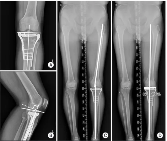
The medial tibial plateau angle (mTPA), the proximal posterior tibial angle (PPTA), the femoral tibial angle (FTA), and the me
chanical axis deviation (MAD) were also evaluated (Fig. 3). The MAD was measured by assessing the location of the mechanical axis crossing through the articular surface of the tibial plateau17). Authors also measured the mTPA and the PPTA of the uninjured limb preoperatively and calculated differences in the mTPA and the PPTA between the postoperative radiograph and the pre
A B C
Fig. 2. Radiographs of case no. 19 presented in Table 1. (A) A 60yearold male patient was injured in a pedestrian traffic accident and suffered a tibial plateau fracture (Schatzker type VI) as well as a fracture of the fibula. (B) A temporary external fixator was immediately applied after injury. (C) Six days after the first operation, dual plating using medial and lateral approaches was applied.
0%
0% 100%100%
A
B C D
Fig. 3. Radiological evaluation. (A) The me
dial tibial plateau angle was measured be
tween the axis of the articular surface of the tibial plateau and the anatomical axis of the proximal tibia on the anteroposterior view of the knee. (B) The proximal posterior tibi
al angle was measured between the articular surface of the medial tibial plateau and the perpendicular line to the anterior cortical margin of the proximal tibia on the lateral view of the knee. (C) The femoral tibial an
gle was measured between the anatomical axes of the femur and tibia. Genu valgum was given a positive angle. (D) Mechanical axis and deviation of the mechanical axis.
The mechanical axis was defined as a line connecting the center of the hip and the center of the ankle. Mechanical axis devia
tion was measured by assessing the location of the mechanical axis crossing through the articular surface of the tibial plateau.
operative radiograph of the uninjured limb. Clinical results and radiological results were evaluated with regard to the correlation between the results. Furthermore, correlation between preopera
tive prognostic factors and clinical outcomes were evaluated.
The means and ranges for all continuous variables were ob
tained with IBM SPSS ver. 23.0 (IBM Co., Armonk, NY, USA).
MannWhitney Utest, Spearman correlation analysis, Kruskal
Wallis test, and Fisher exact test were used. A pvalue <0.05 was considered to be statistically significant.
Results
Demographic characteristics of 24 patients are presented in Table 2. At the final followup, the mean AKSS was 85.3±6.2
(range, 68 to 93), the WOMAC was 11.2±6.2 (range, 1.0 to 21.3), and the ROM was 123.4°±10.0° (range, 101° to 142°). The bone union time at the final followup was 16.5±4.6 weeks (range, 10.9 to 26.1 weeks). In case of metal failure (case no. 10), bone union time was measured from revision surgery (Table 2).
The mean mTPA at the final followup was 88.3°±1.9° (range, 83.3° to 91.3°) and the PPTA was 8.4°±5.9° (range, 0.8° to 22.1°).
The mean FTA at the final followup was 4.53°±1.9° (range, –3.3° to 10.6°) and the MAD was 44.9%±17.5% (range, 9.6% to 70.6%). Compared with the uninjured limb, the mean difference of mTPA (DmTPA) was 1.5°±1.1° (range, 0° to 4.6°) and that of PPTA (DPPTA) was 4.0°±2.8° (range, 0.1° to 10.7°). The mean difference of FTA (DFTA) was 3.3°±2.0° (range, 0.5° to 8.0°), and that of MAD (DMAD) was 12.3%±10.4% (range, 0.1% to
Table 2. Clinical and Radiological Results of the Patients
Case AKSS WOMAC ROM
(°) Bone union
time (wk) mTPA
(°) PPTA
(°) DmTPA
(°) DPPTA
(°) FTA
(°) MAD
(%) DFTA
(°) DMAD
(%)
1 88 5.2 123 8.4 87.7 3.3 1.6 7.9 6.3 50.0 2.5 16.6
2 78 13.5 120 11.4 88.0 13.7 1.7 4.0 3.5 36.5 0.5 1.4
3 81 21.3 142 14.4 89.7 4.2 3.2 4.2 5.9 50.9 4.8 25.0
4 93 17.7 130 18.9 89.5 22.1 0.1 2.7 –3.3 10.1 2.9 4.9
5 78 17.7 130 20.2 87.7 15.4 0.3 4.3 5.6 58.0 1.8 16.1
6 83 6.2 135 18.6 91.3 4.9 1.8 9.5 2.6 37.1 2.9 18.7
7 90 12.5 114 11.4 86.8 5.1 1.4 1.3 3.9 19.8 2.9 3.7
8 91 9.3 125 23.3 89.6 5.5 1.4 2.0 3.5 56.2 1.1 4.1
9 81 8.3 130 13.6 88.2 5.8 0.7 9.7 6.0 41.2 1.4 0.1
10 93 5.2 115 22.9 84.2 1.2 4.6 0.1 3.1 19.8 8.0 15.9
11 93 3.1 115 15.3 89.9 6.8 1.4 2.2 1.8 60.3 4.7 4.0
12 68 21.3 133 14.4 91.0 19.8 1.3 5.0 6.0 69.5 0.7 15.0
13 83 9.3 115 15.6 88.2 7.8 0.0 4.9 9.9 60.9 7.8 39.5
14 81 9.3 122 26.1 90.5 10.2 1.8 0.8 10.6 70.6 6.0 27.3
15 91 7.2 125 16.3 88.6 3.5 0.1 0.7 5.4 45.5 1.6 5.0
16 83 17.7 110 19.3 88.2 6.4 1.3 1.4 1.0 23.5 3.0 3.6
17 80 21.3 114 11.7 88.7 0.8 1.2 10.7 –3.1 9.6 2.5 18.8
18 88 7.2 120 24.7 88.8 4.7 1.1 1.9 10.3 61.0 3.0 2.5
19 88 10.4 130 13.9 86.4 7.9 0.8 3.3 5.0 46.2 1.8 0.4
20 88 5.2 130 13.1 83.3 6.0 2.1 2.5 1.7 47.5 3.1 7.8
21 78 21.3 110 19.3 86.6 17.4 2.9 4.0 4.3 46.4 2.9 17.0
22 91 1 135 10.9 90.0 17.9 3.1 4.5 9.9 59.7 5.6 25.0
23 90 10.4 137.2 15.1 89.1 5.3 1.7 3.9 8.4 62.2 3.1 22.2
24 89 7.2 101 17.3 86.3 5.8 0.9 3.9 –1.4 36.0 5.2 0.3
AKSS: American Knee Society score, WOMAC: Western Ontario and McMaster Universities Osteoarthritis index, ROM: range of motion (flexion/
extension arc), mTPA: medial tibial plateau angle, PPTA: proximal posterior tibial angle, DmTPA: mean difference of mTPA, DPPTA: mean difference of PPTA, FTA: femoral tibial angle, MAD: deviation of the mechanical axis, DFTA: mean difference of FTA, DMAD: mean difference of MAD.
39.5%) (Table 2).
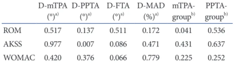
The DPPTA and the AKSS demonstrated negative correlation, which was statistically significant (p=0.007; Spearman correla
tion coefficient, –0.538). As in previous studies on comparison between the normal and abnormal groups18,19), patients with nor
mal mTPA showed better ROM than those with abnormal mTPA (p=0.041). However, other radiological outcomes were not in correlation with the clinical outcomes (Table 3).
Although it was not statistically significant, when the bicondy
lar fracture had open wounds, the ROM was worse (p=0.060).
Other prognostic factors, for example, type of fracture, preopera
tive arthritic changes, compartment syndrome, and clinical out
comes showed no strong correlation (Table 4). Unlike in previous publications, the incidence of compartment syndrome in open fractures (25%) was relatively high; however, no statistically sig
nificant correlation was found between open fracture and com
partment syndrome (p=0.388). By contrast, the incidence among closed fractures (50%) was typical (Fig. 4).
There were two cases of complications. Case no. 10 (Table 1) required revision surgery due to metal failure 1 month after first fixation even though the patient did not start early weight bear
ing. Case no. 12 (Table 1) had total knee arthroplasty due to post
operative arthritis 2 years after first fixation. No other incidences of complications such as infection, knee joint stiffness, and mal
union were seen.
Discussion
TPFs are generally caused by high energy trauma such as traffic accident or falling down. This intraarticular fracture is divided into many subtypes according to mechanisms of injury20). Re
duction strategy and prognosis vary according to fracture types such as simple or complex. Nevertheless, the main goals of treat
ment of bicondylar TPFs are to recover the articular surface and alignment of the lower extremity and to maintain the length of legs21,22).
There are several fixation methods of bicondylar TPFs such as conventional ORIF, hybrid external fixation, and staged treat
ment using a temporary external fixator. Lee et al.23) reported a series of 45 bicondylar TPFs in 45 patients using conventional dual plating. The mean WOMAC was 34.1±4.91 (range, 0 to worst 96), and one case of infection and two cases of nonunion were noted. Chae et al.24) also reported a series of 12 Schatzker type VI TPFs in 11 patients using conventional dual plating. The mean AKSS was 85.0±8.6, and there was one case of joint stiff
ness and one case of varus malalignment.
Stamer et al.11) reported a series of 22 patients with Schatzker
Open Closed
0 18 16 14 12 10 8 6 4 2
No.ofpatients
p=0.388a) Non-compartment syndrome
Compartment syndrome
Fig. 4. Correlation between open fracture and compartment syndrome.
a)Fisher exact test.
Table 3. Correlation between Clinical and Radiological Results DmTPA
(°)a) DPPTA (°)a) DFTA
(°)a) DMAD (%)a) mTPA
groupb) PPTA
groupb)
ROM 0.517 0.137 0.511 0.172 0.041 0.536
AKSS 0.977 0.007 0.086 0.471 0.431 0.637
WOMAC 0.420 0.376 0.066 0.779 0.225 0.252
DmTPA: mean difference of medial tibial plateau angle, DPPTA: mean difference of proximal posterior tibial angle, DFTA: mean difference of femoral tibial angle, DMAD: mean difference of deviation of the mechanical axis, mTPAgroup: group difference between normal mTPA group and abnormal mTPA group, PPTAgroup: group difference between normal PPTA group and abnormal PPTA group, ROM: range of motion (flexion/extension arc), AKSS: American knee society score, WOMAC:
Western Ontario and McMaster Universities Osteoarthritis index.
a)Spearman correlation analysis.
b)MannWhitney Utest.
Table 4. Correlation between Clinical Results and Prognostic Factors
Variable ROM AKSS WOMAC
Schatzker typea) 0.881 0.631 0.810
AO classificationb) 0.596 0.592 0.367
Preoperative OA changea) 0.536 0.637 0.289
Opena) 0.060 0.644 0.601
Compartment syndromea) 0.637 0.118 0.601
ROM: range of motion (flexion/extension arc), AKSS: American Knee Society score, WOMAC: Western Ontario and McMaster Universities Osteoarthritis index, AO: the AO Foundation and Orthopaedic Trauma Association classification, OA: osteoarthritis.
a)MannWhitney Utest.
b)KruskalWallis test.
type IV TPFs treated with a hybrid ring external fixator using tensioned wires proximally and halfpins distally. The average AKSS was 84.7, and there was one case of pin tract infection, three cases of deep infection, and one case of malunion. Babis et al.12) also described 33 cases of bicondylar TPFs, which were treated by minimal intervention and hybrid external fixation. Ac
cording to AKSS criteria25), the results were evaluated as excellent in 18 patients (55%), good in 10 patients (30%), fair in 4 patients (12%), and poor in 1 patient (3%).
Egol et al.14) described staged management of highenergy prox
imal TPFs. The mean WOMAC was 95±55 (range, 0 to worst 240), the mean ROM was 106°±15°, and there were two cases of infection. Many other authors have also reported good clinical outcomes of dual plating using medial and lateral approaches af
ter temporary external fixation1315). Our study also demonstrated favorable clinical and radiological outcomes with staged treat
ment using a temporary external fixator. According to Chang et al.26), compartment syndrome can occur in 30% of bicondylar TPFs. When compartment syndrome is suspected, emergent fas
ciotomy is essential and subsequent temporary fixation is recom
mended.
To our knowledge, there was no published report of compari
son with uninjured limbs in TPFs. In our study, although not all radiological outcomes were statistically correlated to clinical outcomes, it was observed that patients with fewer differences with uninjured limbs on plain radiographs showed a tendency to have better clinical outcomes. In particular, the correlation was statistically significant for the PPTA. Some authors have reported that the reduction status on plain radiographs can affect clinical outcomes27). In our study, it was observed that when mTPA and PPTA were within normal range, the clinical outcomes were bet
ter, and especially, mTPA showed statistical significance. Authors propose that all patients should be evaluated not only for the in
jured limb but also for the uninjured limb to have better clinical outcome by referring to the angles intraoperatively. We did make an effort not only to recover mTPA and PPTA within normal limits using fluoroscopy intraoperatively but also to refer to the data of the uninjured limb in all patients.
According to Egol et al.14), there was a significant association between the presence of external wounds and the need for a sec
ondary surgery because of complications. In our study, although it was not statistically significant, clinical outcomes were not good in the presence of external wounds. Therefore, it is proposed that surgeons should warn the patients adequately about the possibil
ity of worsening of clinical outcomes. Other prognostic factors such as the type of fracture, preoperative arthritic change, and
compartment syndrome were not statistically correlated to the clinical outcomes. Although compartment syndrome occurs fre
quently in closed fractures, two patients (25%) of open fractures were accompanied by compartment syndrome in this study. Ac
cordingly, primary physicians should do careful physical exami
nation in cases of bicondylar TPFs.
Conclusions
Staged treatment using a temporary external fixator in bicon
dylar TPFs showed good clinical and radiological outcomes due to appropriate soft tissue management. Furthermore, excellent results could be obtained by radiological evaluation of not only the injured limb but also the uninjured limb. Moreover, it is im
portant to warn bicondylar TPF patients with external wounds about the risk of worsening of clinical outcomes before surgery.
Conflict of Interest
No potential conflict of interest relevant to this article was re
ported.
References
1. Whiteside LA, Lesker PA. The effects of extraperiosteal and subperiosteal dissection: II. On fracture healing. J Bone Joint Surg Am. 1978;60:2630.
2. Lachiewicz PF, Funcik T. Factors influencing the results of open reduction and internal fixation of tibial plateau frac
tures. Clin Orthop Relat Res. 1990;(259):2105.
3. Littenberg B, Weinstein LP, McCarren M, Mead T, Swiont
kowski MF, Rudicel SA, Heck D. Closed fractures of the tibial shaft: a metaanalysis of three methods of treatment. J Bone Joint Surg Am. 1998;80:17483.
4. Apley AG. Fractures of the tibial plateau. Orthop Clin North Am. 1979;10:6174.
5. Hohl M. Tibial condylar fractures. J Bone Joint Surg Am.
1967;49:145567.
6. Ibsen JG, Mossing N. Conservative treatment of tibial con
dylar fractures. Acta Orthop Scand. 1971;42:4312.
7. Sarmiento A, Kinman PB, Latta LL, Eng P. Fracutres of the proximal tibia and tibial condyles: a clinical and laboratory comparative study. Clin Orthop Relat Res. 1979;(145):136
45.
8. Lin S, Mauffrey C, Hammerberg EM, Stahel PF, Hak DJ.
Surgical site infection after open reduction and internal fixa
tion of tibial plateau fractures. Eur J Orthop Surg Traumatol.
2014;24:797803.
9. Moore TM, Patzakis MJ, Harvey JP. Tibial plateau fractures:
definition, demographics, treatment rationale, and long
term results of closed traction management or operative reduction. J Orthop Trauma. 1987;1:97119.
10. Young MJ, Barrack RL. Complications of internal fixation of tibial plateau fractures. Orthop Rev. 1994;23:14954.
11. Stamer DT, Schenk R, Staggers B, Aurori K, Aurori B, Beh
rens FF. Bicondylar tibial plateau fractures treated with a hybrid ring external fixator: a preliminary study. J Orthop Trauma. 1994;8:45561.
12. Babis GC, Evangelopoulos DS, Kontovazenitis P, Nikolo
poulos K, Soucacos PN. High energy tibial plateau fractures treated with hybrid external fixation. J Orthop Surg Res.
2011;6:35.
13. Anglen JO, Aleto T. Temporary transarticular external fixa
tion of the knee and ankle. J Orthop Trauma. 1998;12:4314.
14. Egol KA, Tejwani NC, Capla EL, Wolinsky PL, Koval KJ.
Staged management of highenergy proximal tibia fractures (OTA types 41): the results of a prospective, standardized protocol. J Orthop Trauma. 2005;19:44855.
15. Haidukewych GJ. Temporary external fixation for the man
agement of complex intra and periarticular fractures of the lower extremity. J Orthop Trauma. 2002;16:67885.
16. Sirkin M, Sanders R, DiPasquale T, Herscovici D Jr. A staged protocol for soft tissue management in the treatment of complex pilon fractures. J Orthop Trauma. 1999;13:7884.
17. Honkonen SE. Indications for surgical treatment of tibial condyle fractures. Clin Orthop Relat Res. 1994;(302):199
205.
18. Chao EY, Neluheni EV, Hsu RW, Paley D. Biomechanics of malalignment. Orthop Clin North Am. 1994;25:37986.
19. Paley D, Herzenberg JE, Tetsworth K, McKie J, Bhave A.
Deformity planning for frontal and sagittal plane corrective osteotomies. Orthop Clin North Am. 1994;25:42565.
20. Schatzker J. Anterior approach to the knee with osteotomy of the tibial tubercle for bicondylar tibial fractures. J Bone Joint Surg Am. 1988;70:15756.
21. Marsh JL, Slongo TF, Agel J, Broderick JS, Creevey W, De
Coster TA, Prokuski L, Sirkin MS, Ziran B, Henley B, Audige L. Fracture and dislocation classification compendium: 2007:
Orthopaedic Trauma Association classification, database and outcomes committee. J Orthop Trauma. 2007;21(10 Suppl):S1133.
22. Tejwani NC, Hak DJ, Finkemeier CG, Wolinsky PR. High
energy proximal tibial fractures: treatment options and deci
sion making. Instr Course Lect. 2006;55:36779.
23. Lee MH, Hsu CJ, Lin KC, Renn JH. Comparison of outcome of unilateral locking plate and dual plating in the treatment of bicondylar tibial plateau fractures. J Orthop Surg Res.
2014;9:62.
24. Chae IJ, Park SW, Lee SH, Noh W, Kim HJ, Hahn SB. Treat
ment of Shatzker type VI tibia plateau fracture using lateral and posteromedial dual incision approach and dual plating.
J Korean Fract Soc. 2009;22:2528.
25. Insall JN, Dorr LD, Scott RD, Scott WN. Rationale of the Knee Society clinical rating system. Clin Orthop Relat Res.
1989;(248):134.
26. Chang YH, Tu YK, Yeh WL, Hsu RW. Tibial plateau fracture with compartment syndrome: a complication of higher inci
dence in Taiwan. Chang Gung Med J. 2000;23:14955.
27. Bhattacharyya T, McCarty LP 3rd, Harris MB, Morrison SM, Wixted JJ, Vrahas MS, Smith RM. The posterior shearing tibial plateau fracture: treatment and results via a posterior approach. J Orthop Trauma. 2005;19:30510.