생강나무 추출물의 항산화 활성과 미백효과
방채영·원은경·박권우*·이광원**·정세영#
경희대학교 약학대학 위생약학 및 독성학실, *고려대학교 생명과학대학 채소 및 허브학실,
**고려대학교 생명과학대학 식품생화학 및 독성학실
(Received June 11, 2008; Revised September 27, 2008)
Antioxidant Activities and Whitening Effect from Lindera obtusiloba BL. Extract
Chae Young Bang, Eun Kyung Won, Kuen Woo Park*, Gwang Won Lee** and Se Young Choung#
Department of Hygienic Chemistry, College of Pharmacy, Kyung Hee University, Seoul 130-701, Korea
*Divisions of Bioscience and Technology, Korea University, Seoul 136-701, Korea
**Department of Food Science, College of Life Sciences and Biotechnology, Korea University, Seoul 136-701, Korea
Abstract
— In this study we investigated antioxidant activity of against several free radicals and skin whitening effect of 70% ethanol extract (leaf extracts and branch/stem mixed) of Lindera obtusiloba BL. Antioxidant activity was assessed by DPPH, superoxide radical and hydroxyl radical assays. The Lindera obtusiloba BL. extract had antioxidant activity dose dependently with an IC
50value of 243.14 and 181.10
µg/ml for DPPH, 165.77 and >1500
µg/ml for non-enzymatic system of superoxide radical assay, 35.47 and >100
µg/ml for enzymatic system of superoxide radical assay, 1.21 mg/ml for hydroxyl radical assay. In addition we tested tyrosinase inhibition activity and melanin contents on B16 melanoma F10. B16 melanoma cell was treated by such sample as 1, 5, 10 and 50
µg/ml for 72 hr and tyrosinase inhibition was tested. Melanogenesis was inhibited to 22% at the dose of 50
µg/ml and tyrosinase was inhibited to 45.2% at the same dose. In conclusion Lindera obtusiloba BL had potent antioxidant activity and inhibitory activity of tyrosinase and melanin formation. It could be devel- oped as the health functional food and functional cosmetic resources.
Keywords □
Lindera obtusiloba BL., antioxidant, DPPH, hydroxyl radical, superoxide radical, whitening effect
외부로부터의자극이나산업화에따른환경오염
,
잘못된식습 관등다양한스트레스로인하여인체내에서높은활성산소(reactive oxygen species: ROS)
를발생시킨다.
활성산소에는superoxide radical(O
2.-), hydrogen peroxide(H
2O
2), hydroxyl radical(OH
-), singlet oxygen(
1O
2)
등이있으며대사과정중의부생성물로서발생되고
,
1,2)세포구성성분을비가역적으로파괴한 다고알려져있다.
그러나식물은이러한활성산소에대한방어 기작으로이를제거할수있는tocopherol, ascorbic acid, catechine,
glutathione
등과같은다양한형태의천연항산화제를함유하고있으며
,
3-6)특히,
항산화효소의발현은중요한역할을한다.
7)항 산화효소는superoxide dismutase, catalase, glutathione per- oxidase
등이있는데superxoide dismutase(SOD)
는superoxide
anion radical(O
2-)
을제거하여hydrogen peroxide(H
2O
2)
로전환 시키는촉매효소이고, catalase(CAT)
는주로peroxisom
에존재하며
H
2O
2를물과산소로분해한다.
8)이러한천연항산화제나항산화효소들은독자적인작용이나다른작용을보조하는과 정을통해활성산소로부터생체를보호하는작용을한다
.
활성산소에대한생체방어력에이상이생기거나과도한활성산소에 노출될경우
,
활성산소가인체세포구성성분인지질,
단백질, DNA
등의비선택적
,
비가역적인파괴가유도됨으로써노화는물론암을비롯하여뇌졸중
,
파키슨씨병과같은각종질병을일으키는것으로알려져있다
.
9-12)위에서언급한일반적인노화현상과같이광노화과정에서도 활성산소는중요한원인인자이다
.
광노화의원인이되는요소인 자외선이활성산소의증가를유도하기때문이다.
자외선차단제를사용했을때피부에서
ROS
를감소시킬수있다는보고와자외선의조사량을증가시키면서활성산소생성량을측정한결 과조사량이증가할수록활성산소의생성이증가되었다는보고
#본논문에관한문의는저자에게로
(
전화) 02-961-0372 (
팩스) 02-961-0372
(E-mail) [email protected]
가13,14)자외선과
ROS
생성증가의상관관계를증명하고있다.
자외선에의해증가된활성산소는표피세포에장해를주고또한 표피세포에서의염증성
cytokine
인interleukin(IL)-1
α,
β와IL-6, TNF-
α등의분비를촉진시켜서15,17)melanocyte
에서keratinocyte
로의
melanosome
이송의증가18)와melanocyte
에서의melanin
생성증가
,
19,20)등피부착색과진피에서fibroblast
의collagen
합 성저해21)등주름생성을일으킴으로써광노화의중요한원인이 되고있다.
이와같은산화적스트레스가노화를비롯한각종질병의중 요한원인으로밝혀지면서활성산소를제거하는항산화제에대 한관심이증가하고있다
.
또한생체내활성산소를조절하고항 산화활성이우수하면서보다안전한천연물유래의항산화제의 개발이절실히요구되고있다.
본실험에서사용된생강나무
(
Lindera obtusilobaBL.)
는녹나 무과식물로꽃은3
월에잎보다먼저핀다.
노란색의작은꽃들 이여러개뭉쳐꽃대없이산형꽃차례를이루어달리며그모 양이산수유꽃과비슷하다.
새로잘라낸가지에서알싸한생강 냄새가난다고하여생강나무라하며한국,
일본,
중국등지에분포하고주로산지의계곡이나숲속의냇가에서서식한다
.
연한잎은먹을수있고
,
꽃은관상용이며,
열매는기름을짜서먹을 수있으며머리기름으로22)쓰인다.
한방에서는나무껍질을삼첩풍이라고부르고있다
.
약재로사용하는데타박상에의한어혈과산후에몸이붓고팔다리가아픈증세에효과가있다
.
생강 나무는geranyl acetate, linderic acid, tsudzuic acid,
그리고L- phellandrene
를함유하고23)있는데이들성분들은간보호,
항당뇨효과그리고항산화활성이있다고알려져있다
.
본연구에서는예로부터약용및식용으로사용되는생강나무 를새로운가능성소재로활용하기위해잎추출물과가지
/
줄기혼합추출물에서항산화활성과미백효과를측정하였다
.
재료 및 방법
실험재료
본실험에서사용한생강나무는충청남도천안에서채집하였 다
,
잎과가지줄기부분만을따로분리,
세척하고물기를제거한 다음건조시켜각각의재료100 g
을분쇄하여70% ethanol
을넣고환류하면서
80
oC
에서4
시간씩3
회반복추출하였다.
이를 감압농축,
동결건조하여그분말을실험에사용하였다.
잎의수 득율은26.8%,
가지줄기혼합추출물의수득률은17.8%
였다.
세포및시약
실험에사용한
B16F10
은마우스의melanoma
세포주로미국ATCC
에서 구입하였다. Cell culture
에DMEM(Dulbecco's modified Eagle's medium), 10% FBS, 1% Penicillin-Streptomycin
은
Hyclone Laboratories Inc. USA
를 사용하였고, DPPH(2,2- diphenyl-1-picrylhydrazyl), ascorbic acid, xanthine oxidase, NADH, Phenazine methosulfate, NBT(nitroblue tetrazolium), N,N-dimethyl-p-nitrosoaniline, tyrosinase
는Sigma chemical Co. USA
를사용하였다.
실험 방법
생강나무잎과가지
/
줄기추출물의항산화활성DPPH radical
소거활성에의한항산화효능검색 − 각농도별로조제한생강나무잎추출물
5, 10 50, 100, 500, 1000
µg/
m
l의용액과가지·줄기혼합추출물10, 50, 100, 500
µg/m
l의용액
20
µl와70% ethanol 100
µl, 95% ethanol
에 녹인0.15 mM DPPH(2,2-diphenyl-1-picrylhydrazyl)
용액80
µl를 넣고37
oC
에서30
분동안방치시킨후517 nm
에서흡광도를측정하 였다.
대조 약물로서는ascorbic acid(vitamin C) 0.5, 1.0, 5.0,
10, 50
µg/m
l을사용하였으며결과는시료를처리하지않은군에대한
percentage(%)
로표기하였다.
Superoxide radical(Enzymatic system : xanthine- xanthine oxidase)
소거활성에 의한 항산화 효능 검색 −Xanthine-xanthine oxidase
는superoxide radical(O
2.-)
을발생시키는대표적인실험계이다
.
시험관에55 mM Potassium phosphate buffer(pH 7.5) 1.8 m
l, 0.15 mM xanthine 1 m
l과각농도별로 조제한생강나무잎추출물1.0, 5.0, 10, 50, 100
µg/m
l과가지·줄기 혼합 추출물
1, 10, 100
µg/m
l의 용액0.1 m
l, xanthine oxidase 0.1 m
l를혼합하고실온에서20
분방치시킨후1 N HCl
1 m
l을가하여반응을정지시키고290 nm
에서흡광도를측정하였다
.
대조약물로는xanthine oxidase
억제작용이있는약물로 널리알려진allopurinol 0.68, 1.36, 6.8, 13.36, 136
µg/m
l을사 용하였으며결과는시료를처리하지않은군에대한percentage (%)
로표기하였다.
Superoxide radical(Non-enzymatic system : NADH- PMS)
소거활성에의한항산화효능검색 −24 well plate
에각농도 별로 조제한생강나무 잎추출물
1.0, 5.0, 10, 50, 100, 500
µg/m
l과가지/
줄기혼합추출물1.0, 10, 100, 1000
µg/m
l용 액20
µl에30 mM Tris-HCl buffer(pH 8.0) 100
µl와100
µM PMS(Phenazine methosulfate) 20
µl, 0.5 mM NADH 40
µl, 0.5 mM NBT 20
µl를혼합하여37
oC
에서20
분동안방치한후560 nm
에서흡광도를측정하였다.
대조약물로서는ascorbic acid 0.01, 0.1, 1.0, 10, 100, 1000
µg/m
l를사용하였으며결과는시료 를처리하지않은군에대한percentage(%)
로표기하였다.
Hydroxyl radical
소거활성에의한항산화효능검색 − 시험관에
95% ethanol
에 녹인2.5 mM
β-carotene 200
µl에5.94
mM H
2O
2, 800
µl와26.4 mM FeSO
4의농도가되게녹인정제수
800
µl,
각농도별로조제한생강나무잎추출물0.5, 1.0, 1.1, 1.2, 1.3, 1.4, 1.5 mg/m
l과가지줄기혼합추출물0.5, 1.0, 1.5 mg/m
l용액200
µl를혼합한후곧바로536 nm
에서흡광도 를 측정하였다.
대조 약물로서는mannitol 0.0018, 0.0091, 0.0182, 0.091, 0.182, 0.91, 1.82 mg/m
l을사용하였으며결과는시료를처리하지않은군에대한
percentage(%)
로표기하였다.
생강나무잎추출물의미백효과
B16 melanoma F10 cell culture
−B16 melanoma F10 cell
은10% fetal bovine serum
과penicillin 100 IU/m
l, strep- tomysin 50
µg/m
l을함유한Dulbecco's modified Eagle's medium (DMEM)
용액에서37
oC, 0.5% CO
2조건의CO
2incubator
에서배양하였다
.
Cell viability
−B16 melanoma F10 cell
을24 well
에well
당
10
4cell
씩분주하였다. 17
시간후cell
의부착을확인한후배 지를걷어내고각농도별로배지에녹여조제한생강나무잎추 출물1.0, 5.0, 10, 50, 100
µg/m
l을각well
에1 m
l씩분주하였 다. 72
시간동안CO
2incubator
에서배양한후MTT
용액을넣고
,
다시4
시간동안CO
2incubator
에서배양하였다.
배지를제거하고
, DMSO
로cell
을녹여560 nm
에서흡광도를측정하였다.
결과는시료를처리하지않은군에대한
percentage(%)
로표기하였다
.
Tyrosinase activity inhibition
−B16 melanoma F10 cell
을
24 well
에well
당10
4cell
씩분주하였다. 17
시간후cell
의부착을확인한후배지를걷어내고각농도별로배지에녹여조 제한생강나무잎 추출물
1.0, 5.0, 10, 50
µg/m
l을각well
에1 m
l씩분주하였다. 72
시간동안CO
2incubator
에서배양한후배지를걷어내고
PBS 2 m
l로2
회세척하였다.
세척후1 M tris- HCl
이 포함된lysis buffer
를 이용하여cell
을lysis
시키고×
10,000 g
에서5
분동안원심분리하였다.
상층액의protein
량을측정하고 농도는
lysis buffer
로 맞춰준다.
상층액90
µl와10 mM dihydroxyphenylalanine(DOPA)
용액10
µl를섞어37
oC
에서
20
분동안방치시킨후470nm
에서흡광도를측정하였다.
24,25)Total melanin content
−B16 melanoma F10 cell
을24 well
에well
당10
4cell
씩분주하였다. 17
시간후cell
의부착을 확인한후배지를걷어내고각농도별로배지에녹여조제한생 강나무잎추출물1.0, 5.0, 10, 50
µg/m
l을각well
에1 m
l씩분 주하였다. 72
시간동안CO
2incubator
에서배양한후배지를걷 어내고PBS 2 m
l로2
회세척하였다.
세척후1 N NaOH
를well
당
500
µl씩넣고37
oC
에서20
분간방치한후tube
에옮겨99
oC
에서
30
분간세포를파괴하였다.
이용액을96 well
에200
µl씩분주한후
470 nm
에서흡광도를측정하였다.
표준용액을제조하여
470 nm
에서의흡광도를이용하여standard curve
를구하고 이로부터농도를계산하였다.
자료분석및통계처리 − 모든실험결과는평균±표준편차로표 기하였고
,
통계적유의성은Student's
t-test
로분석하였으며,
p값이
0.01
미만일때통계적으로유의하다고판단하였다.
실험결과 및 고찰
생강나무잎과가지
/
줄기추출물의항산화활성DPPH radical
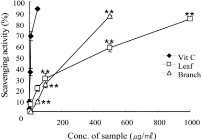
소거활성에의한항산화효능검색 − 생강나무여러부위별
(
잎,
가지/
줄기)
추출물의DPPH radical
소거능은잎 의경우5.0~1000
µg/m
l에서농도의존적인경향을나타냈으며,
가지
/
줄기혼합추출물의경우0.5~500
µg/m
l에서농도의존적 인경향을나타냈다.
잎추출물1000
µg/m
l(84%),
가지/
줄기추출물
500
µg/m
l(87%)
에서나타나는소거능은,
생강나무추출물이가지는고유의짙은색으로인해높은농도에서의흡광도값 이일정하지못하여항산화검색에간섭을최소화할수있는농 도범위에서검색한결과이다
(Fig. 1). IC
50은Vit C
는7.47
µg/
m
l,
잎은243.14
µg/m
l이며,
가지/
줄기는181.10
µg/m
l이다. Superoxide radical(Enzymatic system : xanthine- xanthine oxidase)
소거활성에의한항산화효능검색 − 효소적superoxide radical
생성계인xanthine-xanthine
계에서의super- oxide radical
소거능은Fig. 2
에서 보는 바와같이잎의경우50
µg/m
l에서농도의존적으로소거되는경향을나타냈으며,
가지
/
줄기혼합추출물의경우10
µg/m
l에서농도의존적으로소거 되는경향을보였다. IC
50은allopurinol
은68
µg/m
l,
잎은35.47
µ
g/m
l이며,
가지/
줄기는>100
µg/m
l이었다.
Superoxide radical(Non-enzymatic system : NADH- PMS)
소거활성에의한항산화효능검색 − 비효소적superoxide radical
생성계인NADH/PMS
에서의superoxide radical
소거능은잎의경우
1~500
µg/m
l에서농도의존적으로소거되는경향Fig. 1 −
DPPH radical scavenging activity of Lindera obtusiloba BL.
extract. Values are mean±SD (n=3), **p<0.01 vs positive
CTL.
을나타냈으며
,
가지/
줄기혼합추출물의경우1~1,500
µg/m
l에서농도의존적으로소거되는경향을보였다
(Fig. 3). IC
50은Vit C
는3.42
µg/m
l,
잎은165.77
µg/m
l이며,
가지/
줄기는>1500
µg/
m
l이다.
Hydroxyl radical
소거활성에의한항산화효능검색 − 생강나무잎추출물의
hydroxyl radical
소거능은생강나무잎추출물에서만확인할수있었다
. Fig. 4
에서보는바와같이잎추출물
1.0~1.5 mg/m
l범위에서농도의존적으로소거하였으며,
그 효과는잎추출물의IC
50수치가1.21 mg/m
l로hydroxyl radical
소거제인
mannitol
의IC
50수치0.159 mg/m
l과비교했을때10
배정도의활성산소소거능차이를가짐을알수있었다
.
생강나무잎추출물의미백효과
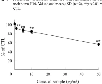
Cell viability
− 세포에서의미백효과확인은위의항산화활성결과를바탕으로잎추출물에서만확인하였다
.
생강나무추 출물의세포독성은잎추출물의경우100
µg/m
l의농도이상에서독성이있음을확인하였다
(Fig. 5).
이후의tyrosinase activity
Fig. 2 −
Superoxide radical scavenging activity (enzymatic system) of Lindera obtusiloba BL. extract. Values are mean±SD (n=3), **p<0.01 vs positive CTL.
Fig. 3
- Superoxide radical scavenging activity (nonenzymatic system) of Lindera obtusiloba BL. extract. Values are mean±SD (n=3), **p<0.01 vs positive CTL.
Fig. 6 −
Tyrosinase inhibition activity of Lindera obtusiloba BL. leaf extract on B16 melanoma F10.
Fig. 4 −
Hydroxyl radical scavenging activity of Lindera obtusiloba BL. extract. Values are mean±SD (n=3), **p<0.01 vs positive CTL.
Fig. 5 −
Cytotoxicity of Lindera obtusiloba BL. leaf extract on B16
melanoma F10. Values are mean±SD (n=3), **p<0.01 vs
CTL.
와
melanin content
측정실험은독성이나타나지않은1, 5, 10, 50
µg/m
l의농도범위안에서실시하였다.
Tyrosinase activity inhibition
− 세포독성이나타나지않는1, 5, 10, 50
µg/m
l농도범위에서tyrosinase
활성저해율은각 각9.8, 14.3, 17.3, 45.2%
를보였다(Fig. 6).
이로부터생강나무잎추출물의
melanin
생성억제가능성이있음을확인할수있었다.
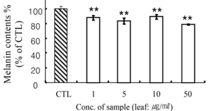
Melanin synthesis inhibition
−B16 melanoma F10
의total melanin content
는잎추출물농도50
µg/m
l에서22%
감소하였다
(Fig. 7).
이는Fig. 5
의결과와종합하여볼때세포의증식을 억제하지않는범위에서세포내total melanin content
가22%
감소한것으로생강나무의미백능을확인할수있다
.
결 론
생강나무잎추출물과가지
/
줄기추출물의항산화효과는가지
/
줄기추출물보다는잎추출물에서높은항산화활성을관찰 할수있었다.
이러한결과를바탕으로세포에서잎추출물을농도별로처리하여
tyrosinase activity
와melanin content
를본 결과미백효과가있음을검증할수있었다.
생강나무추출물특 히잎추출물은항산화능력에비해미백효과가더강하게나타 나새로운미백소재로서개발가능성이충분할것으로확인할 수있었다.
감사의 말씀
본연구는농림수산식품부농림기술관리센터농림기술개발사
업과제
(20060411)
의연구비에의해지원되었습니다.
참고문헌
1) Badwey, J. A. and Karnovsky, M. L. : Active oxygen species
and the functions of phagocytic leucocytes. Ann. Intern. Med.
93
, 480 (1980).
2) Babior, B. M. : Phagocytes and oxidative stress. Am. J. Med.
109
, 33 (2000).
3) Namiki, M. O. : Antioxidants/Antimutagens in Food. Crit. Rev.
Food Sci. Nutr.
29, 273 (1990).
4) Ames, B. N., Cahcart, R., Schwiers, E. and Hochstein, P. : Uric acid provides an antioxidant defense in himan against oxidant amd radical-caused aging and cancer. Proc. Natl., Acad. Sci.
78, 6858 (1981).
5) Frankel, N. : Antioxidants in lipid food and their on food quality.
Food Chem.
75, 51 (1996).
6) Giese, J. : Antioxidants tools for prevention lipid oxidation.
Food Technol.
5, 73 (1996).
7) Kang, N. J., Kwon, J. G., Lee, H. C., Jeong, H. B. and Kim, H. T. : Antioxidant enzymes as defense mechanism against oxidative stress induced by chilling in cucurbita ficifolia leeaves. J. Kor. Sod. Sci.
44, 605 (2003).
8) Bowler, C., Van Montagu, M. and Inze, D. : Superoxide dismutases and stress tolerance. Annu. Rev. Plant Physiol.
Plant Mol. Biol.
43, 83 (1992).
9) Heinonen, I. M., Meyer, A. S. and Frankel, E .N. : Antioxidant activity of berry phenolic on human low-density lipoprotein and liposome oxidation. J. Agric. Food Chem.
46, 4107 (1998).
10) Rice-Evans, C. A. and Miller, N. J. : Antioxidant activities of flavonoids as bioactive components of food. Biochemical Soc.
Trans.
24, 790 (1996).
11) Satue-Garcia, M. T., Heinonen, I. M. and Frankel, E. N. : Anthocyanins as antioxidants on human low-density lipoprotein and lecithin-liposome system. J. Agric. Food Chem.
45, 3362 (1997).
12) Miquel, J. : An update on the oxygen stress-mitochondrial mutation theory of aging: genetic and evolutionary implications.
Exp. Gerontol.
33, 113 (1998).
13) Yasui, H. and Sakurai, H. : Age-dependent generation of reactive oxygen species in the skin of live hairless rats exposed to UVA light. Exp. Dermatol.
12, 655 (2003).
14) Herrling, T., Fuchs, J., Rehberg, J and Groth, N. : UV-induced free radicals in the skin detected by ESR spectroscopy and imaging using nitroxides. Free Radic. Biol. Med.
35, 59 (2003).
15) Ansel, J. C., Luger, T. A., Lowry, D., Perry, P., Roop, D. R. and Mountz, J. D. : The expression and modulation of IL-1
αin murine keratinocytes. J. Immunol.
140, 2274 (1988).
16) Kirnbaner, R., Kock, A., Neuner, P., Forster, E., Krutmann, J., Urbanski, A., Schauer, E., Ansel, J. C., Schwarz, T. and Luger T. A. : Regulation of epidermal cell interleukin-6 production by UV light and corticosteroids. J. Invest. Dermatol.
96, 484 (1991).
17) Oxholm, A., Oxholm, P., Staberg, B. and Bendtzen, K. :
Fig. 7 −