http://dx.doi.org/10.14405/kjvr.2015.55.4.221
221
<Original Article>
Effects of alfaxalone on echocardiographic examination in healthy dogs
Ye-Won Kim, Tae-Jun Kim, Changbaig Hyun*
Section of Small Animal Internal Medicine, College of Veterinary Medicine, Kangwon National University, Chuncheon 24341, Korea
(Received: August 4, 2015; Revised: October 15, 2015; Accepted: October 16, 2015)
Abstract : This study evaluated the effects of alfaxalone (3 mg/kg, intravenously) on echocardiographic examination in healthy dogs using echocardiography. Six adult Beagle dogs were used for this study. Left ventricular dimensions with systolic indexes, trans-blood flow at all cardiac valvular annulus and trans-mitral tissue Doppler values were measured from routine transthoracic echocardiography. Although the changes were not statistically significant, heart rate, left ventricular end-systolic diameter, left ventricular end-diastolic diameter, peak velocities of tricuspid A-wave and transpulmonic flow were increased after alfaxalone induction, while systolic blood pressure, fractional shortening, left ventricular ejection fraction, peak velocities of mitral E-wave, mitral A wave, tricuspid E-wave, transaortic flow and medial e’-, a’- and s’-peaks decreased after alfaxalone induction. No dogs showed hypoxemia during sedation, regardless of intubation and oxygen supply. Although alfaxalone showed mild cardiovascular depression, this protocol could be a good alternative sedative protocol for echocardiographic examination in healthy dogs because the cardiovascular depression was statistically and clinically insignificant. However, further studies in dogs with heart diseases should be conducted to confirm these findings after alfaxalone induction.
Keywords : alfaxalone, canine, cardiac function, cardiovascular depression, left ventricle
Introduction
Alfaxalone (3 α-hydroxy-5α-pregnane-11,20-dione) is a recently developed neurosteroid injectable anesthetic agent that is gaining popularity for anesthetic induction in dogs and cats [3, 4, 14]. Alfaxalone binds to the gamma amino butyric acid sub-type A receptor in the central nervous system and exerts anesthetic action by modulation of chloride ion trans- port [4, 14]. Alfaxalone has been safely administered con- comitantly with a range of drugs commonly used perio- peratively, in order to improve quality of anesthesia and to increase duration of anesthesia in dogs [1, 2, 5-9]. Further- more, alfaxalone is known to have little or no cardiovascular effects when given at clinical dose rates [15], although pre- medication with α
2-adrenoceptor agonists (e.g., medetomi- dine and dexmedetomidine) can markedly increase the duration of anesthesia in a dose-dependent fashion [2, 10].
Although the pharmacological effect of alfaxalone has been extensively investigated in dogs, studies on its effects on echocardiographic parameters in dogs are still insufficient.
Therefore, in this study, we evaluated the effect of alfaxa- lone on echocardiographic measurement in healthy dogs.
Materials and Methods
Animals
Approval from the animal ethics committee of Kangwon National University was obtained for this experiment prior to the commencement of this study. Six adult Beagle dogs (three males and three females in mean body weight 8.3 ± 3.1 kg and mean age 4.1 ± 1.7 yr) were used for this study.
All dogs were healthy based upon physical examination, an electrocardiogram, serum chemistry, hematologic analyses, and diagnostic imaging studies including thoracic radiogra- phy and echocardiography (data not shown). Systolic arterial blood pressure was measured at right cranial limb between carpal and elbow joints using a Doppler flow detector (811B;
Parks Medical Electronics, USA). Saturation of peripheral oxygen (SpO
2) was monitored using a hand-held pulse oximeter (MP110P; MEK-ICS, Korea) during sedation.
Echocardiographic examination
A complete routine transthoracic echocardiographic exam- ination was performed on each dog with an ultrasound machine (X-300; Siemens, Germany) equipped with a 3.0- 9.0 MHz sector transducer, as described in elsewhere [15].
*Corresponding author
Tel: +82-33-250-8681, Fax: +82-33-244-2367 E-mail: [email protected]
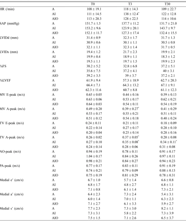
Table 1. Changes of echocardiographic measurement before (T0), 3 min after (T3) and after recovery (T30) of anesthesia with 3 mg/
kg of alfaxalone (A), alfaxalone with intubation (AI), and alfaxalone with intubation supplied with 100% oxygen (AIO) ) in healthy dogs (n = 6)
T0 T3 T30
HR (/min) A 108± 19.1 118± 14.3 109± 22.7
AI 111± 14.5 138± 12.4* 122± 12.8
AIO 113± 20.3 128± 22.5 114± 10.6
SAP (mmHg) A 151.7± 1.5 137.7± 11.2 131.7± 21.0
AI 153.2± 9.6 123.9± 20.1 143.7± 9.7
AIO 152.1± 11.7 127.3± 17.4 132.4± 15.5
LVIDd (mm) A 31.4± 0.9 32.3± 1.7 31.7± 1.3
AI 30.9± 0.6 30.1± 1.1 30.5± 0.8
AIO 32.1± 1.1 32.3± 1.4 31.7± 0.5
LVIDs (mm) A 19.4± 1.2 21.7± 2.3 19.9± 2.1
AI 19.9± 0.4 18.9± 1.1 18.3± 1.2
AIO 19.5± 1.1 19.7± 1.5 19.9± 2.3
%FS A 38.2± 5.2 32.8± 6.8 37.2± 5.1
AI 35.6± 7.5 37.2± 4.1 40± 3.1
AIO 39.2± 3.5 39± 3.7 37.2± 2.1
%LVEF A 61.9± 9.4 57.3± 18.9 62.7± 20.3
AI 66.4± 7.1 64.3± 13.2 67.1± 9.1
AIO 62.3± 11.6 60.7± 8.8 61.1± 12.3
MV E-peak (m/s) A 0.65± 0.05 0.44± 0.16 0.59± 0.13
AI 0.63± 0.06 0.53± 0.17 0.62± 0.21
AIO 0.64± 0.03 0.54± 0.11 0.54± 0.19
MV A-peak (m/s) A 0.49± 0.20 0.39± 0.27* 0.41± 0.29
AI 0.53± 0.17 0.55± 0.21 0.51± 0.11
AIO 0.51± 0.12 0.54± 0.18 0.40± 0.24
TV E-peak (m/s) A 0.24± 0.11 0.21± 0.11 0.18± 0.09
AI 0.22± 0.14 0.27± 0.17 0.28± 0.10
AIO 0.20± 0.04 0.25± 0.14 0.28± 0.16
TV A-peak (m/s) A 0.26± 0.02 0.37± 0.05* 0.20± 0.08
AI 0.27± 0.10 0.35± 0.08* 0.34± 0.11*
AIO 0.24± 0.14 0.28± 0.06 0.31± 0.08
AO-peak (m/s) A 0.94± 0.19 0.78± 0.11 0.91± 0.17
AI 1.04± 0.17 0.84± 0.26 0.97± 0.11
AIO 0.98± 0.21 0.84± 0.27 0.94± 0.23
PA-peak (m/s) A 0.77± 0.17 0.83± 0.11 0.91± 0.19
AI 0.74± 0.21 0.79± 0.09 0.88± 0.13
AIO 0.75± 0.19 0.81± 0.29 0.78± 0.31
Medial e’ (cm/s) A 6.7± 1.0 5.7± 1.4 6.6± 0.8
AI 6.8± 1.7 4.8± 2.7 6.8± 1.1
AIO 7.1± 0.8 6.1± 1.4 7.3± 2.1
Medial a’ (cm/s) A 6.4± 2.3 7.3± 2.4 5.4± 3.1
AI 6.0± 1.4 7.0± 1.1 6.3± 2.3
AIO 7.1± 2.7 6.1± 3.3 5.9± 2.7
Medial s’ (cm/s) A 7.7± 2.3 7.3± 3.0 8.2± 1.1
AI 7.3± 3.1 5.8± 2.2 7.3± 3.9
AIO 7.5± 1.5 7.1± 2.6 6.5± 3.7
*Statistically different from baseline T0, (p < 0.05). HR, heart rate; LVIDs, left ventricular end-systolic diameter; LVIDd, left ventricular end-diastolic diameter; %LVEF, left ventricular ejection fraction; %FS, fractional shortening; MV E-peak, peak velocity of mitral E-wave;
MV A-peak, peak velocity of mitral A wave; TV E-peak, peak velocity of tricuspid E-wave; TV A-peak, peak velocity of tricuspid A wave;
AO-peak, peak velocity of transaortic flow; PA-peak, peak velocity of transpulmonic flow.
Measurements were taken by using two-dimensional guided M-mode on the standard right parasternal short-axis view at the level of the chordae tendineae for measurement of the left ventricular (LV) dimension. Heart rate (HR) was also calcu- lated based on R-R interval determined from a simultaneous lead II electrocardiogram. All M-mode measurements were performed in accordance with the recommendations of the American Society of Echocardiography [13] and the Echo- cardiography Committee of the Specialty of Cardiology, American College of Veterinary Internal Medicine [16]. Dop- pler studies including tissue Doppler imaging were per- formed from either the left apical five-chamber view (mitral and aortic) or the left cranial parasternal location (pulmonic).
Transmitral peak velocities of early (E wave) and late (A wave) diastole were acquired at the tips of the mitral valve leaflets, while transtricuspid peak velocities of early (E wave) and late (A wave) diastole were acquired at the tips of the tri- cuspid valve leaflets, using left apical long axis planes. Aor- tic peak velocity was also measured at left apical 5 chamber plane, while pulmonic peak velocity was measured at right parasternal short axis of pulmonic valve location. To assess LV diastolic function, tissue Doppler imaging velocities were obtained on the septal (medial) corner of the mitral annulus, including the peak systolic (s’), and early (e’) and late annu- lar velocities (a’). Before administration of drugs, a catheter (22 or 24 gauge, BD Angiocath; Becton, Dickinson and Com- pany, USA) was placed in a cephalic vein in each dog. All echocardiographic measurements were recorded in the con- scious animal (baseline; T0), at 3 min after administration of alfaxalone (3 mg/kg, intravenously, Alfaxan; Jurox, Austra- lia; T3; slowly bolus injection), and after recovery from anes- thesia (T30). After induction, all dogs breathed room air without an endotracheal intubation (experiment A). After one-day washout period, the experiment was repeated with the previous method, except that all dogs were intubated without oxygen supply (experiment AI), and intubated with 100% oxygen supply (experiment AIO). All dogs were extu- bated, if dogs showed either gagging or swallowing. Tra- cheas were desensitized with 2% lidocaine spray when intubated. Duration of sedation was recorded as the time from lateral recumbency to extubation, as described in else- where [14]. Time to recovery was calculated as the time from lateral recumbency to standing, as described in elsewhere [14].
Statistical analysis
Statistical analyses were performed using commercially available a statistical software. Normal distribution was con- firmed by Kolmogorov–Smirnov test. Continuous variables were presented as mean ± SD. A repeated measurement via one-way ANOVA was used to investigate differences from baseline values with a post hoc Tukey–Kramer multiple com- parisons test. For all tests, p < 0.05 was considered signifi- cant.
Results
Sedation duration was 13.8 ± 4.6 min, while recovery time was 17.7 ± 5.6 min calculated as the time from lateral recum- bency to standing. SpO
2was maintained 97-99% in all dogs in this experiment. HR, left ventricular end-systolic diameter (LVIDs), left ventricular end-diastolic diameter (LVIDd), peak velocities of tricuspid A-wave (TV A-peak) and transpul- monic flow (PA-peak) were increased after alfaxalone induc- tion (T3) and returned to baseline value after recovery (T30) in all experiments, although there were no significant differ- ences among 3 experiments except HR in AI group at T3 (p = 0.021), TV A peaks in A and AI groups at T3 (p = 0.011) and AI group at T30 (p = 0.025), and before and after alfaxalone administration. The systolic blood pressure (SAP), fractional shortening (%FS), left ventricular ejection fraction (%LVEF), peak velocities of mitral E-wave (MV E-peak), mitral A wave (MV A-peak), tricuspid E-wave (TV E-peak), transaortic flow (AO-peak), medial e’-, a’- and s’-peaks were decreased after alfaxalone induction (T3) and returned to baseline value after recovery (T30) in all experiments, although there were no significant differences among 3 experiments except MV A peak in A group at T3 (p < 0.05), and before and after alfaxalone administration (T3; Table 1).
Discussion
Although sedation is not necessary for echocardiographic examination in most cases, light sedation is often required for furious or un-cooperated dogs. According to Veterinary Infor- mation Networks (VIN), several sedation protocols in dogs included 0.3 mg/kg of butorphanol and 0.3 mg/kg of diaz- epam intravenous (IV) combination, 0.005 mg/kg of acepro- mazine and 0.01 mg/kg of buprenorphine IV combination, 0.3 mg/kg of butorphanol and 0.15 mg/kg of midazolam IV combination, and 0.05 mg/kg of acepromazine and 0.05 mg/
kg of butorphanol combination have been suggested for echo- cardiographic examination [11]. Durations of sedative effect in most protocols described in VIN was 15-20 min. The sed- ative effect of alfaxalone in dogs of our study was 12-17 min as similar to other sedative protocols, suggesting a possible alternative sedative protocols for echocardiographic examination.
Several studies found that alfaxalone produced good to excellent short-term anesthesia even in unpremedicated dogs with minimal adverse effects [1, 2, 7, 8]. One study also found cardiorespiratory effects of alfaxalone were dose dependent, with high doses producing increased cardiopulmonary depres- sion in dogs [6]. Furthermore, the recommended dose of alfaxalone rarely induced cardiopulmonary suppression in dogs [5, 6, 12, 14]. Prior to this study, we performed a pilot study with 3 toy breed dogs. One dog showed a significant reduction in SAP and HR after alfaxalone administration.
The LV diastolic and systolic performances were signifi-
cantly decreased (data not shown). Since these dogs were not
intubated during echocardiographic examination, we suspected
the hypoventilation and hypoxemia might be the cause of car- diorespiratory depression. Several studies also found dose- dependent respiratory depression after alfaxalone administra- tion [6, 9]. Hypoxemia could induce a significant reduction of myocardial contractibility. We presumed myocardial depres- sion in only one dog in our pilot study due to hypoxemia (because all dogs breathed room air without intubation dur- ing anesthesia) with dose–dependent respiratory depression from alfaxalone administration. Since intubation supplied with 100% oxygen could be a good option to reduce hypoxemia, we evaluated the effect of alfaxalone in dogs with/without intubation and oxygen supply.
Our study also found that 3 mg/kg of alfaxalone (recom- mended dose for unpremedicated dogs) did not induce a sig- nificant cardiovascular depression, although the SAP and LV systolic and diastolic performance was slightly reduced. Muir et al. [6] found that cardiac output did not change or increased minimally after the IV administration of 6 mg kg of alfaxa- lone (higher than our study dose). Authors presumed that this effect might be due to a transient increase in HR and small decreases in peripheral vascular resistance (decreased after- load) secondary to peripheral vasodilation as suggested by other [5]. Our study showed mild increased TV A-peak and PA-peak along with mild decreased LVIDd and AO-peak, suggesting increased venous return in the right cardiac cham- ber. Alfaxalone might induce sympathetic activation of veins, causing to decrease venous compliance along with increas- ing vasomotor tone. This effect might promote venous return indirectly by augmenting cardiac output through the Frank- Starling mechanism. To summarize the effect of alfaxalone on echocardiographic examination, TV-A peak and PA-peak were mildly increased, possibly due to increased right ven- tricular preload from increased venous return. Mildly increased LVIDs and LVIDd might be due to increased LV preload from increased pulmonic flow, while mildly decreased AO- peak, and medial e’-, a’- and s’-peaks on tissue Doppler might be due to increased LV afterload and wall stress by vasoconstriction.
No study evaluated LV diastolic function in dogs after alfaxalone administration, to date. Reduced medial e’, a’ and s’-peaks on tissue Doppler in this study suggested decreased LV diastolic function by alfaxalone, although it was transient and was clinically insignificant. Increased LV afterload and wall stress by vasoconstriction might be the cause for impair- ment of LV diastolic function.
In the present study, unlike our expectation, no dogs showed hypoxemia during sedation, regardless of intubation and oxy- gen supply. Therefore minimally suppressed cardiovascular function might be due to direct effect from alfaxalone. Increased sympathetic tone evidenced by HR acceleration after alfaxa- lone administration might induce vasoconstriction via an adr- energic effect, and might be responsible minimal cardiovascular depression in this study.
There are several limitations to the present study. Firstly, the study population was limited to a small number of
healthy colony dogs and could not achieve sufficient statisti- cal power to prove minimal cardiovascular detrimental effects.
Secondly, the study dogs were healthy, and thus the study result may be differed in dogs with heart diseases. Thirdly, because the full echocardiography generally took to finish within 10-15 min, we were unable to check the time point between the induction and recovery (e.g., 5-7 min after alfax- alone injection). Lastly, the LV systolic and diastolic func- tion has only been assessed by echocardiography, although this methodology has been associated with potential error in measurement of LV function.
Although our sedative protocol showed mild cardiovascu- lar depression, this protocol might be a good alternative sed- ative protocol in echocardiographic examination in dog, because the cardiovascular depression was statistically and clinically insignificant.
Acknowledgments
This work was supported by Kangwon National University.
References
1. Ambros B, Duke-Novakovski T, Pasloske KS. Com- parison of the anesthetic efficacy and cardiopulmonary effects of continuous rate infusions of alfaxalone-2-hydroxy- propyl-β-cyclodextrin and propofol in dogs. Am J Vet Res 2008, 69, 1391-1398.
2. Herbert GL, Bowlt KL, Ford-Fennah V, Covey-Crump GL, Murrell JC. Alfaxalone for total intravenous anaesthesia in dogs undergoing ovariohysterectomy: a comparison of premedication with acepromazine or dexmedetomidine. Vet Anaesth Analg 2013, 40, 124-133.
3. Karas AZ. Sedation and chemical restraint in the dog and cat. Clin Tech Small Anim Pract 1999, 14, 15-26.
4. Lambert JJ, Belelli D, Peden DR, Vardy AW, Peters JA.
Neurosteroid modulation of GABAA receptors. Prog Neurobiol 2003, 71, 67-80.
5. Maney JK, Shepard MK, Braun C, Cremer J, Hofmeister EH. A comparison of cardiopulmonary and anesthetic effects of an induction dose of alfaxalone or propofol in dogs. Vet Anaesth Analg 2013, 40, 237-244.
6. Muir W, Lerche P, Wiese A, Nelson L, Pasloske K, Whittem T. Cardiorespiratory and anesthetic effects of clinical and supraclinical doses of alfaxalone in dogs. Vet Anaesth Analg 2008, 35, 451-462.
7. O’Hagan B, Pasloske K, McKinnon C, Perkins N, Whittem T. Clinical evaluation of alfaxalone as an anaesthetic induction agent in dogs less than 12 weeks of age. Aust Vet J 2012, 90, 346-350.
8. Pasloske K, Sauer B, Perkins N, Whittem T. Plasma pharmacokinetics of alfaxalone in both premedicated and unpremedicated Greyhound dogs after single, intravenous administration of Alfaxan at a clinical dose. J Vet Pharmacol Ther 2009, 32, 510-513.
9. Psatha E, Alibhai HI, Jimenez-Lozano A, Armitage- Chan E, Brodbelt DC. Clinical efficacy and cardiorespiratory effects of alfaxalone, or diazepam/fentanyl for induction of
anaesthesia in dogs that are a poor anaesthetic risk. Vet Anaesth Analg 2011, 38, 24-36.
10. Quirós Carmona S, Navarrete-Calvo R, Granados MM, Domínguez JM, Morgaz J, Fernández-Sarmiento JA, Muñoz-Rascón P, Gómez-Villamandos RJ. Cardiorespiratory and anaesthetic effects of two continuous rate infusions of dexmedetomidine in alfaxalone anaesthetized dogs. Res Vet Sci 2014, 97, 132-9.
11. Rishniw M. Sedation protocols for echocardiography. Veterinary Information Networks, Davis, 2010.
12. Rodríguez JM, Muñoz-Rascón P, Navarrete-Calvo R, Gómez-Villamandos RJ, Domínguez Pérez JM, Fernández Sarmiento JA, Quirós Carmona S, Granados Machuca MM. Comparison of the cardiopulmonary parameters after induction of anaesthesia with alphaxalone or etomidate in dogs. Vet Anaesth Analg 2012, 39, 357-65.
13. Sahn DJ, DeMaria A, Kisslo J, Weyman A. Recommen- dations regarding quantitation in M-mode echocardiography:
results of a survey of echocardiographic measurements.
Circulation 1978, 58, 1072-1083.
14. Seo JI, Han SH, Choi R, Han J, Lee L, Hyun C.
Cardiopulmonary and anesthetic effects of the combination of butorphanol, midazolam and alfaxalone in Beagle dogs.
Vet Anaesth Analg 2015, 42, 304-308.
15. Sousa MG, Carareto R, De-Nardi AB, Brito FL, Nunes N, Camacho AA. Effects of isoflurane on echocardiographic parameters in healthy dogs. Vet Anaesth Analg 2008, 35, 185-190.
16. Thomas WP, Gaber CE, Jacobs GJ, Kaplan PM, Lombard CW, Moise NS, Moses BL. Recommendations for standards in transthoracic two-dimensional echocardiography in the dog and cat. J Vet Intern Med 1993, 7, 247-252.