ENHANCED T CELL PROLIFERATIVE RESPONSE TO TYPE II COLLAGEN AND SYNTHETIC PEPTIDE CII (255–274) IN PATIENTS
WITH RHEUMATOID ARTHRITIS
HO-YOUN KIM, WAN-UK KIM, MI-LA CHO, SUK KYEONG LEE, JEEHEE YOUN, SUNG-IL KIM, WAN-HEE YOO, JAE-HO PARK, JUN-KI MIN, SANG-HEON LEE,
SUNG-HWAN PARK, and CHUL-SOO CHO
Objective. To determine the presence of specific
immune recognition of type II collagen (CII) and its immunodominant epitope CII (255–274) in patients with rheumatoid arthritis (RA).
Methods. T cell proliferative responses to bovine
CII and a synthetic peptide encompassing CII (255–274) in peripheral blood mononuclear cells (PBMC) and synovial fluid mononuclear cells (SFMC) from RA patients, and in PBMC from osteoarthritis (OA) pa- tients and healthy controls were assayed by mixed lymphocyte culture.
Results. The stimulation index (SI) and the num-
ber of positive (SI >2) T cell responses to CII were higher in RA patients (n 5 106) than in OA patients (n 5 26) and healthy controls (n 5 34). T cell responses to CII (255–274) were also enhanced in RA patients and correlated well with those to CII. In SFMC, positive responses to CII or CII (255–274) were detected in 61.9% of 42 RA patients. T cell responses to CII in SFMC were stronger and more prevalent than peri- pheral responses. The SI and positive responses to CII were higher in early RA than in late RA. Levels of IgG
antibodies to CII in synovial fluid inversely correlated with T cell responses to CII.
Conclusion. T cell responses to CII or CII (255–
274) were enhanced in RA, especially in early disease.
Synthetic peptide CII (255–274), as well as native CII, could be recognized as immunogenic antigens by T cells, particularly in the synovial fluid. These observations suggest that CII-reactive T cells play an important role in the pathogenesis of RA. Peripheral tolerance induc- tion using CII (255–274) might be useful in the treat- ment of RA.
Rheumatoid arthritis (RA) is a T cell–mediated autoimmune disease that is characterized by destructive polyarthritis. Although the etiologic agent of RA re- mains unclear, there is some evidence that type II collagen (CII) might act as an autoantigen (1–4). Immu- nization of susceptible strains of mice with CII leads to the development of an autoimmune polyarthritis and the production of autoantibodies to CII (1,2). CII-reactive CD4 1 T cell lines have been reported to transfer disease to naive mice, indicating that autoreactive CD41 T cells responding to CII may be capable of initiating or perpetuating autoimmune arthritis (3,4). However, stud- ies of T cell responses to CII in human RA have yielded inconsistent results (5–9). Thus, it is unclear whether autoreactive T cells responding to CII could play a role in the pathogenesis of human RA.
It has been documented that 3 portions of the human CII chain, CII (74–93), CII (254–273), and CII (924–943), contain major immunodeterminants of T cells in an animal experimental model of RA (10), and that human and bovine CII (254–273) share a common important epitope. Recently, it has been demonstrated that HLA–DR restricted T cell responses to human CII are often directed against an immunodominant determi-
Supported by grants from the Ministry of Health and Welfare of the Republic of Korea (no. G035649) and the Catholic Research Institutes of Medical Science.
Ho-Youn Kim, MD, Wan-Uk Kim, MD, Mi-La Cho, MS, Suk Kyeong Lee, PhD, Jeehee Youn, PhD, Jun-Ki Min, MD, Sang-Heon Lee, MD, Sung-Hwan Park, MD, Chul-Soo Cho, MD: Catholic University of Korea, Seoul, Republic of Korea; Sung-IL Kim, MD:
Pusan National University, Pusan, Republic of Korea; Wan-Hee Yoo, MD: ChonBuk National University, ChunBuk, South Korea; Jae-Ho Park, MD: KeiMyung University, TaeGu, South Korea.
Address reprint requests to Ho-Youn Kim, MD, Division of Rheumatology, Department of Internal Medicine, School of Medicine, The Catholic University of Korea, The Center for Rheumatic Diseases in Kang-Nam St. Mary’s Hospital, # 505 Banpo-Dong, Seocho-Ku, Seoul, 137-040, South Korea.
Submitted for publication March 8, 1999; accepted in revised form June 3, 1999.
nant within CII (263–270) (FKGEQGPK) (11). To- gether, these findings suggest that CII and CII-derived peptides could be recognized as autoantigens by rheu- matoid T cells in an HLA–DR restricted manner (12,13). Unfortunately, no reports have appeared to date in which the T cells obtained from RA patients have been tested for the ability to proliferate on culture with synthetic peptides containing CII (263–270).
Antibody and delayed inflammatory reactions can be dissociated during an immune response. This can be explained, in part, by the cross-inhibition of type 1 T helper (Th1) and Th2 cells (14–16). IgG antibodies to CII are commonly detected in RA (17,18) and are of potential pathologic relevance in the destruction of cartilage (19,20). Studies on the association between antibody responses and T cell responses to CII have been limited in RA. In a recent report, positive T cell responses to CII by peripheral blood mononuclear cells (PBMC) were more frequently found in patients with antibodies to CII in the sera (9), a finding which is difficult to interpret in light of current concepts of the functional diversity of T lymphocytes. Moreover, there are presently no reports concerning the association of T cell responses with antibody responses to CII within the joints, the actual site of the pathologic immune process.
We have, therefore, examined T cell proliferative responses to CII or a synthetic peptide encompassing the immunodominant epitope, CII (255–274), in PBMC obtained from patients with RA, compared the T cell responses between paired PBMC and synovial fluid mononuclear cells (SFMC), and determined the rela- tionship between the T cell responses and antibody responses to CII.
PATIENTS AND METHODS
Patients. One hundred six patients who fulfilled the revised criteria for the classification of RA of the American College of Rheumatology (formerly, the American Rheuma- tism Association) (21) were studied. The mean age of the RA patients (20 male and 86 female) was 52.6 years (range 22–84 years). The mean disease duration was 83.4 months (range 1–396 months). All medications were stopped 48 hours before study entry. Comparisons were made with 26 patients with osteoarthritis (OA) (5 male and 21 female) and with 34 healthy controls (7 male and 27 female) who had no rheumatic diseases. The mean ages of the OA patients and healthy controls were 56.3 years (range 38–72 years) and 47.6 years (range 21–63 years), respectively. No difference was found in age and sex between RA and OA patients or healthy controls.
Antigens. Lyophilized bovine CII and human CII, generous gifts from Dr. Andrew H. Kang and Dr. Michael A.
Cremer (University of Tennessee, Memphis, TN), were dis- solved in 0.1N acetic acid at 1 mg/ml, dialyzed against 50 mM
Tris, 0.2M NaCl, and then sterilized by filtering through a 0.2-mm micropore filter. Forty micrograms/well of ovalbumin and 1 mg/well of phytohemagglutinin (PHA; Gibco BRL, Grand Island, NY) were used as the negative and positive controls, respectively. Synthetic peptide CII (255–274) (255TGEBGIAGFKGEQGPKGEBG274; Emory University Microchemical Facility, Atlanta, GA) was used as the immu- nodominant peptide, and CII (233–252) (233FBGPRGPBG- PQGATGPLGPK252; Emory University Microchemical Facil- ity), which is known to be a nonimmunodominant region of CII (10–12), was used as the negative control peptide.
Cell isolations.Heparinized peripheral blood was col- lected under sterile conditions and diluted 1:1 with RPMI 1640. Synovial fluid (SF) from RA patients with joint effusions was collected by arthrocentesis into sterile tubes, diluted 1:5 with phosphate buffered saline immediately after collection, and passed through sterile gauze, as previously described (22).
Mononuclear cells in peripheral blood or SF were isolated by density gradient centrifugation on Ficoll-Hypaque (SG 1077).
Cell viability was.95% by trypan blue exclusion.
T cell assay.Cells were resuspended in complete me- dium, which consisted of RPMI 1640 supplemented with 10%
fetal calf serum, 100 units/ml penicillin, 100 mg/ml streptomycin, and 2 mML-glutamine. Mononuclear cells were then separated immunomagnetically into T cells and non–T cells using anti-CD3 microbeads (Miltenyi Biotec, Auburn, CA). Non–T cells were g-irradiated with 3,000 rad and used as antigen-presenting cells (APC). Each culture was performed in triplicate at a density of 13 105/well for T cells and 13 105/well for non–T cells in 96-well round-bottom microtiter plates (Nunc, Roskilde, Denmark).
Variable concentrations of antigens or buffer alone were added to the wells. The plates were incubated at 37°C in 5% CO2 for different culture times. Before the last 12 hours of culture, 0.5mCi of3H-thymidine (NEN Life Science Products, Boston, MA) was added to each well.
Cells were harvested onto nitrocellulose, and the ra- dioactivity incorporated was counted in a scintillation counter.
The data are presented as stimulation indices (SI), calculated as the ratio of counts per minute in the presence of antigens to cpm without antigens. T cell proliferative responses were considered positive if the SI was$2 and if the increase in cpm (D cpm) was .1,000. T cell proliferative responses with anti-CD3 microbeads showed similar results as those without the microbeads, in 8 RA patients and 2 healthy controls, so that the possibility that anti-CD3 microbeads may further activate T cells could be excluded (data not shown).
Assay for IgG antibodies to CII.All patients’ sera and SF were obtained at the same time blood samples were collected for T cell studies, and were stored at 220°C until assayed. IgG antibodies to bovine native CII (IgG anti-CII) in sera and SF were measured using enzyme-linked immunosor- bent assay (ELISA), as described earlier (17,23). Briefly, 96-well microtiter plates (Nunc) were coated overnight at 4°C with bovine native CII (5mg/ml). After washing, sera and SF were diluted 1:100 in 100% normal goat serum (Jackson ImmunoResearch, Westgrove, PA) buffered with crystalline Tris (0.1M) and NaCl (0.15M), pH 8.0, and then added to the microtiter plates for reaction with CII for 2 hours at room temperature. Biotinylated anti-human IgG F(ab9)2 (Jackson ImmunoResearch) diluted 1:5,000 was then added and reacted with the plate for 2 hours at room temperature. After washing,
avidin-conjugated peroxidase (Jackson ImmunoResearch) di- luted 1:8,000 was added to react with the plate at room temperature for 2 hours. Color reaction was induced by the addition of o-phenylenediamine–H2O2dissolved in phosphate citrate buffer, pH 5.0, and was stopped 30 minutes later by adding 50ml of 2.5N H2SO4.
An automated microplate reader (Vmax; Molecular Devices, Palo Alto, CA) was used to measure the optical density at a wavelength of 490/650. A patient’s serum with the highest optical density was selected as a standard and the optical density of a 1:800 dilution was considered as 100 arbitrary units. The 1:800-diluted standard serum was then diluted 2-fold serially, and the optical densities of the other patients’ sera are presented as relative values (arbitrary units) in comparison with that of the serially diluted standard serum.
Positive levels of IgG anti-CII were defined as values that were .2 SD (6.6 arbitrary units) above the mean level in sera from the 58 healthy controls.
HLA typing.Each HLA–DR allele was genotyped by reverse dot hybridization. The primers and probes for geno- typing of generic DR types were designed according to the Eleventh International Histocompatibility Workshop and Conference (24).
Statistical analysis. Since the various data sets were not normally distributed, results were expressed as medians (minimum, maximum). Comparisons of numerical data be- tween groups were performed by the Mann-Whitney rank sum test or Kruskal-Wallis test, and of categorical data by a chi-square test or Fisher’s exact probability test, when appro- priate. Correlation between 2 variables was performed using
Spearman’s rank correlation coefficient. P values less than 0.05 were considered statistically significant.
RESULTS
Optimal concentrations of antigens and culture times. To determine the optimal culture conditions, T cell responses to CII or CII (255–274) were tested at various concentrations and times using PBMC from 3 patients with RA and 3 healthy controls. As shown in Figure 1, 5 days of culture was optimal for the PBMC from both patients and controls, and the optimal con- centrations of CII and CII (255–274) were 40 mg/well and 10 mg/well, respectively. Two additional studies yielded similar results. Consequently, these doses were utilized for the remainder of the studies.
T cell proliferative responses to CII or CII (255–
274) in PBMC. Comparisons of T cell responses be- tween RA, OA, and healthy controls showed that the magnitude of the proliferative responses to PHA tended to be lower in RA patients than in OA patients and healthy controls (Table 1). In contrast, the number of positive T cell responses (SI $2, D cpm .1,000) and median SI with bovine CII were significantly higher in RA patients (n 5 106) than in OA patients (n 5 26) and
Figure 1. T cell responses to type II collagen (CII) or CII (255–274) in various concentrations of antigens (3 rheumatoid arthritis [RA] patients and 3 healthy controls) and culture times (2 RA patients and 2 healthy controls). Solid circles denote patients with RA; open triangles denote healthy controls. Solid lines indicate T cell responses with CII; broken lines indicate responses with CII (255–274). The time course of the T cell response (A) and the dose-dependent T cell response at 5 days (B) are shown. Values are the mean counts per minute (CPM) of triplicate cultures.
healthy subjects (n 5 34) (34.9% with positive T cell responses, median SI 1.70 with bovine CII in RA patients versus 3.8% positive, median SI 1.32 in OA patients and 2.9% positive, median SI 1.45 in healthy controls; P , 0.001 for both). Positive T cell responses and the median SI with CII (255–274) were also higher in patients with RA (n 5 87) than in OA patients (n 5 24) and healthy controls (n 5 33) (37.9% positive, median SI 1.73 in RA patients versus 4.2% positive, median SI 1.38 in OA patients and 12.1% positive, median SI 1.44 in healthy controls; P , 0.001 and P 5 0.002, respectively).
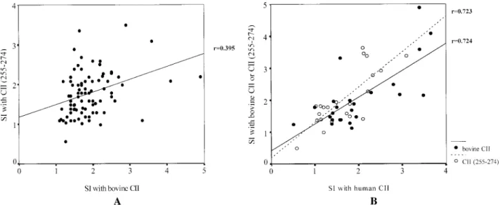
Forty-seven (54%) of 87 RA patients had positive T cell responses to CII and/or CII (255–274) (Table 1 and Figure 2). T cell responses to bovine CII (n 5 87) or human CII (n 5 32) strongly correlated with those to CII (255–274) (r 5 0.395 [P , 0.001] and r 5 0.723 [P , 0.001], respectively) (Figures 3A and B). T cell re- sponses to bovine CII also strongly correlated with those to human CII (r 5 0.724, P , 0.001) (Figure 3B).
However, no correlation was found between the SI with PHA and that with CII or CII (255–274) peptide.
T cell proliferative responses of paired SFMC and PBMC to CII and CII (255–274) peptide. SFMC were obtained from 42 patients with knee joint effusions.
Twenty-six (61.9%) of 42 SFMC samples responded positively to CII and/or CII (255–274); good correlations were noted between the SI induced with CII and that with CII (255–274) peptide (r 5 0.705, P , 0.001).
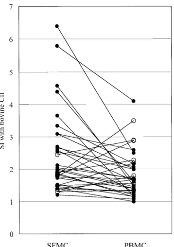
Comparisons of T cell responses in paired SFMC and PBMC (n 5 34) showed that positive T cell responses to CII were found more frequently in SFMC than in PBMC (58.8% versus 35.3%; P 5 0.032). The SI with CII in SFMC was significantly higher than in PBMC (P 5 0.003), yet correlated well with PBMC responses (r 5 0.385, P 5 0.024). Twenty-two (64.7%) of 34 RA patients had positive T cell responses to CII in PBMC and/or SFMC when measured simultaneously (Figure 4). However, no differences were found in the median SI with PHA, ovalbumin, or CII (233–252) peptide between SFMC and PBMC (data not shown).
Association of T cell responses to CII with dis- ease duration. In 95 patients in whom disease duration could be documented, those with a positive T cell
Table 1. T cell proliferative responses to type II collagen (CII) and its immunodominant epitope CII (255–274) in peripheral blood mononuclear cells obtained from patients with rheumatoid arthritis (RA), osteoarthritis (OA), and healthy controls*
Group tested, antigen response RA OA Healthy controls P
No. of patients 106 26 34
Medium only
Median cpm 6,881 8,032 6,050 NS
Range 2,371–28,000 4,500–10,500 3,135–21,340
PHA response
Median SI 7.64 11.4 8.32 NS
Range 2.41–25.2 4.94–26.24 3.04–41.0
Ovalbumin
Median SI 1.10 1.08 1.20 NS
Range 0.50–2.41 0.90–1.70 0.81–1.90
Bovine CII
Median SI 1.70 1.32 1.45 ,0.001
Range 1.00–12.40 0.89–2.05 0.96–2.20
Positive response, no. (%)† 37 (34.9) 1 (3.8) 1 (2.9) ,0.001
No. of patients 87 24 33
CII (255–274)
Median SI 1.73 1.38 1.44 0.002
Range 0.58–3.50 0.78–2.05 0.90–2.60
Positive response, no. (%)† 33 (37.9) 1 (4.2) 4 (12.1) ,0.001
CII (233–252)‡
Median SI 1.10 1.14 1.10 NS
Range 0.80–2.10 0.80–1.79 0.95–1.90
Bovine CII or CII (255–274)
Positive response, no. (%)† 47 (54.0) 2 (8.3) 4 (12.1) ,0.001
* The stimulation index (SI) was calculated as the counts per minute (cpm) in wells containing antigen divided by the cpm in wells without antigen.
Significance of differences were between RA patients and OA patients and healthy controls, by Kruskal-Wallis test. NS5 not significant; PHA 5 phytohemagglutinin.
† A positive T cell response was defined as an SI$2 and an increase in cpm (D cpm) .1,000.
‡ Control peptide of a T cell non-reactive sequence.
response to CII (n 5 35) often had a shorter disease duration than those (n 5 60) with negative responses (median 84 months [range 12–331] versus 103.5 months [range 4–359]; P 5 0.021). These 95 patients with RA were divided into 2 groups based on disease duration, early (#3 years) and late (.3 years) RA. T cell re-
sponses to CII were then compared between the 2 groups. Positive T cell responses and the median SI in PBMC were significantly higher in early RA patients (n 5 19) than in late RA patients (n 5 76) (68.4%
positive and median SI 2.12 for the early group versus 28.9% positive and median SI 1.62 for the late group;
P
5 0.001 and P 5 0.021, respectively). In SFMC (n 5 42), positive T cell responses and the median SI also tended to be higher in early RA patients (n 5 8) than in late RA patients (n 5 34), but these differences were not statistically significant (Table 2). A trend for positive T cell responses to decrease over time is shown in Figure 5.
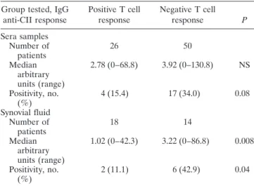
Correlation of T cell response with antibody response to CII. IgG anti-CII were measured in sera and SF obtained simultaneously from patients with RA (Table 3). Twenty-one (27.6%) of 76 patients with RA tested positive for circulating IgG anti-CII. In the sera, patients with negative T cell responses to bovine CII (n 5 50) had a higher frequency of IgG anti-CII positivity (34% versus 15.4%; P 5 0.08) and had a higher median level of anti-CII (3.92 versus 2.78 arbitrary units;
P
5 not significant) than did patients with positive T cell responses (n 5 26). In the SF, the positivity and median level of IgG anti-CII were significantly higher in patients with negative T cell responses (n 5 14) than in those with positive T cell responses (n 5 18) (42.9% positive and median 3.22 arbitrary units versus 11.1% positive and median 1.02 arbitrary units; P 5 0.04 and P 5 0.008, respectively).
Figure 2. T cell proliferative responses to bovine type II collagen (CII), ovalbumin (Oval), CII (255–274) (immunodominant peptide, or IP), and CII (233–252) (control peptide, or CP) of a T cell non-reactive sequence in patients with rheumatoid arthritis (RA), osteoarthritis (OA), and healthy controls. Broken line indicates the cutoff value for positive T cell responses. Bars represent the median. Findings in 1 RA patient (stimulation index 12.4) are not expressed in this figure.
Figure 3. Correlations of T cell responses to bovine type II collagen (CII) (A) with those to human CII or CII (255–274) (B) in peripheral blood mononuclear cells from rheumatoid arthritis patients. Values are presented as stimulation indices (SI).
HLA–DR association. HLA–DR types were ex- amined in 61 patients with RA. No association was found between HLA–DR4 or –DR1 positivity and pos- itive T cell responses (Table 4).
DISCUSSION
The hypothesis that RA is a T cell–mediated autoimmune disease has been widely held by a number of investigators (5–9), although not by all (25). One obstacle to confirming that T cells play a specific role in the pathogenesis of RA has been the identification of autoantigens specific to the joint, where immune- mediated inflammation and injury are most intense. One candidate autoantigen relevant to the pathogenesis of
RA is CII. Type II collagen is a strong candidate because of its abundance in cartilage, an immunologically privi- leged tissue, and because of its ability to induce destruc- tive immune-mediated polyarthritis in rodents and higher primates (1–4,10–12). Reports describing T cell immunity to CII in RA are few (5–9), and the conclu- sions are not entirely uniform. Trentham et al (5) reported that rheumatoid PBMC cultured with CII produced increased amounts of a chemotactic factor in the majority of RA patients. Similar conclusions were reached by others using a variety of assay systems (6–8).
Figure 4. Comparison of T cell responses to bovine type II collagen (CII) in paired synovial fluid mononuclear cells (SFMC) and peri- pheral blood mononuclear cells (PBMC) (n5 34) obtained simulta- neously from patients with rheumatoid arthritis. Solid circles denote cases where the stimulation index (SI) was higher in SFMC than in PBMC (P5 0.003), and open circles denote the opposite.
Table 2. Comparison of T cell responses to CII between early (#3 years) and late (.3 years) RA*
Group tested,
T cell response Early RA Late RA P
PBMC studies Number of
patients 19 76
Positive response,
no. (%) 13 (68.4) 22 (28.9) 0.001
Median SI
(range) 2.12 (1.08–12.39) 1.62 (1.00–5.60) 0.021 SFMC studies
Number of
patients 8 34
Positive response,
no. (%) 5 (62.5) 17 (50.0) NS
Median SI
(range) 2.27 (1.20–3.87) 1.93 (1.30–6.40) NS
* PBMC 5 peripheral blood mononuclear cells; SFMC 5 synovial fluid mononuclear cells (see Table 1 for other definitions).
Figure 5. The frequency of positive T cell responses to type II collagen (CII) in peripheral blood mononuclear cells over time.
To the contrary, Snowden et al (9) recently reported that increased PBMC T cell responses to CII were present in only a minority of patients with RA. This difference may reflect variability in the sensitivities or specificities of the assay systems used.
To address these problems, we conducted a series of studies using T cells isolated from the PBMC and SFMC of RA patients, and cultured the cells with irradiated APC and optimal concentrations of heterolo- gous CII, homologous CII, and synthetically prepared CII oligopeptides. In using this protocol, we demon- strated that positive T cell responses to CII and/or CII (255–274) peptide could be detected in the majority (61.9%) of RA patients, and that T cell responses to CII are more prevalent and stronger using SFMC than PBMC. In contrast, little activity was detected using PBMC T cells from patients with OA or healthy subjects.
Importantly, T cell responses to bovine CII correlated strongly with responses to human CII, which contains the same CII (255–274) sequence. Earlier reports using DR1- and DR4-transgenic mice have shown that CII (255–274) contains the immunodominant T cell deter- minant of CII. Moreover, by using a synthetically pre- pared antigen, we were able to eliminate concerns that the responses to tissue-derived CII were against contam- inants, e.g., proteoglycan, pepsin, or a matrix protein.
Collectively, these observations underscore the impor- tance of T cells in maintaining RA, and suggest that CII-reactive T cells in the joints could play a critical role in the pathogenesis of RA.
Recently, antigen-specific tolerance induction has been utilized as a treatment for various autoimmune diseases (26–29). Oral administration of CII has been reported to be effective for the treatment of some patients with RA (30). In an animal experimental model of RA, it is documented that oral administration of immunodominant peptide CII (250–270) can modulate collagen-induced arthritis, and that the HLA–DR re- stricted T cell response to human CII is focused on an immunodominant determinant within CII (263–270) (11,31). Our observations, together with previous find- ings, provide strong evidence that synthetic peptide CII (255–274) might be useful in the treatment of human RA, with benefit possibly being derived from the mech- anism of antigen-driven active suppression of the auto- reactive T cell response to CII.
Our finding that T cell responses to CII were more frequent and vigorous in SFMC suggests either an influx of CII-reactive T cells into the joint, or their local expansion in response to autologous CII exposed during cartilage degradation; given that positive PBMC T cell responses to CII were detected in a number of patients whose SFMC responses were negative, and that 9 of 32 patients showed stronger responses to CII in PBMC than in SFMC, our data tend to favor the former possibility. However, because these 2 mechanisms are not mutually exclusive, local expansion of T cells in the joint may occur in some patients or may not have been reflected by the study of SF.
Notwithstanding these caveats, the response of T cells recovered from SFMC to CII generally correlated well with the response in PBMC.
Another variable possibly influencing the T cell proliferative response, in addition to the tissue source of T cells, was the duration of RA. T cells collected from patients with RA of ,3 years’ duration frequently responded better to CII than those obtained from patients with disease of longer duration. This finding suggests that autoimmune T cell responses may be intrinsic to the pathogenesis of RA, rather than conse- quent to cartilage destruction and CII breakdown and release. However, positive PBMC T cell responses were still found in about one-third of patients with longstand-
Table 4. Association of HLA–DR types with T cell responses to CII*
DR type Positive T cell
response (n5 26) Negative T cell response (n5 35) P
DR4 positive 15 (57.7) 19 (54.3) NS
DR4 or DR1
positive 19 (73.1) 24 (68.6) NS
* Values are the number (%) of patients. See Table 1 for definitions.
Table 3. Inverse correlation of T cell responses with antibody responses to CII in serum and synovial fluid*
Group tested, IgG
anti-CII response Positive T cell
response Negative T cell
response P
Sera samples Number of
patients 26 50
Median arbitrary units (range)
2.78 (0–68.8) 3.92 (0–130.8) NS
Positivity, no.
(%) 4 (15.4) 17 (34.0) 0.08
Synovial fluid Number of
patients 18 14
Median arbitrary units (range)
1.02 (0–42.3) 3.22 (0–86.8) 0.008
Positivity, no.
(%) 2 (11.1) 6 (42.9) 0.04
* Positive levels of IgG antibodies were defined as a value .6.6 arbitrary units, which was.2 SD above the mean level in sera from 58 healthy controls. See Table 1 for definitions.
ing RA ( .10 years). Moreover, one-half of patients with RA lasting .3 years, but ,10 years, yielded positive SFMC T cell responses to CII. These observations suggest that abnormal T cell responses to CII might also perpetuate chronic arthritis. If this assumption is cor- rect, CII-reactive T cells could be a good marker for progressive cartilage destruction and poor prognosis, in that few activated T cells are needed to induce a profound effect on tissues.
The separation of immune responses into delayed-type hypersensitivity and antibody responses has been well documented. These 2 reactions can be dissociated, which has led to the hypothesis that, in vivo, they are reciprocally regulated (14–16). In this regard, RA is not unique. Pathogenic T cell responses prevailing over antibody responses have been described in other autoimmune diseases, such as insulin-dependent diabe- tes mellitus (IDDM) and experimental allergic enceph- alomyelitis (EAE). In IDDM, antibodies to glutamic acid decarboxylase (GAD) are usually demonstrable at the time of clinical diagnosis in most patients, but rapidly decline over time (32), and correlate inversely with the presence of GAD-specific T cell responses (33). In an animal model of EAE, anti-IgD peptide conjugate pre- treatment protects rats from encephalomyelitis by induc- ing a Th2 response and suppressing the Th1 reaction to the autoantigen (34). RA resembles the aforementioned diseases in showing that antibodies to CII also decline over time, and that loss of IgG anti-CII, which indicates a decrease in the Th2-type response, is associated with severe disease and the early development of tissue erosions (18).
The inverse correlation between the T cell and B cell responses to CII in RA is a new observation and was best demonstrated by the study of SF. This finding is in contrast with the observations reported by Snowden et al (9). Reports describing circulating IgG anti-CII antibodies in RA vary widely among laboratories, with positive rates ranging from 3% to 63% (17,18,23,35,36). Some of this disparity undoubtedly reflects differences in the assay systems used. The sensitivity and specificity of the ELISA for IgG anti-CII has been improved considerably by the use of heterologous serum as a blocking agent and by the use of the avidin–biotin capture system (17,23). We and others have shown that levels of IgG anti-CII antibodies can fluctuate with time (17,36–38). Therefore, the number of IgG anti-CII antibody–positive sera can differ depending on the time, as well as the method used, to detect the antibody. These factors may explain the difference between the findings of Snowden et al and ours.
Increased risk for RA is associated with the
inheritance of certain DR1 and subtypes of DR4 that share a conserved amino acid sequence (39,40). In type II collagen–induced arthritis, an animal model of RA, DR4 and DR1, expressed as transgenes, can confer susceptibility to an otherwise-resistant strain of mice by influencing T cell immunity to CII (11). In human RA, it is reported that T cell responses to CII are inhibited by DR-specific antibodies (9), and that augmented cell- mediated responsiveness to CII is associated with DR4 (41). However, we did not find an association between positive T cell responses to CII and the presence of HLR–DR4 or –DR1 allotypes. Since we did not analyze our patients for the DR4 subtype or RA susceptibility motifs, QKRAA and QRRAA, it is uncertain whether the T cell responses detected may or may not be associated with either motif.
In summary, our studies show that T cell re- sponses to tissue-derived CII and the synthetic CII (255–274) peptide are increased in the majority of patients with RA as compared with patients with OA or healthy subjects. Importantly, CII (255–274) contains the immunodominant epitope of CII that is recognized by HLA–DR1 and –DR4 molecules which share the
“susceptibility sequence” associated with RA. Positive responses were detected most often in early RA, espe- cially when the T cells used were recovered from the SF of inflamed joints. T cell responses to CII were found to correlate inversely with antibody responses, suggesting that sensitization to CII with subsequent expansion of CII-reactive T cells may lead to progressive joint de- struction. The identification of a peptide sequence com- monly recognized in patients with RA raises the possi- bility that it can be utilized to develop a way to suppress autoreactive T cells in the RA joint in an antigen-specific manner and to reduce the severity of the disease.
ACKNOWLEDGMENTS
The authors are grateful to Dr. Andrew H. Kang and Dr. Michael A. Cremer for their advice.
REFERENCES
1. Trentham DE, Townes AS, Kang AH. Autoimmunity to type II collagen: an experimental model of arthritis. J Exp Med 1977;146:
857–68.
2. Courtenay JS, Dallman MJ, Dayan AD, Martin A, Mosedale B.
Immunisation against heterologous type II collagen induces arthri- tis in mice. Nature 1980;283:666–8.
3. Holmdahl R, Klareskog L, Rubin K, Larsson E, Wigzell H. T lymphocytes in collagen II-induced arthritis in mice: characteriza- tion of arthritogenic collagen II-specific T-cell lines and clones.
Scand J Immunol 1985;22:295–306.
4. Seki N, Sudo Y, Yoshioka T, Sugihara S, Fujitsu T, Sakuma S, et al. Type II collagen-induced murine arthritis. I. Induction and
perpetuation of arthritis require synergy between humoral and cell-mediated immunity. J Immunol 1988;140:1477–84.
5. Trentham DE, Dynesius RA, Rocklin RE, David JR. Cellular sensitivity to collagen in rheumatoid arthritis. N Engl J Med 1978;299:327–32.
6. Stuart JM, Postlethwaite AE, Townes AS, Kang AH. Cell mediated immunity to collagen and collagen alpha chains in rheumatoid arthritis and other rheumatic disease. Am J Med 1980;69:13–8.
7. Londei M, Savil CM, Verhoef A, Brennan F, Leech ZA, Duance V, et al. Persistence of collagen type II specific T-cell clones in synovial membrane of a patient with rheumatoid arthritis. Proc Natl Acad Sci U S A 1989;86:636–40.
8. Desai BV, Dixit S, Pope RM. Limited proliferative response to type II collagen in rheumatoid arthritis. J Rheumatol 1989;16:1310–4.
9. Snowden N, Reynolds I, Morgan K, Holt L. T cell responses to human type II collagen in patients with rheumatoid arthritis and healthy controls. Arthritis Rheum 1997;40:1210–8.
10. Krco CJ, Pawelski J, Harders J, McCormick D, Griffiths M, Luthra HS, et al. Characterization of the antigenic structure of human type II collagen. J Immunol 1996;156:2761–8.
11. Rosloniec EF, Brand DD, Myers LK, Esaki Y, Whittington KB, Zaller DM, et al. Induction of autoimmune arthritis in HLA-DR4 (DRB1*0401) transgenic mice by immunization with human and bovine type II collagen. J Immunol 1998;160:2573–8.
12. Brand DD, Myers LK, Terato K, Whittington KB, Stuart JM, Kang AH, et al. Characterization of the T cell determinants in the induction of autoimmune arthritis by bovine alpha 1(II)-CB11 in H-2q mice. J Immunol 1994;152:3088–97.
13. Myers LK, Miyahara H, Terato K, Seyer JM, Stuart JM, Kang AH.
Collagen-induced arthritis in B10.RIII mice (H-2r): identification of an arthritogenic T-cell determinant. Immunology 1995;84:509–13.
14. Parish CR. The relationship between humoral and cell mediated immunity. Transplant Rev 1972;13:35–66.
15. Katsura Y. Cell mediated and humoral immune responses in mice.
III. Dynamic balance between delayed type hypersensitivity and antibody response. Immunology 1977;32:227–35.
16. Heinzel FP, Sadick MD, Holaday BJ, Coffman RL, Locksley RM.
Reciprocal expression of interferon gamma or IL-4 during the resolution or progression of murine leishmaniasis: evidence for expansion of distinct helper T cell subsets. J Exp Med 1989;169:
59–72.
17. Terato K, DeArmey DA, Ye XY, Griffiths MM, Cremer MA. The mechanism of autoantibody formation to cartilage in rheumatoid arthritis: possible cross-reaction of antibodies to dietary collagens with autologous type II collagen. Clin Immunol Immunopathol 1997;79:142–54.
18. Cook AD, Rowley MJ, Mackay IR, Gough A, Emery P. Antibod- ies to type II collagen in early rheumatoid arthritis: correlation with disease progression. Arthritis Rheum 1996;39:1720–7.
19. Stuart JM, Cremer MA, Townes AS, Kang AH. Type II collagen induced arthritis in rats: passive transfer with serum and evidence that IgG anticollagen antibodies can cause arthritis. J Exp Med 1982;155:1–16.
20. Terato K, Hasty KA, Reife RA, Cremer MA, Kang AH, Stuart JM. Induction of arthritis with monoclonal antibodies to collagen.
J Immunol 1992;148:2103–8.
21. Arnett FC, Edworthy SM, Bloch DA, McShane DJ, Fries JF, Cooper NS, et al. The American Rheumatism Association 1987 revised criteria for the classification of rheumatoid arthritis.
Arthritis Rheum 1988;31:315–24.
22. Wahl SM, Malone DG, Wilder RL. Spontaneous production of fibroblast-activating factor(s) by synovial inflammatory cells: a potential mechanism for enhanced tissue destruction. J Exp Med 1985;161:210–22.
23. Fujii K, Tsuji M, Murota K, Terato K, Shimozuru Y, Nagai Y.
An improved enzyme-linked immunosorbent assay of anti-
collagen antibodies in human serum. J Immunol Methods 1989;124:63–70.
24. Tsuji K, Aizawa M, Sasazuki T, editors. HLA 1991: proceedings of the eleventh International Histocompatibility Workshop and Con- ference. Oxford: Oxford University Press; 1992. p. 83–108.
25. Firestein GS, Zvaifler NJ. How important are T cells in chronic rheumatoid synovitis? Arthritis Rheum 1990;33:768–73.
26. Weiner HL, Mackin GA, Matsui M, Orav EJ, Khoury SJ, Dawson DM, et al. Double-blind pilot trial of oral tolerization with myelin antigens in multiple sclerosis. Science 1993;259:1321–4.
27. Trentham DE, Dynesius-Trentham RA, Orav EJ, Combitchi D, Lorenzo C, Sewell KL, et al. Effects of oral administration of type II collagen on rheumatoid arthritis. Science 1993;261:
1727–30.
28. Zhang ZJ, Davidson L, Eisenbarth G, Weiner HL. Suppression of diabetes in nonobese diabetic mice by oral administration of porcine insulin. Proc Natl Acad Sci U S A 1991;88:10252–6.
29. Nussenblatt RB, Caspi RR, Mahdi R, Chan CC, Roberge F, Lider O, et al. Inhibition of S-antigen induced experimental autoimmune uveoretinitis by oral induction of tolerance with S-antigen. J Im- munol 1990;144:1689–95.
30. Barnett ML, Kremer JM, St. Clair EW, Clegg DO, Furst D, Weisman M, et al. Treatment of rheumatoid arthritis with oral type II collagen: results of a multicenter, double-blind, placebo- controlled trial. Arthritis Rheum 1998;41:290–7.
31. Khare SD, Krco CJ, Griffiths MM, Luthra HS, David CS. Oral administration of an immunodominant human collagen peptide modulate collagen induced arthritis. J Immunol 1995;155:3653–9.
32. De Aizpurua HJ, Wilson YM, Harrison LC. Glutamic acid decar- boxylase autoantibodies in preclinical insulin-dependent diabetes.
Proc Natl Acad Sci U S A 1992;89:9841–5.
33. Harrison LC, Honeyman MC, DeAizpurua HJ, Schmidli RS, Colman PG, Tait BD, et al. Inverse relation between humoral and cellular immunity to glutamic acid decarboxylase in subjects at risk of insulin-dependent diabetes. Lancet 1993;341:1365–9.
34. Saoudi A, Simmonds S, Huitinga I, Mason D. Prevention of experimental allergic encephalomyelitis in rats by targeting au- toantigen to B cells: evidence that the protective mechanism depends on changes in the cytokine response and migratory properties of the autoantigen-specific T cells. J Exp Med 1995;
182:335–44.
35. Greenbury CL, Skingle J. Anti-collagen antibody. J Clin Pathol 1979;32:826–31.
36. Cook AD, Rowley MJ, Stockman A, Muirden KD, Mackay IR.
Specificity of antibodies to type II collagen in early rheumatoid arthritis. J Rheumatol 1994;21:1186–91.
37. Kim W-U, Lee S-S, Joo Y-S, Min J-K, Hong Y-S, Lee S-H, et al.
IgG antibodies to native bovine type II collagen reflect disease activity in rheumatoid arthritis [abstract]. Arthritis Rheum 1998;41 Suppl 9:S313.
38. Morgan K, Clague RB, Collins I, Ayad S, Phinn SD, Holt PJL. A longitudinal study of anticollagen antibodies in patients with rheumatoid arthritis. Arthritis Rheum 1989;32:139–45.
39. Gregersen PK, Shen M, Song QL, Merryman P, Degar S, Seki T, et al. Molecular diversity of HLA-DR4 haplotypes. Proc Natl Acad Sci U S A 1986;83:2642–6.
40. Wordsworth BP, Lanchbury JS, Sakkas LI, Welsh KI, Panayi GS, Bell JI. HLA-DR4 subtype frequencies in rheumatoid arthritis indicate that DRB1 is the major susceptibility locus within the HLA class II region. Proc Natl Acad Sci U S A 1989;86:10049–53.
41. Solinger AM, Stobo JD. Immune response gene control of colla- gen reactivity in man: collagen unresponsiveness in HLA-DR4 negative nonresponders is due to the presence of T-dependent suppressive influences. J Immunol 1982;129:1916–20.
![Figure 1. T cell responses to type II collagen (CII) or CII (255–274) in various concentrations of antigens (3 rheumatoid arthritis [RA] patients and 3 healthy controls) and culture times (2 RA patients and 2 healthy controls)](https://thumb-ap.123doks.com/thumbv2/123dokinfo/4990941.303561/3.921.114.843.131.496/responses-collagen-concentrations-antigens-rheumatoid-arthritis-patients-controls.webp)