The authors’ full names, academic de- grees, and affiliations are listed in the Appendix. Address reprint requests to Dr.
Davies at Hammersmith Hospital, Impe- rial College London, London W12 OHS, United Kingdom, or at justindavies@
heart123 . com.
This article was published on March 18, 2017, at NEJM.org.
N Engl J Med 2017;376:1824-34.
DOI: 10.1056/NEJMoa1700445 Copyright © 2017 Massachusetts Medical Society.
BACKGROUND
Coronary revascularization guided by fractional flow reserve (FFR) is associated with better patient outcomes after the procedure than revascularization guided by angiography alone. It is unknown whether the instantaneous wave-free ratio (iFR), an alternative measure that does not require the administration of adenosine, will offer benefits similar to those of FFR.
METHODS
We randomly assigned 2492 patients with coronary artery disease, in a 1:1 ratio, to undergo either iFR-guided or FFR-guided coronary revascularization. The primary end point was the 1-year risk of major adverse cardiac events, which were a com- posite of death from any cause, nonfatal myocardial infarction, or unplanned re- vascularization. The trial was designed to show the noninferiority of iFR to FFR, with a margin of 3.4 percentage points for the difference in risk.
RESULTS
At 1 year, the primary end point had occurred in 78 of 1148 patients (6.8%) in the iFR group and in 83 of 1182 patients (7.0%) in the FFR group (difference in risk,
−0.2 percentage points; 95% confidence interval [CI], −2.3 to 1.8; P<0.001 for noninferiority; hazard ratio, 0.95; 95% CI, 0.68 to 1.33; P = 0.78). The risk of each component of the primary end point and of death from cardiovascular or noncar- diovascular causes did not differ significantly between the groups. The number of patients who had adverse procedural symptoms and clinical signs was signifi- cantly lower in the iFR group than in the FFR group (39 patients [3.1%] vs. 385 patients [30.8%], P<0.001), and the median procedural time was significantly shorter (40.5 minutes vs. 45.0 minutes, P = 0.001).
CONCLUSIONS
Coronary revascularization guided by iFR was noninferior to revascularization guided by FFR with respect to the risk of major adverse cardiac events at 1 year. The rate of adverse procedural signs and symptoms was lower and the procedural time was shorter with iFR than with FFR. (Funded by Philips Volcano; DEFINE-FLAIR ClinicalTrials.gov number, NCT02053038.)
ABS TR ACT
Use of the Instantaneous Wave-free Ratio or Fractional Flow Reserve in PCI
J.E. Davies, S. Sen, H.-M. Dehbi, R. Al-Lamee, R. Petraco, S.S. Nijjer, R. Bhindi, S.J. Lehman, D. Walters, J. Sapontis, L. Janssens, C.J. Vrints, A. Khashaba, M. Laine, E. Van Belle, F. Krackhardt, W. Bojara, O. Going, T. Härle, C. Indolfi, G. Niccoli, F. Ribichini, N. Tanaka, H. Yokoi, H. Takashima, Y. Kikuta, A. Erglis,
H. Vinhas, P. Canas Silva, S.B. Baptista, A. Alghamdi, F. Hellig, B.-K. Koo, C.-W. Nam, E.-S. Shin, J.-H. Doh, S. Brugaletta, E. Alegria-Barrero, M. Meuwissen,
J.J. Piek, N. van Royen, M. Sezer, C. Di Mario, R.T. Gerber, I.S. Malik, A.S.P. Sharp, S. Talwar, K. Tang, H. Samady, J. Altman, A.H. Seto, J. Singh, A. Jeremias, H. Matsuo, R.K. Kharbanda, M.R. Patel, P. Serruys, and J. Escaned
Original Article
F
or the past 20 years, physiological measurements obtained during invasive pro- cedures have been used to guide coronary revascularization. Pioneering work supported the use of flow measurements to make safe decisions about revascularization,1,2 but this approach was soon superseded by the use of fractional flow reserve (FFR), which measures pressure as a sur- rogate of flow to estimate the severity of steno- sis.3-5 FFR was successful largely because of its technical simplicity and because clinical trials showed that it was associated with improved clinical outcomes after percutaneous coronary intervention (PCI).6,7 Consequently, FFR is now included in the appropriate-use criteria for coro- nary angiography and in the American College of Cardiology–American Heart Association–European Society of Cardiology guidelines; despite these recommendations, its adoption remains limited.8-10FFR must be measured during maximal hyper- emia, which is typically induced with the admin- istration of a potent intravenous or intracoronary vasodilator, such as adenosine.11 Several studies have questioned the need for the administration of a vasodilator to assess stenosis severity.12-14 In these studies, investigators found that in deter- mining stenosis severity, FFR was not superior to the instantaneous wave-free ratio (iFR), a pressure- derived index of stenosis severity that is not ob- tained with the administration of a vasodilator.
We aimed to determine the efficacy and safety of an iFR-guided strategy versus an FFR-guided strategy for coronary revascularization.
Methods Trial Design and Management
DEFINE-FLAIR (Functional Lesion Assessment of Intermediate Stenosis to Guide Revascularisation) is a multicenter, international, randomized, blinded trial in which iFR is being compared with FFR for physiologically guided coronary revascularization. The trial, which is ongoing, is being performed at 49 interventional sites across 19 countries on 4 continents. The 1-year out- comes, on which the primary trial analysis is based, are reported here.
The trial was designed by the steering com- mittee (for a list of committee members, see the Supplementary Appendix, available with the full text of this article at NEJM.org). Central ethics approval was granted by the National Research Ethics Service Committee London, and local ethics
approval was granted at each participating site.
The trial is funded by an unrestricted educa- tional grant from Philips Volcano, which had no role in the design of the trial, the collection or analysis of the data, the writing of the manu- script, or the decision to submit the manuscript for publication.
Trial management and oversight were per- formed by personnel at the Imperial College Trials Unit, Imperial College London, who main- tained the clinical database and conducted all the data analyses independent of the funder. A risk assessment established that the trial was of low risk to the patients; therefore, no data and safety monitoring board was established. The first draft of the manuscript was written by the first author, and all the authors participated in trial oversight, approved all subsequent drafts of the manuscript, and made the decision to sub- mit the manuscript for publication. The steering committee and all the authors vouch for the ac- curacy and completeness of the data and analy- ses and for the fidelity of the study to the trial protocol and statistical analysis plan, which are available at NEJM.org.
Population
Patients who had undergone coronary angiogra- phy were assessed for trial eligibility. Patients were eligible for inclusion in the trial if they had coronary artery disease with at least one native artery in which the stenosis was of questionable physiological severity (typically, an artery with 40 to 70% stenosis of the diameter on visual as- sessment). Patients with tandem stenoses (i.e., stenoses separated by more than 10 mm within a single vessel) that would require independent evaluation and treatment were excluded. A full list of inclusion and exclusion criteria is provided in Table S1 in the Supplementary Appendix. No exclusions were made on the basis of heart rate or rhythm. Written informed consent was ob- tained from all the patients before their enroll- ment in the trial.
Randomization
Eligible patients were randomly assigned to un- dergo revascularization guided by either FFR or iFR. Randomization was performed with the use of an automated and validated online random- ization tool (SRUB, Imperial College London).
During the trial procedures, investigators were allowed to obtain FFR or iFR measurements only
in accordance with group assignment. Verification of the data was performed in each patient with the use of the electronic physiology record, which was uploaded directly from the physiolog- ical console for each patient into the electronic clinical record (Fig. S1 in the Supplementary Appendix). During the procedure, patients were not told which technique was used for physio- logical assessment, and they remained unaware of their group assignment throughout the entire course of the trial. The research nurses and doc- tors who were responsible for the follow-up visits were also unaware of the group assignments.
Procedure
Before the FFR or iFR measurement was ob- tained, intracoronary nitrates were administered to control vasomotor tone. The physiological measurements were obtained in the routine manner with the use of a coronary-pressure guidewire (Philips Volcano) (Figs. S1 and S2 in the Supplementary Appendix). Physiological as- sessment was performed in all vessels with ques- tionable stenosis severity. In patients with an acute coronary syndrome, physiological assess- ment was performed in only nonculprit vessels, after the culprit vessel had been revascularized.
Prespecified treatment thresholds were an FFR of 0.80 and an iFR of 0.89 (Fig. S3 in the Supple- mentary Appendix). When the FFR or iFR for a given stenosis was equal to or lower than the prespecified threshold, the stenosis was revascu- larized with a drug-eluting stent or a bioresorb- able vascular scaffold or by coronary-artery by- pass grafting (CABG). When the FFR or iFR was higher than the prespecified threshold, treatment was deferred. When multivessel revasculariza- tion was attempted, investigators could choose to prespecify a staged treatment plan, with the staged procedure performed within 60 days. Ad- verse procedural signs and symptoms were docu- mented.
Routine clinical follow-up assessments were performed at 30 days and at 1 year, and follow- up by telephone was conducted at 6 months.
Complete monitoring of every electronic clinical record was performed, and the data were con- firmed by on-site source-document verification in a randomly selected 30% of patients.
End Points
The primary end point was the 1-year risk of major adverse cardiac events, which were a com-
posite of death, nonfatal myocardial infarction, or unplanned revascularization. Death was con- sidered to be from cardiovascular causes unless an unequivocal noncardiovascular cause was es- tablished. Myocardial infarction was classified as either spontaneous or periprocedural and as either ST-segment elevation myocardial infarction (STEMI) or non-STEMI (NSTEMI). Revasculariza- tion was considered to be unplanned when it was not the index procedure and was not identified at the time of the index procedure as a staged procedure to occur within 60 days. Detailed end- point definitions, which did not change after the commencement of the trial, are provided in the Supplementary Appendix.
End-point events were adjudicated with the use of anonymized source documentation by a com- mittee of international experts who were not part of the steering committee. A consensus decision was made on the basis of prespecified end-point definitions. Members of the events committee remain unaware of the identities of the patients and their group assignments.
Statistical Analysis
The prespecified trial hypothesis was that iFR would be noninferior to FFR with respect to the risk of major adverse cardiac events at 1 year among patients undergoing physiologically guid- ed revascularization. We based the sample size on an assumed annual rate of primary end-point events of 8.5% in a population that includes a mix of patients with either stable coronary dis- ease or acute coronary syndromes15; given this rate, we calculated that a sample size of 2305 patients would provide the trial with 90% power to detect the noninferiority of iFR to FFR, with the use of a noninferiority margin of 3.4 percent- age points for the difference in risk, at a type I error rate of 5%. To allow for attrition, the target sample size was set at 2500 patients.
Both a risk-difference analysis and a time-to- event analysis were performed. The time-to-event analysis was conducted with the use of the Kaplan–Meier method. A Cox survival model was used to derive hazard ratios. For the results of both analyses, two-sided 95% confidence in- tervals (whose upper limits correspond to the upper limits of one-sided 97.5% confidence in- tervals) and two-sided 99% confidence intervals (whose upper limits correspond to the upper limits of one-sided 99.5% confidence intervals) are reported. The validity of the proportional-
hazards assumption was tested with Schoenfeld residuals. There were no signs of violation of the proportional-hazards assumption.
Patients who withdrew from the study before they reached 1 year of follow-up and who were event-free at their last visit were excluded from the risk-difference analysis for the primary end point and its components. Data for these pa- tients were censored at the time of withdrawal for the time-to-event analysis. Patients who had a myocardial infarction or an unplanned revas- cularization before withdrawing from the study were included in the risk-difference analysis.
R esults Patients and Procedures
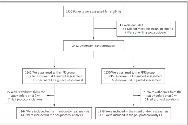
During the recruitment period (January 2014 to December 2015), a total of 2535 patients who underwent coronary angiography were assessed for trial eligibility. Of the 2492 patients who met the enrollment criteria, 1242 were assigned to the iFR group and 1250 to the FFR group (Fig. 1).
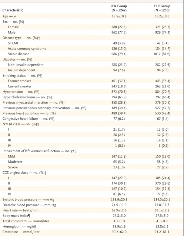
The baseline demographic characteristics of the patients are shown in Table 1. The mean age of the patients was 65 years, 76% were men, and 80% had stable coronary artery disease.
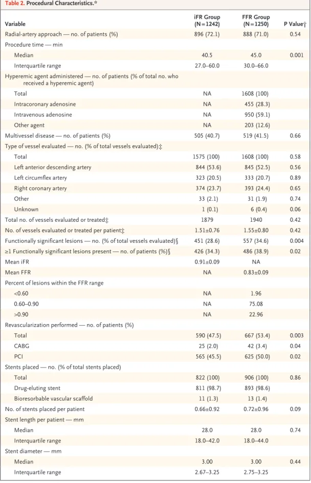
Procedural characteristics for the two trial groups are shown in Figure 1 and Table 2. A total of 99.4% of the patients assigned to the iFR group and 99.6% of those assigned to the FFR group underwent the assigned procedure. Cross- over, which represented a deviation from the protocol, occurred in 13 cases and was due to profound early adenosine-induced bradycardia and hypotension in 1 case and to site errors in the remaining 12 cases. There were no cases in which heart-rhythm disturbances or lack of electrocar- diographic assessment prevented FFR or iFR measurements from being obtained.
The number of vessels evaluated did not dif- fer significantly between the iFR group and the FFR group (total number assessed, 1575 and 1608, respectively; mean [±SD] number evaluated per patient, 1.27±0.61 and 1.29±0.63; P = 0.58).
The mean iFR and FFR measurements were close to their respective thresholds (mean iFR, 0.91±0.09;
mean FFR, 0.83±0.09); these findings suggest that most of the assessed vessels had stenosis of intermediate severity (Figs. S4 and S5 in the Supplementary Appendix). The number of func- tionally significant stenoses (i.e., stenoses with an iFR or FFR below the treatment threshold) was significantly lower in the iFR group than in the
Figure 1. Enrollment, Randomization, Follow-up, and Analysis.
FFR denotes fractional flow reserve, and iFR instantaneous wave-free ratio.
2492 Underwent randomization 2535 Patients were assessed for eligibility
43 Were excluded
39 Did not meet the inclusion criteria 4 Were unwilling to participate
1242 Were assigned to the iFR group 1234 Underwent iFR-guided assessment
8 Underwent FFR-guided assessment
1250 Were assigned to the FFR group 1245 Underwent FFR-guided assessment
5 Underwent iFR-guided assessment
95 Were withdrawn from the study before or at 1 yr 7 Had protocol violations
71 Were withdrawn from the study before or at 1 yr 6 Had protocol violations
1147 Were included in the intention-to-treat analysis
1140 Were included in the per-protocol analysis 1179 Were included in the intention-to-treat analysis 1173 Were included in the per-protocol analysis
Characteristic iFR Group
(N = 1242) FFR Group
(N = 1250)
Age — yr 65.5±10.8 65.2±10.6
Sex — no. (%)
Female 280 (22.5) 321 (25.7)
Male 962 (77.5) 929 (74.3)
Disease type — no. (%)†
STEMI 49 (3.9) 42 (3.4)
Acute coronary syndrome 186 (15.0) 184 (14.7)
Stable disease 986 (79.4) 1012 (81.0)
Diabetes — no. (%)
Non–insulin dependent 288 (23.2) 282 (22.6)
Insulin dependent 94 (7.6) 94 (7.5)
Smoking status — no. (%)
Former smoker 461 (37.1) 443 (35.4)
Current smoker 243 (19.6) 262 (21.0)
Hypertension — no. (%) 873 (70.3) 884 (70.7)
Hypercholesterolemia — no. (%) 794 (63.9) 792 (63.4)
Previous myocardial infarction — no. (%) 358 (28.8) 376 (30.1)
Previous percutaneous coronary intervention — no. (%) 489 (39.4) 527 (42.2)
Previous heart condition — no. (%) 489 (39.4) 530 (42.4)
Congestive heart failure — no. (%) 77 (6.2) 67 (5.4)
NYHA class — no. (%)‡
I 21 (1.7) 13 (1.0)
II 28 (2.3) 32 (2.6)
III 16 (1.3) 14 (1.1)
IV 1 (0.1) 3 (0.2)
Impairment of left ventricular function — no. (%)
Mild 147 (11.8) 150 (12.0)
Moderate 65 (5.2) 58 (4.6)
Severe 23 (1.9) 27 (2.2)
CCS angina class — no. (%)§
I 347 (27.9) 305 (24.4)
II 374 (30.1) 370 (29.6)
III 127 (10.2) 154 (12.3)
IV 81 (6.5) 72 (5.8)
Systolic blood pressure — mm Hg 133.9±20.3 134.3±20.1
Diastolic blood pressure — mm Hg 74.9±11.9 75.0±11.8
Heart rate — beats/min 68.9±12.6 69.1±12.8
Body-mass index¶ 27.8±5.0 27.5±5.0
Total cholesterol — mmol/liter 4.1±1.0 4.1±0.9
Hemoglobin — mg/dl 13.9±1.6 13.8±1.6
Creatinine — mmol/liter 90.2±62.0 93.2±81.1
* Plus–minus values are means ±SD. There were no significant differences between the two groups in baseline character- istics. To convert the values for cholesterol to milligrams per deciliter, divide by 0.02586. FFR denotes fractional flow reserve, iFR instantaneous wave-free ratio, and STEMI ST-segment elevation myocardial infarction.
† In patients with STEMI or an acute coronary syndrome, only nonculprit lesions were evaluated. Patients with STEMI were evaluated more than 48 hours after the event occurred.
‡ In the New York Heart Association (NYHA) functional classification system, classes range from I to IV, with higher classes indicating greater limitations of physical activity owing to heart disease.
§ In the Canadian Cardiovascular Society (CCS) functional classification system, classes range from I to IV, with higher classes indicating greater limitations of physical activity owing to angina.
¶ The body-mass index is the weight in kilograms divided by the square of the height in meters.
Table 1. Baseline Characteristics of the Patients.*
FFR group (451 vs. 557 [28.6% vs. 34.6% of total vessels evaluated], P = 0.004).
In both the iFR group and the FFR group, the number of patients who underwent PCI (565 and 625, respectively) was greater than the number who had functionally significant stenoses (426 and 486, respectively). This is because PCI pro- cedures that were performed in culprit vessels of patients with an acute coronary syndrome and in angiographically significant stenoses (neither of which required physiological assessment) were included in the totals. The median procedure time was significantly shorter in the iFR group than in the FFR group (40.5 minutes [interquar- tile range, 27.0 to 60.0] vs. 45.0 minutes [inter- quartile range, 30.0 to 66.0], P = 0.001).
Outcomes
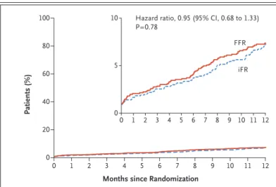
At 1 year, the primary end point (a composite of major adverse cardiac events) had occurred in 78 of 1148 patients (6.8%) in the iFR group and in 83 of 1182 patients (7.0%) in the FFR group (Fig. 2). The hazard ratio was 0.95 (95% confi- dence interval [CI], 0.68 to 1.33; P = 0.78), and the difference in risk was −0.2 percentage points (95% CI, −2.3 to 1.8; 99% CI, −2.9 to 2.5;
P = 0.83) (Table 3, and Table S2 in the Supple- mentary Appendix). The upper limits of the two- sided 95% and 99% confidence intervals were within the prespecified noninferiority margin of 3.4 percentage points (P<0.001 for noninferior- ity). The risks of each individual component of the primary end point and of death from cardio- vascular or noncardiovascular causes did not differ significantly between the two groups.
The noninferiority of iFR to FFR was also confirmed in the per-protocol analysis (Tables S4 and S5 in the Supplementary Appendix). In the per-protocol analysis, the hazard ratio for major adverse cardiac events was 0.94 (95% CI, 0.67 to 1.31; P = 0.72), and the difference in risk was −0.3 percentage points (95% CI, −2.4 to 1.8;
99% CI, −3.0 to 2.4; P = 0.77). The risk of each individual component of the composite end point did not differ significantly between the two groups in the per-protocol analyses.
Procedural Signs and Symptoms
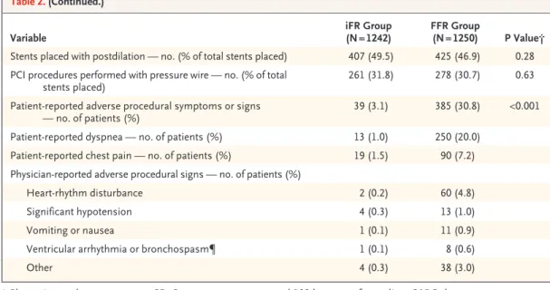
In the iFR group, 39 patients (3.1%) reported adverse procedural symptoms or signs, includ- ing 19 who reported chest pain and 13 who re- ported dyspnea (Table 2). In the FFR group, 385
patients (30.8%) reported adverse procedural symptoms or signs, including 250 who reported dyspnea and 90 who reported chest pain. The difference between the two groups in the num- ber of patients with adverse procedural symp- toms or signs was significant (P<0.001) (Fig. S9 in the Supplementary Appendix). Serious adverse events (bronchospasm and ventricular arrhyth- mias) were reported in 8 patients in the FFR group (after hyperemia) and in 1 patient in the iFR group.
Discussion
In the DEFINE-FLAIR trial, we found that iFR- guided coronary revascularization was noninfe- rior to FFR-guided revascularization with respect to the risk of major adverse cardiac events. The use of iFR was also associated with a lower rate of procedural signs and symptoms and with a shorter procedural time than the use of FFR.
There were no significant differences between the trial groups in the rates of death from any cause, death from cardiovascular causes, non- fatal myocardial infarction, and unplanned re- vascularization. These results suggest that the benefits of physiologically guided coronary re- vascularization with FFR can also be achieved with iFR. Our principal findings are similar to those now reported in the Journal by Götberg et al.16
It has previously been proposed that a hybrid iFR–FFR approach might be advantageous for the detection of functionally significant steno- ses, with iFR used as the initial measure and FFR used only to evaluate stenoses that were of intermediate severity on iFR-guided assess- ment.17,18 However, the results of our trial sug- gest that iFR alone can effectively identify steno- ses that require intervention. Our trial also provides clinical evidence that there is no sig- nificant advantage to the administration of a hyperemic agent — a finding consistent with results of studies in which iFR and FFR were compared with other reference standards.13,14,19,20
Although evidence supporting the benefits of physiologically guided revascularization has ac- cumulated over the past decade, adoption of this approach in clinical practice has lagged. There are many reasons for this, including equipment and drug costs, inadequate reimbursement, phy- sician preferences, patient symptoms, and addi-
Variable iFR Group
(N = 1242) FFR Group
(N = 1250) P Value†
Radial-artery approach — no. of patients (%) 896 (72.1) 888 (71.0) 0.54
Procedure time — min
Median 40.5 45.0 0.001
Interquartile range 27.0–60.0 30.0–66.0
Hyperemic agent administered — no. of patients (% of total no. who received a hyperemic agent)
Total NA 1608 (100)
Intracoronary adenosine NA 455 (28.3)
Intravenous adenosine NA 950 (59.1)
Other agent NA 203 (12.6)
Multivessel disease — no. of patients (%) 505 (40.7) 519 (41.5) 0.66
Type of vessel evaluated — no. (% of total vessels evaluated)‡
Total 1575 (100) 1608 (100) 0.58
Left anterior descending artery 844 (53.6) 845 (52.5) 0.56
Left circumflex artery 323 (20.5) 333 (20.7) 0.89
Right coronary artery 374 (23.7) 393 (24.4) 0.65
Other 33 (2.1) 31 (1.9) 0.74
Unknown 1 (0.1) 6 (0.4) 0.06
Total no. of vessels evaluated or treated‡ 1879 1940 0.42
No. of vessels evaluated or treated per patient‡ 1.51±0.76 1.55±0.80 0.42 Functionally significant lesions — no. (% of total vessels evaluated)§ 451 (28.6) 557 (34.6) 0.004
≥1 Functionally significant lesions present — no. of patients (%)§ 426 (34.3) 486 (38.9) 0.02
Mean iFR 0.91±0.09 NA
Mean FFR NA 0.83±0.09
Percent of lesions within the FFR range
<0.60 NA 1.96
0.60–0.90 NA 75.08
>0.90 NA 22.96
Revascularization performed — no. of patients (%)
Total 590 (47.5) 667 (53.4) 0.003
CABG 25 (2.0) 42 (3.4) 0.04
PCI 565 (45.5) 625 (50.0) 0.02
Stents placed — no. (% of total stents placed)
Total 822 (100) 906 (100) 0.86
Drug-eluting stent 811 (98.7) 893 (98.6)
Bioresorbable vascular scaffold 11 (1.3) 13 (1.4)
No. of stents placed per patient 0.66±0.92 0.72±0.96 0.09
Stent length per patient — mm
Median 28.0 28.0 0.74
Interquartile range 18.0–42.0 18.0–44.0
Stent diameter — mm
Median 3.00 3.00 0.44
Interquartile range 2.67–3.25 2.75–3.25
Table 2. Procedural Characteristics.*
tional procedural burden. Although adenosine is a generally safe drug that is used in millions of diagnostic procedures annually, its risks are well documented21,22 and it is not suitable for every patient; therefore, avoiding the use of adenosine is preferable.11,23,24 In addition, adenosine contrib- utes substantially to the cost of physiological
stenosis assessment, and its use is hampered in many countries because it is unavailable or not indicated for this purpose. Thus, the ability to perform physiological assessments of coronary- artery stenoses without the use of adenosine may increase the use of such assessments in clinical practice.
Variable iFR Group
(N = 1242) FFR Group
(N = 1250) P Value†
Stents placed with postdilation — no. (% of total stents placed) 407 (49.5) 425 (46.9) 0.28 PCI procedures performed with pressure wire — no. (% of total
stents placed) 261 (31.8) 278 (30.7) 0.63
Patient-reported adverse procedural symptoms or signs
— no. of patients (%) 39 (3.1) 385 (30.8) <0.001
Patient-reported dyspnea — no. of patients (%) 13 (1.0) 250 (20.0) Patient-reported chest pain — no. of patients (%) 19 (1.5) 90 (7.2) Physician-reported adverse procedural signs — no. of patients (%)
Heart-rhythm disturbance 2 (0.2) 60 (4.8)
Significant hypotension 4 (0.3) 13 (1.0)
Vomiting or nausea 1 (0.1) 11 (0.9)
Ventricular arrhythmia or bronchospasm¶ 1 (0.1) 8 (0.6)
Other 4 (0.3) 38 (3.0)
* Plus–minus values are means ±SD. Percentages may not total 100 because of rounding. CABG denotes coronary-artery bypass grafting, NA not applicable, and PCI percutaneous coronary intervention.
† P values that compare distributions were calculated by means of the Wilcoxon rank-sum test. P values that compare percentages were calculated by means of a test for proportions.
‡ Evaluated vessels are vessels that underwent physiological assessment. Treated vessels are vessels that underwent PCI.
§ Functionally significant lesions are lesions with an iFR or FFR equal to or lower than the treatment threshold (0.89 and 0.80, respectively).
¶ Serious adverse events included ventricular arrhythmias and bronchospasm; one case of ventricular arrhythmia occurred in the iFR group, and one case of ventricular arrhythmia and seven cases of bronchospasm occurred in the FFR group.
Table 2. (Continued.)
Outcome iFR Group FFR Group Difference in Risk P Value
no./total no. (%) percentage points
(95% CI) percentage points (99% CI) Primary end point: death from any cause,
nonfatal myocardial infarction, or unplanned revascularization
78/1148 (6.8) 83/1182 (7.0) −0.2 (−2.3 to 1.8)† −0.2 (−2.9 to 2.5) 0.83
Unplanned revascularization 46/1147 (4.0) 63/1181 (5.3) −1.3 (−3.0 to 0.4) −1.3 (−3.1 to 1.9) 0.13 Nonfatal myocardial infarction 31/1148 (2.7) 28/1180 (2.4) 0.3 (−1.0 to 1.6) 0.3 (−1.4 to 2.0) 0.62 Death from cardiovascular causes 7/1147 (0.6) 4/1179 (0.3) 0.3 (−0.3 to 0.8) 0.3 (−0.5 to 1.0) 0.34 Death from noncardiovascular causes 15/1147 (1.3) 9/1179 (0.8) 0.5 (−0.3 to 1.4) 0.5 (−0.5 to 1.6) 0.19 Death from any cause 22/1147 (1.9) 13/1179 (1.1) 0.8 (−0.2 to 1.8) 0.8 (−0.5 to 2.1) 0.11
* Patients who had a myocardial infarction or an unplanned revascularization before withdrawing from the study were included in the analyses.
† For the primary end point, the upper limit of the 95% confidence interval was 1.8 percentage points, which was within the prespecified non- inferiority margin of 3.4 percentage points.
Table 3. Outcomes for Difference in Risk at 1 Year.*
Although the patients were not informed of their group assignments, adverse procedural symptoms or signs occurred in 30.8% of the patients in the FFR group, as compared with 3.1% of the patients in the iFR group. This dif- ference is most likely due to the side effects of adenosine. It is therefore possible that at least some patients in the FFR group became aware of their group assignment. Such unblinding could have led to bias in the rates of unplanned revas- cularization, especially if patients discussed these symptoms with their physicians.
The number of functionally significant steno- ses was lower in the iFR group than in the FFR group. This difference could be a consequence of dissimilar thresholds for the two measures.
In addition, iFR has been shown to be more closely linked to coronary flow reserve than FFR, and a previous study has shown higher revascularization rates associated with assess- ment guided by FFR than with assessment guided by coronary flow reserve.25 Regardless of the explanation, the results of our trial suggest that the use of iFR can lead to outcomes similar
to those associated with FFR and to the place- ment of fewer (potentially unnecessary) stents.
The clinical population in our trial differed from the population in the FAME trial (Fractional Flow Reserve Versus Angiography for Multivessel Evaluation), in which all the patients had multi- vessel disease and were scheduled for revascular- ization.6 In DEFINE-FLAIR, only 41% had multi- vessel disease. Although the benefit of coronary revascularization in patients with single-vessel disease is likely to be more uncertain, our trial population is probably similar to the population that would be seen in current clinical practice.
Given the clinical evidence in support of physi- ologically guided revascularization, it was con- sidered unethical to repeat a study similar to FAME, in which iFR-guided revascularization was compared with angiography-guided revascular- ization.
In our trial, the noninferiority margin for the difference in risk was set at 3.4 percentage points, which meant that the upper limit of the hazard ratio could have been as high as 1.40 while still allowing a claim of noninferiority.
Although this noninferiority margin is wide, it is similar to margins used in other major clinical trials in cardiology.26-32 The event rates were lower than had been expected, because the num- ber of patients with an acute coronary syndrome who were enrolled in the trial was lower than had been anticipated. However, when we used the pre- specified noninferiority margin to test the ac- tual event rate among the prespecified number of patients, we found that iFR was noninferior to FFR even when the upper limit of a one-sided 99.5% confidence interval was used.
In conclusion, we found that coronary revascu- larization guided by iFR was noninferior to re- vascularization guided by FFR with respect to major adverse cardiac events at 1 year. The rate of adverse procedural signs or symptoms was lower and the procedure time was shorter in the iFR group than in the FFR group.
Supported by an unrestricted educational grant from Philips Volcano to Imperial College London.
Disclosure forms provided by the authors are available with the full text of this article at NEJM.org.
We thank the staff of the Biomedical Research Centre at Im- perial College London and Oxford University, the events com- mittee, and the trial-management team.
Figure 2. Cumulative Risk of the Primary End Point.
Shown is the cumulative risk of the composite of death from any cause, nonfatal myocardial infarction, or unplanned revascularization at 1 year.
The inset shows the same data on an enlarged y axis.
Patients (%)
100 80 60 40 20
0
0 1 2 3 4 5 6 7 8 9 10 11 12
Months since Randomization
Hazard ratio, 0.95 (95% CI, 0.68 to 1.33) P=0.78
No. at Risk iFR FFR
1242 1250
1149 1169
1131 1156
1122 1149
1118 1144
1111 1141
1088 1119
1052 1081
1037 1066
1027 1055
1019 1046
995 1017
764 793 10
5
00 1 2 3 4 5 6 7 8 9 10 11 12 FFR
iFR
Appendix
The authors’ full names and academic degrees are as follows: Justin E. Davies, M.D., Ph.D., Sayan Sen, M.D., Ph.D., Hakim-Moulay Dehbi, Ph.D., Rasha Al-Lamee, M.D., Ricardo Petraco, M.B., B.S., Ph.D., Sukhjinder S. Nijjer, M.B., B.S., Ph.D., Ravinay Bhindi, M.B., B.S., Ph.D., Sam J. Lehman, M.B., B.S., Ph.D., Darren Walters, M.B., B.S., James Sapontis, M.B., B.S., Luc Janssens, M.D., Christiaan J.
Vrints, M.D., Ph.D., Ahmed Khashaba, M.D., Mika Laine, M.D., Ph.D., Eric Van Belle, M.D., Ph.D., Florian Krackhardt, M.D., Walde- mar Bojara, M.D., Olaf Going, M.D., Tobias Härle, M.D., Ciro Indolfi, M.D., Giampaolo Niccoli, M.D., Ph.D., Flavo Ribichini, M.D., Nobuhiro Tanaka, M.D., Ph.D., Hiroyoshi Yokoi, M.D., Hiroaki Takashima, M.D., Ph.D., Yuetsu Kikuta, M.D., Andrejs Erglis, M.D., Ph.D., Hugo Vinhas, M.D., Pedro Canas Silva, M.D., Sérgio B. Baptista, M.D., Ali Alghamdi, M.D., Farrel Hellig, M.B., B.S., Bon-Kwon Koo, M.D., Ph.D., Chang-Wook Nam, M.D., Ph.D., Eun-Seok Shin, M.D., Joon-Hyung Doh, M.D., Ph.D., Salvatore Brugaletta, M.D., Ph.D., Eduardo Alegria-Barrero, M.D., Ph.D., Martijin Meuwissen, M.D., Ph.D., Jan J. Piek, M.D., Ph.D., Niels van Royen, M.D., Ph.D., Murat Sezer, M.D., Carlo Di Mario, M.D., Ph.D., Robert T. Gerber, Ph.D., Iqbal S. Malik, Ph.D., Andrew S.P. Sharp, M.D., Suneel Talwar, M.B., B.S., M.D., Kare Tang, M.D., Habib Samady, M.D., John Altman, M.D., Arnold H. Seto, M.D., Jasvindar Singh, M.D., Allen Jeremias, M.D., Hitoshi Matsuo, M.D., Ph.D., Rajesh K. Kharbanda, M.D., Ph.D., Manesh R. Patel, M.D., Patrick Serruys, M.D., Ph.D., and Javier Escaned, M.D., Ph.D.
The authors’ affiliations are as follows: Hammersmith Hospital (J.E.D., S.S., R.A.-L., R.P., S.S.N., I.S.M., P.S.) and Royal Brompton Hospital (C.D.M.), Imperial College London, Cancer Research UK and University College London Cancer Trials Centre (H.-M.D.), London, Conquest Hospital, St. Leonards-on-Sea (R.T.G.), Royal Devon and Exeter Hospital and University of Exeter, Exeter (A.S.P.S.), Royal Bournemouth General Hospital, Bournemouth (S.T.), Essex Cardiothoracic Centre, Basildon (K.T.), Anglia Ruskin University, Chelmsford (K.T.), and John Radcliffe Hospital, Oxford University Hospitals Foundation Trust, Oxford (R.K.K.) — all in the United Kingdom; Royal North Shore Hospital, Sydney (R.B.), Flinders University, Adelaide, SA (S.J.L.), Prince Charles Hospital, Brisbane, QLD (D.W.), and MonashHeart and Monash University, Melbourne, VIC (J. Sapontis) — all in Australia; Imelda Hospital, Bonheiden (L.J.), and Antwerp University Hospital, Antwerp (C.J.V.) — both in Belgium; Ain Shams University, Cairo (A.K.); Helsinki University Hospital, Helsinki (M.L.); Institut Coeur Poumon, Lille University Hospital, and INSERM Unité 1011, Lille, France (E.V.B.); Charite Campus Virchow Klinikum, Universitaetsmedizin, Berlin (F.K.), Gemeinschaftsklinikum Mittelrhein, Kemperhof Koblenz, Koblenz (W.B.), Sana Klinikum Lichtenberg, Lichtenberg (O.G.), and Klinikum Oldenburg, European Medical School, Carl von Ossietzky University, Olden- burg (T.H.) — all in Germany; University Magna Graecia, Catanzaro (C.I.), Catholic University of the Sacred Heart, Rome (G.N.), University Hospital Verona, Verona (F.R.), and University of Florence, Florence (C.D.M.) — all in Italy; Tokyo Medical University, Tokyo (N.T.), Fukuoka Sannou Hospital, Fukuoka (H.Y.), Aichi Medical University Hospital, Aichi (H.T.), Fukuyama Cardiovascular Hospital, Fukuyama (Y.K.), and Gifu Heart Center, Gifu (H.M.) — all in Japan; Pauls Stradins Clinical University Hospital, Riga, Latvia (A.E.);
Hospital Garcia de Horta (H.V.) and Hospital Santa Maria (P.C.S.), Lisbon. and Hospital Prof. Doutor Fernando Fonseca, Amadora (S.B.B.) — all in Portugal; King Abdulaziz Medical City Cardiac Center, Riyadh, Saudi Arabia (A.A.); Sunninghill Hospital, Johannesburg (F.H.); Seoul National University Hospital, Seoul (B.-K.K.), Keimyung University Dongsan Medical Center, Daegu (C.-W.N.), Ulsan University Hospital, University of Ulsan College of Medicine, Ulsan (E.-S.S.), and Inje University Ilsan Paik Hospital, Daehwa-Dong (J.-H.D.) — all in South Korea; Cardiovascular Institute, Hospital Clinic, Institut d’Investigacions Biomèdiques August Pi i Sunyer (IDIBAPS), Barcelona (S.B.); Hospital Universitario de Torrejón and Universidad Francisco de Vitoria (E.A.-B.) and Hospital Clinico San Carlos and Universidad Complutense de Madrid (J.E.), Madrid; Amphia Hospital, Breda (M.M.), and AMC Heart Center, Academic Medical Center (J.J.P.), and VU University Medical Center (N.R.), Amsterdam — all in the Netherlands; Istanbul University, Istanbul Faculty of Medicine, Istanbul, Turkey (M.S.); Emory University, Atlanta (H.S.); Colorado Heart and Vascular, Lakewood (J.A.); Veterans Affairs Long Beach Healthcare System, Long Beach, CA (A.H.S.); Washington University School of Medicine, St. Louis (J. Singh); Stony Brook University Medical Center, New York (A.J.); and Duke University, Durham, NC (M.R.P.).
References
1. Di Mario C, Moses JW, Anderson TJ, et al. Randomized comparison of elective stent implantation and coronary balloon angioplasty guided by online quantitative angiography and intracoronary Doppler.
Circulation 2000; 102: 2938-44.
2. Kern MJ, Donohue TJ, Aguirre FV, et al.
Clinical outcome of deferring angioplas- ty in patients with normal translesional pressure-flow velocity measurements. J Am Coll Cardiol 1995; 25: 178-87.
3. Serruys PW, Di Mario C, Meneveau N, et al. Intracoronary pressure and flow ve- locity with sensor-tip guidewires: a new methodologic approach for assessment of coronary hemodynamics before and after coronary interventions. Am J Cardiol 1993;
71: 41D-53D.
4. Pijls NH, van Son JA, Kirkeeide RL, De Bruyne B, Gould KL. Experimental basis of determining maximum coronary, myo- cardial, and collateral blood flow by pres-
sure measurements for assessing func- tional stenosis severity before and after percutaneous transluminal coronary an- gioplasty. Circulation 1993; 87: 1354-67.
5. Gould KL, Kirkeeide RL, Buchi M.
Coronary flow reserve as a physiologic measure of stenosis severity. J Am Coll Cardiol 1990; 15: 459-74.
6. Tonino PAL, De Bruyne B, Pijls NHJ, et al. Fractional flow reserve versus angi- ography for guiding percutaneous coro- nary intervention. N Engl J Med 2009; 360:
213-24.
7. Fearon WF, Shilane D, Pijls NHJ, et al.
Cost-effectiveness of percutaneous coro- nary intervention in patients with stable coronary artery disease and abnormal fractional flow reserve. Circulation 2013;
128: 1335-40.
8. Patel MR, Dehmer GJ, Hirshfeld JW, et al. ACCF/SCAI/STS/AATS/AHA/ASNC/
HFSA/SCCT 2012 appropriate use criteria
for coronary revascularization focused up- date: a report of the American College of Cardiology Foundation Appropriate Use Criteria Task Force, Society for Cardiovas- cular Angiography and Interventions, Soci- ety of Thoracic Surgeons, American Asso- ciation for Thoracic Surgery, American Heart Association, American Society of Nuclear Cardiology, and the Society of Car- diovascular Computed Tomography. J Tho- rac Cardiovasc Surg 2012; 143: 780-803.
9. Patel MR, Dehmer GJ, Hirshfeld JW, Smith PK, Spertus JA. ACCF/SCAI/STS/
AATS/AHA/ASNC/HFSA/SCCT 2012 appro- priate use criteria for coronary revascular- ization focused update: a report of the American College of Cardiology Founda- tion Appropriate Use Criteria Task Force, Society for Cardiovascular Angiography and Interventions, Society of Thoracic Surgeons, American Association for Tho- racic Surgery, American Heart Associa-
TRACK THIS ARTICLE’S IMPACT AND REACH
Visit the article page at NEJM.org and click on the Metrics tab for a dashboard that logs views, citations, media references, and commentary, with easy linking.
Learn more at www.nejm.org/page/article-metrics-faq.
tion, American Society of Nuclear Cardi- ology, and the Society of Cardiovascular Computed Tomography. J Am Coll Cardiol 2012; 59: 857-81.
10. Windecker S, Kohl P, Alfonso S, et al.
2014 ESC/EACTS guidelines on myocar- dial revascularization. Eur Heart J 2014;
35: 2541-619.
11. Pijls NHJ, Tonino PAL. The crux of maximum hyperemia: the last remaining barrier for routine use of fractional flow reserve. JACC Cardiovasc Interv 2011; 4:
1093-5.
12. Sen S, Escaned J, Malik IS, et al. Devel- opment and validation of a new adenosine- independent index of stenosis severity from coronary wave-intensity analysis:
results of the ADVISE (ADenosine Vasodi- lator Independent Stenosis Evaluation) study. J Am Coll Cardiol 2012; 59: 1392-402.
13. Sen S, Asrress KN, Nijjer S, et al. Di- agnostic classification of the instanta- neous wave-free ratio is equivalent to frac- tional flow reserve and is not improved with adenosine administration: results of CLARIFY (Classification Accuracy of Pres- sure-Only Ratios Against Indices Using Flow Study). J Am Coll Cardiol 2013; 61:
1409-20.
14. Petraco R, van de Hoef TP, Nijjer S, et al. Baseline instantaneous wave-free ratio as a pressure-only estimation of underly- ing coronary flow reserve: results of the JUSTIFY-CFR Study (Joined Coronary Pres- sure and Flow Analysis to Determine Di- agnostic Characteristics of Basal and Hyperemic Indices of Functional Lesion Severity-Coronary Flow Reserve). Circ Car- diovasc Interv 2014; 7: 492-502.
15. Layland J, Oldroyd KG, Curzen N, et al.
Fractional flow reserve vs. angiography in guiding management to optimize out- comes in non-ST-segment elevation myo- cardial infarction: the British Heart Foun- dation FAMOUS-NSTEMI randomized trial.
Eur Heart J 2015; 36: 100-11.
16. Götberg M, Christiansen EH, Gud- mundsdottir IJ, et al. Instantaneous wave-
free ratio versus fractional flow reserve to guide PCI. N Engl J Med 2017; 376:1813-23.
17. Petraco R, Park JJ, Sen S, et al. Hybrid iFR-FFR decision-making strategy: impli- cations for enhancing universal adoption of physiology-guided coronary revasculari- sation. EuroIntervention 2013; 8: 1157-65.
18. Escaned J, Echavarría-Pinto M, Garcia- Garcia HM, et al. Prospective assessment of the diagnostic accuracy of instanta- neous wave-free ratio to assess coronary stenosis relevance: results of ADVISE II in- ternational, multicenter study (ADenosine Vasodilator Independent Stenosis Evalua- tion II). JACC Cardiovasc Interv 2015; 8:
824-33.
19. Sen S, Nijjer S, Petraco R, Malik IS, Francis DP, Davies J. Instantaneous wave- free ratio: numerically different, but diag- nostically superior to FFR? Is lower always better? J Am Coll Cardiol 2013; 62: 566.
20. van de Hoef TP, Meuwissen M, Es- caned J, et al. Head-to-head comparison of basal stenosis resistance index, instanta- neous wave-free ratio, and fractional flow reserve: diagnostic accuracy for stenosis- specific myocardial ischaemia. EuroInter- vention 2015; 11: 914-25.
21. Dilsizian V, Gewirtz H, Paivanas N, et al. Serious and potentially life threaten- ing complications of cardiac stress testing:
physiological mechanisms and manage- ment strategies. J Nucl Cardiol 2015; 22:
1198-213.
22. Cerqueira MD, Verani MS, Schwaiger M, Heo J, Iskandrian AS. Safety profile of adenosine stress perfusion imaging:
results from the Adenoscan Multicenter Trial Registry. J Am Coll Cardiol 1994; 23:
384-9.
23. Kern MJ, Seto AH. On the search for an “easy” FFR: submaximal hyperemia and NTG-induced translesional pressure drop (Pd/Pa-NTG). Catheter Cardiovasc Interv 2016; 87: 270-2.
24. Mallet ML. Proarrhythmic effects of adenosine: a review of the literature.
Emerg Med J 2004; 21: 408-10.
25. van de Hoef TP, van Lavieren MA, Damman P, et al. Physiological basis and long-term clinical outcome of discor- dance between fractional flow reserve and coronary flow velocity reserve in coronary stenoses of intermediate severity. Circ Cardiovasc Interv 2014; 7: 301-11.
26. Stone GW, Sabik JF, Serruys PW, et al.
Everolimus-eluting stents or bypass sur- gery for left main coronary artery disease.
N Engl J Med 2016; 375: 2223-35.
27. Mohr FW, Morice MC, Kappetein AP, et al. Coronary artery bypass graft sur- gery versus percutaneous coronary inter- vention in patients with three-vessel dis- ease and left main coronary disease:
5-year follow-up of the randomised, clini- cal SYNTAX trial. Lancet 2013; 381: 629-38.
28. Mäkikallio T, Holm NR, Lindsay M, et al. Percutaneous coronary angioplasty versus coronary artery bypass grafting in treatment of unprotected left main steno- sis (NOBLE): a prospective, randomised, open-label, non-inferiority trial. Lancet 2016; 388: 2743-52.
29. Patel MR, Mahaffey KW, Garg J, et al.
Rivaroxaban versus warfarin in nonvalvu- lar atrial fibrillation. N Engl J Med 2011;
365: 883-91.
30. Granger CB, Alexander JH, McMurray JJV, et al. Apixaban versus warfarin in pa- tients with atrial fibrillation. N Engl J Med 2011; 365: 981-92.
31. Holmes DR Jr, Kar S, Price MJ, et al.
Prospective randomized evaluation of the Watchman Left Atrial Appendage Closure device in patients with atrial fibrillation versus long-term warfarin therapy: the PREVAIL trial. J Am Coll Cardiol 2014; 64:
1-12.
32. von Birgelen C, Basalus MWZ, Tand- jung K, et al. A randomized controlled trial in second-generation zotarolimus- eluting Resolute stents versus everolimus- eluting Xience V stents in real-world pa- tients: the TWENTE trial. J Am Coll Cardiol 2012; 59: 1350-61.
Copyright © 2017 Massachusetts Medical Society.