M.D. , Ki Ouk Min, M.D.
Departments of
1Internal Medicine, and
2Pathology, The Catholic University of Korea College of Medicine, Seoul, Korea
Herein we report the case of a 71-year-old woman who complained of fatigue and enlarged right axillary lymph nodes for 18 months. At her first visit, her chest X-ray showed diffuse nodular opacities in both lung fields. Initial excisional biopsy of the axillary lymph nodes showed granulomatous lesions and acid fast bacilli were seen on Ziehl-Neelsen staining. However, even after 15 months of anti-tuberculosis (TB) medication, her right axillary lymph nodes were enlarged. We re-performed an excisional biopsy of the nodes, which showed Hodgkin's lymphoma (HL). A retrograde review of the biopsy before anti-tuberculous medication, revealed HL coexisting with TB. HL and TB cause difficulties in differential diagnosis due to similarities in clinical course, imaging procedures and histopathological analysis of the involved tissue. Therefore, it is important to consider the possibility of concurrent HL and TB when patients who undergo treatment for TB or chemotherapy for lymphoma complain of persistent systemic symptoms or enlarged lymph nodes.Key Words: Tuberculosis; Hodgkin Disease; Lymphadenitis
Address for correspondence: Sang Haak Lee, M.D.
Department of Internal Medicine, St. Paul's Hospital, The Catholic University of Korea College of Medicine, 620-56, Jeonnong 1-dong, Dongdaemun-gu, Seoul 130-709, Korea Phone: 82-2-961-4500, Fax: 82-2-968-7250
E-mail: [email protected] Received: Oct. 14, 2010 Accepted: Jan. 23, 2011
서 론
호지킨 림프종은 조직 소견에서 리드-슈테른베르크 (Reed-Sternberg)세포를 특징으로 하는 림프종의 한 형태 로 악성 림프종의 약 8.2%를 차지하는 것으로 보고되고 있다1. 호지킨 림프종은 유병률이 서구에 비해 낮으며, 대 부분의 환자에서 동통을 수반하지 않는 림프절을 촉지할 수 있는데, 1/3의 환자에서 발열, 야간 발한, 체중 감소 등의 B 증상을 호소하여 임상적으로 결핵과의 감별을 어
렵게 한다2,3. 영상학적 소견 역시 결핵과 유사하고4 조직 병리학적으로도 결핵의 특징적인 소견인 건락성 육아종 을 관찰할 수 있기 때문에 두 질환의 감별 진단은 쉽지
않다3,5,6. 또한 조직 소견에서 소수의 종양세포와 다수의
반응성 세포로만 관찰되는 경우도 많아 위음성률이 높은 편이다2.
저자 등은 액와부 림프절 조직에서 육아종성 병변과 항 산성 염색 및 결핵배양 양성 소견을 보여, 항결핵제 치료 를 시작하였으나, 림프절 종대가 진행하여 다시 시행한 조직 생검에서 결핵에 동반된 호지킨 림프종을 추후 진단 한 예를 경험하였기에 문헌고찰과 함께 보고하는 바이다.
증 례
환 자: 71세, 여자
주 소: 우측 액와부의 다발성 종괴
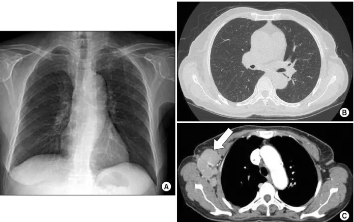
Figure 1. Initial chest X-ray (A) and computed tomography (B) shows multiple fine nodular opacities in both lung fields and multiple enlarged axillary lymph nodes (C, arrow).
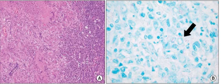
Figure 2. The initial right axillary lymph node biopsy shows multiple granulomatous lesions with giant cell and diffused small lymphocytes (A, H&E stain, ×100). Acid fast bacilli (arrow) are seen on Ziehl-Neelsen stain (B, AFB stain, ×1,000).
현병력: 환자는 내원 15개월 전 단순 흉부 방사선 및 흉부 전산화 단층촬영에서 양측 미만성 다발성 결절 (Figure 1A, B) 및 우측 액와부 림프절 종대(Figure 1C) 소견으로 액와부 림프절 절제 생검을 시행하였으며, 특징
적인 결핵 조직 소견과 항산균 염색 양성(Figure 2), 결핵 균 배양 검사 양성 소견이 나와 항결핵제를 복용 중이었 다. 당시 시행한 두 차례의 객담 결핵균 도말 검사 및 배양 검사에서는 항산균이 관찰되지 않았으나 기관지 세척액
Figure 3. Chest X-ray (A) and computed tomography (B) taken 15 months after anti-tuberculosis medication shows decreased infiltration in both lung fields, but axillary lymph nodes (arrow) are increased in number and size (C).
가족력 및 사회력: 특이 소견은 없었다.
이학적 소견: 내원 시 활력 징후는 혈압 120/60 mm Hg, 체온 36.6oC, 맥박수 72회/분, 호흡수 20회/분이었고 전신상태는 양호하였다. 흉부 청진에서 호흡음은 깨끗하 였다. 우측 액와부에서 압통을 동반하지 않는 다양한 크 기의 다발성 종물이 만져졌다. 그 외 특이 소견은 없었다.
검사실 소견: 말초 혈액 검사에서 백혈구 5,600/mm3, 중성구 59.8%, 혈색소 10.7 g/dL, 혈소판 82,000/mm3이
여 크기 및 개수가 증가된 소견이었다(Figure 3C).
병리학적 소견: 액와부 림프절 절제 생검에서 육안상으 로는 절단면이 회백색을 띠며 매끄러웠으며 중심부는 연 한 갈색을 띠었다. 광학현미경상 저배율에서는 산재된 림 프구 및 건락성 육아종을 확인할 수 있었으며(Figure 4A), 고배율에서는 호지킨 림프종의 특징인 단핵 이형성의 리 드-슈테른베르크 세포가 다수 관찰되어 림프구 우세 호지 킨 림프종으로 진단할 수 있었다(Figure 4B). 이후 15개월
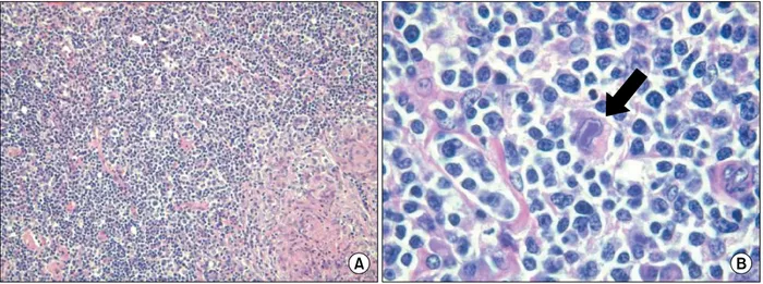
Figure 4. Biopsy taken after 15 months of antituberculosis medication shows multiple granulomatous lesions with diffused small lymphocytes and scattered macrophages (A, H&E stain, ×40), and mononuclear Reed-Stenberg cells (B, arrow, H&E stain, ×1,000).
전 시행한 림프절 절제 생검의 조직 소견을 재고찰한 결과 호지킨 림프종이 동반됨을 확인할 수 있었다.
치료 및 경과: 환자는 호지킨 림프종에 대한 치료를 거 부하고 퇴원한 상태이다.
고 찰
호지킨 림프종은 조직 소견에서 리드-슈테른베르크 세 포를 특징으로 하는 림프종의 한 형태이다1. 인구 10,000 명 당 약 3명의 비율로 발생하는 드문 질환이며, 모든 연 령대에서 발생할 수 있으나 20대와 50대에서 빈도가 증가 하는 특징을 보인다7.
호지킨 림프종 환자들은 세포성 면역결핍으로 인해 결 핵균(Mycobacterium tuberculosis), Ebstein-Barr Virus, 거대세포바이러스(cytomegalovirus), 단순포진 바이러스 (herpes simplex), 사람폐포자충(pneumocystis jirovecii) 등에 감염될 가능성이 높고, 이들 병원체에 의한 감염이 호지킨 림프종의 발생에도 기여할 수 있다1,3. 또한 이러한 감염 자체가 체내에서 호지킨 림프종의 발생과 성장에 기 여하는 것으로 알려져 있다3. 본 증례에서는 호지킨 림프 종과 결핵이 동반되어 있었으나 발생의 선후관계를 명확 히 밝힐 수는 없었다.
결핵성 림프절염은 진단 시에 많은 감별 진단을 요하는 데, 다른 감염(세균, 바이러스, 진균), 종양(림프종, 육종, 전이성 암), 비특이적 반응성 증식증(non-specific re- active hyperplasia), 유육종(sarcoidosis), 톡소포자충증
(toxoplasmosis), 기타 결체조직 질환 등이 이에 포함된 다8. 특히 이 중에서 호지킨 림프종은 림프절 종대, 발열, 야간 발한, 체중감소 등 결핵과 유사한 증상을 보여 병력 및 임상 양상만으로 두 질환을 감별하기는 어렵다3. 또한 단순 방사선촬영 및 전산화 단층촬영 등의 영상학적 검사 도 유사하기 때문에 두 질환을 감별하는데 있어 조직병리 학적 검사가 매우 중요하다9. 그러나, 호지킨 림프종에서 도 병리학적으로 결핵의 특징적인 소견인 건락성 괴사 및 육아종을 관찰할 수 있어서 리드-슈테른베르크 세포를 관 찰할 수 없다면 두 질환을 감별하는 것은 매우 어렵다5,6. 호지킨 림프종과 결핵의 감별을 위해서는 항산균 염색 및 배양, 결핵균에 대한 역전사 중합효소 연쇄반응(reverse transcriptase-polymerase chain reaction)과 함께 리드-슈 테른베르크 세포의 CD15, CD30 항원에 대한 면역 형광 검사를 시행하는 것이 도움이 된다1,10. 본 증례에서는 조 직에서 항산균 염색 및 결핵 배양 양성 소견과 더불어 리 드-슈테른베르크 세포를 확인하여 결핵에 동반된 호지킨 림프종으로 진단할 수 있었다.
Centkowski 등2은 경부 림프절 조직 검사에서 호지킨 림프종으로 진단받고 항암화학요법으로 치료하였으나 임 상 양상의 호전이 없는 환자에서, 처음에 시행했던 조직 검사를 다시 고찰하여 호지킨 림프종에 동반된 결핵을 추 후 진단하였던 증례를 보고한 바 있다. 국내에서는 Lee 등11이 결핵성 림프절염으로 오진된 자연살해세포 백혈병 1예를 보고한 바 있다. 본 증례는 결핵성 림프절염 및 폐 결핵으로 확진 후 항결핵제를 투여하여 폐결핵 소견은 호
문이다.
호지킨 림프종과 결핵은 임상 양상과 조직 소견으로 감 별이 어려울 수 있다. 따라서, 결핵이 호발하는 지역이라 할지라도 진단 당시 림프종의 가능성을 염두에 두고 조직 소견을 면밀히 검토하는 것이 필요하며, 항결핵 치료에 대한 반응이 좋지 않을 때에는 초기 조직 소견의 고찰 혹 은 재 생검을 통해 림프종을 감별하고자 하는 노력이 중요 할 것으로 생각된다.
참 고 문 헌
1. Poppema S. Immunobiology and pathophysiology of Hodgkin lymphomas. Hematology Am Soc Hematol Educ Program 2005:231-8.
2. Centkowski P, Sawczuk-Chabin J, Prochorec M, Warzocha K. Hodgkin's lymphoma and tuberculosis coexistence in cervical lymph nodes. Leuk Lymphoma 2005;46:471-5.
nodes with simultaneous sarcoidosis-like granuloma- tosis in the intrathoracic lymph nodes and liver.
Zentralbl Pathol 1992;138:292-7.
7. Urba WJ, Longo DL. Hodgkin's disease. N Engl J Med 1992;326:678-87.
8. Mohapatra PR, Janmeja AK. Tuberculous lymphaden- itis. J Assoc Physicians India 2009;57:585-90.
9. Yang ZG, Min PQ, Sone S, He ZY, Liao ZY, Zhou XP, et al. Tuberculosis versus lymphomas in the abdominal lymph nodes: evaluation with contrast-enhanced CT.
AJR Am J Roentgenol 1999;172:619-23.
10. Rüdiger T, Jaffe ES, Delsol G, deWolf-Peeters C, Gascoyne RD, Georgii A, et al. Workshop report on Hodgkin's disease and related diseases ('grey zone' lymphoma). Ann Oncol 1998;9 Suppl 5:S31-8.
11. Lee AJ, Kim SG, Jeon CH, Suh HS, Yoon GS, Seo AN.
A case of natural killer cell leukemia misdiagnosed as tuberculous lymphadenopathy. Korean J Lab Med 2009;29:194-8.