DOI : 10.3341/jkos.2009.50.4.542
= 증례보고 = 접수번호 : 09-14
Visante OCT를 이용한 한국 성인의 전안부 계측
이덕구⋅최시환 충남대학교 의과대학 안과학교실
목적: Visante OCT를 이용하여 한국 성인의 중심각막두께를 포함한 전안부 구조들의 평균 계측치를 측정하고, 계측치와 성별, 연령간 의 관계를 알아보며, A-scan 안구초음파로 측정한 중심각막두께와 비교하고자 하였다.
대상과 방법: 성인 185명 298안을 대상으로 Visante OCT를 이용하여 전안부를 촬영한 후, 4방향(수직, 수평, 45도, 135도)에서 중심각 막두께, 전방깊이, 전방각, 전방각간거리 등을 계측하였다. 그 후 A-scan 안구초음파를 이용하여 중심각막두께를 측정하였다.
결과: Visante OCT로 측정한 중심각막두께가 A-scan 안구초음파를 이용한 값보다 작게 나타났다(p=0.000). 중심각막두께, 전방각, 전방각간거리는 남자보다 여자에서 유의하게 측정값이 작았다(p<0.05). 남녀 모두 전방깊이, 전방각, 전방각간거리가 연령에 따라 유 의하게 감소하는 경향을 보였다(p<0.05).
결론: Visante OCT를 이용한 한국 성인의 전안부 계측치와 연령 및 성별에 따른 변화를 파악할 수 있었다. 이는 각종 전안부 수술시의 참고치 및 전안부 질환의 진단 및 추적관찰에 있어서 표준적인 값으로 유용하게 사용될 수 있으리라 생각한다.
<대한안과학회지 2009;50(4):542-550>
■ 접 수 일: 2008년 9월 18일 ■ 심사통과일: 2008년 11월 25일
■ 통 신 저 자: 최 시 환
대전시 중구 대사동 640 충남대학교병원 안과
Tel: 042-280-7609, Fax: 042-255-3745 E-mail: [email protected]
* 본 논문의 요지는 2007년 대한안과학회 제97회 춘계학술대회에서 구연으로 발표되었음.
* 본 논문의 요지는 2007년 ESCRS 제25회 Stockholm, Sweden에서 포스터로 발표되었음.
* 본 논문의 요지는 2007년 APACRS 제20회 Hanoi, Vietnam에서 구연으로 발표되었음.
전안부 구조물의 형태, 크기, 위치에 대한 고해상도의 영 상을 얻고 정확한 계측을 시행하는 것은 형태학적인 연구 및 전안부의 종양, 각막 질환, 녹내장 등의 질환을 진단하고 추적관찰 하는 데 있어서 중요한 가치를 지니고 있어 이를 위한 장비들이 지속적으로 개발되어 왔다. 그동안 전안부 구조의 정량적인 계측은 Scheimpflug 카메라를 비롯한 각 종 화상 계측방법들과 함께 A-scan 안구초음파, 초음파생 체현미경(UBM), OrbscanTM IIz 각막지형도 검사 등에 의 존하여 왔다.
Visante OCT는 10 µm의 해상력으로 한번에 4방향의 단면상을 얻을 수 있으며, 촬영된 영상 내의 임의의 두 지 점 사이를 계측해주는 측경(caliper) 기능을 내장하고 있어, 각막 질환, 녹내장 등에서 좀 더 정확하고 객관적인 미세 계 측이 가능할 것으로 생각되나,1-5국내에서는 아직 Visante OCT를 이용한 연구 보고가 없었다.
이에 본 연구에서는 Visante OCT를 이용하여 한국 성인
군의 성별 및 연령에 따른 전안부 계측치의 평균 정량 분석 값을 구하고, 각각의 계측치와 성별, 연령 간의 관계를 알아 보고자 하였으며, 이 중 중심각막두께는 A-scan 안구초음 파로 측정한 값과 비교하였다.
대상과 방법
2006년 12월부터 2007년 1월까지 내원한 환자 185명 298안을 대상으로 하였다. 모두 유수정체 안으로 경미한 백내장 이외의 특별한 안과적 질환이 없고, 당뇨, 고혈압 및 다른 전신질환이 없으며, 안과적 수술 및 외상의 기왕력이 없었다. 대상자를 남녀 성별에 따라 나누고, 나이에 따라 20~29세, 30~39세, 40~49세, 50~59세, 60세 이상의 연 령 군으로 나누었다. 최대교정시력, 안압, 굴절이상, 세극등 검사 및 안저 검사를 시행하였다.
실내조명 아래에서 산동하지 않은 대상안에 대해 동일 검사자가 Visante OCT를 이용하여 전안부 영상을 촬영한 후, 4방향(수직, 수평, 45도, 135도)에서의 중심각막두께, 전방깊이, 전방각, 전방각간거리 등을 계측하였다. 또한 Visante OCT의 촬영 후에 국소마취제로 점안 마취한 후, A-scan 안구초음파를 이용하여 중심각막두께를 측정하 였다. 각각의 영상은 동일 검사자가 3회씩 반복 촬영하여 계측치를 구한 후 평균값을 선택하였다. 계측은 Visante OCT 내의 측경 기능을 이용하였다(Fig. 1, 2).
4개의 계측치는 다음과 같다.
1) 중심각막두께(central corneal thickness, CCT): 각막
Iris recess
Internal anterior chamber diameter
Anterior chamber depth
Figure 1.Optical coherence tomography image (Visante OCT) with graphic tools for measurement of different anterior chamber dimensions. A caliper for anterior chamber depth and internal anterior chamber diameter measurements is used. Anterior chamber depth is measured between the corneal endothelium and a line joining the two opposite iris recesses. Internal anterior chamber diameter is measured between corresponding iris recesses.
Scleral spur ACA 500 µm
Figure 2.Optical coherence tomography (Visante OCT) cross-sectional view through the anterior chamber angle region. Anterior chamber angle (ACA) is measured with the apex in the iris recess and the arms of the angle passing through a point on the trabecular meshwork at 500 µm from the scleral spur and the point on the iris perpendicularly opposite.
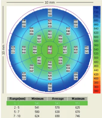
Figure 3. An example of pachymetry mode of Visante OCT. Every sector has three values indicating minimum, average, and maximum corneal thickness in sequence from the above. Average value is the representative of the sector.
내피의 내측경계면에서 각막상피의 외측 경계면까지 의 거리로서 Visante OCT의 각막두께 측정 기능을 이용하여 측정(Fig. 3).
2) 전방깊이(anterior chamber depth): 중심각막내피의 내측경계면에서 양측 iris recess를 연결한 선까지의 거리(Fig. 1).
3) 전방각(anterior chamber angle, ACA): 공막극에서 500 µm 떨어진 섬유주의 한 점과 그 점에서 그은 수
직선이 반대편 홍채와 만나는 지점과 iris recess가 만드는 각도(Fig. 2).
4) 전방각간 거리(internal anterior chamber diameter,
Central corneal thickness (Visante OCT, μm)
[r=-0.069, p=0.235]
Age [years]
700 600 500 400 300 200
100
10 20 30 40 50 60 70 80 90
Central corneal thickness (Ultrasonic pachymeter, μm)
[r=-0.098, p=0.092]
Age [years]
700 600 500 400 300 200
100
10 20 30 40 50 60 70 80 90
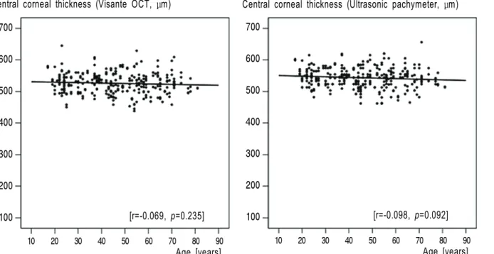
Figure 4. Graphs show the results that there is no statistically significant relationship between central corneal thickness and age.
IACD): 양측 iris recess 사이의 거리(Fig. 1).
측정된 4개의 항목에 대하여 연령 및 성별에 따른 경향을 조사하였다. 또한 Visante OCT와 A-scan 안구초음파를 이용하여 측정한 중심각막두께의 값을 비교하였다.
통계적 검정은 SPSS 11.5 통계 프로그램을 사용하였다.
연령, 성별에 따른 각 측정값들의 평균을 비교하기 위하여 Student t-test, Welch t-test를 사용하였고, 측정방향(수 직, 수평, 45도, 135도)에 따른 전방각의 차이를 비교하기 위해 paired t-test를 사용하였으며, 연령과 각 측정값들의 상관관계를 파악하기 위해 산포도 작성 및 선형회귀분석을 시행하였다. Visante OCT 측정치와 A-scan 안구초음파 측정치와의 일치도를 알아보기 위하여 paired t-test를 시 행하였다. p값이 0.05 미만인 경우를 통계학적으로 유의한 것으로 간주하였다.
결 과
대상안은 남자 145안, 여자 153안, 총 298안이었다 (Table 1). Visante OCT를 이용한 중심각막두께(central corneal thickness)는 전체 연령의 평균값을 구하였을 때 남자 533.0±32.2 µm, 여자 518.8±32.0 µm로 여자의 중 심각막두께가 통계적으로 유의하게 얇았고(p=0.000) 각 연령대별로 나누어 남녀를 비교하였을 때는 여자가 남자보 다 중심각막두께가 얇은 것이 관찰되었지만 통계적으로 유
의하지는 않았다(Table 2). 또한 연령이 증가할수록 중심 각막두께가 감소하는 것이 관찰되었으나 통계적으로 유의 한 상관관계는 보이지 않았다(r=-0.069, p=0.235)(Fig.
4). A-scan 안구초음파를 이용한 중심각막두께(central corneal thickness)는 전체 연령의 평균값을 구하였을 때 남자 549.5±30.2 µm, 여자 537.8±33.5 µm로 여자의 중 심각막두께가 통계적으로 유의하게 얇았고(p=0.002) 각 연령대별로 나누어 남녀를 비교하였을 때는 여자가 남자보 다 중심각막두께가 얇은 것이 관찰되었지만 통계적으로 유 의하지는 않았다(Table 2). 또한 연령이 증가할수록 중심 각막두께가 감소하는 것이 관찰되었으나 통계적으로 유의 한 상관관계는 보이지 않았다(r=-0.098, p=0.092) (Fig.
4). Visante OCT를 이용한 값과 A-scan 안구초음파를 이 용한 값 사이의 상관관계를 보기 위하여 paired t-test 및 상관분석을 시행하였다. 두 계측방법 사이에는 통계적으로 유의하게 강한 상관관계가 존재하였으나(r=0.921), Visante OCT를 이용한 값(525.7±32.8 µm)이 A-scan 안구초음파를 이용한 값(543.5±32.4 µm) 보다 작게 나타났다(p=0.000).
전방깊이(anterior chamber depth)는 전체 연령의 평균 값을 구하였을 때 남자는 3.06±0.26 mm, 여자는 3.05±
0.30 mm로 여자가 남자보다 전방깊이가 얕게 나타났지만 통계적으로 유의하지는 않았다(p=0.665). 각 연령대별로 나누어 남녀를 비교하였을 때는 남녀 간에 유의한 차이는 없었다(Table 3). 또한 연령의 증가에 따라 유의하게 전방
B A
C D
Anterior chamber depth (mm) Anterior chamber angle (degree)
Internal anterior chamber diameter (mm) Spherical equivalent (diopter)
[r=-0.494, p=0.000] [r=-0.508, p=0.000]
[r=-0.549, p=0.000] [r=-0.614, p=0.000]
Age [years]
Age [years] Age [years]
Age [years]
4.0
3.6
3.2
2.8
2.4
2.0
1.6
1.2
0.8
0.4
10 20 30 40 50 60 70 80 90
70
60
50
40
30
20
10
10 20 30 40 50 60 70 80 90
14 13 12 11 10 9 8 7 6 5 4 3 2 1
10 20 30 40 50 60 70 80 90
5.000
0.000
-5.000
-10.000
-15.000
10 20 30 40 50 60 70 80 90
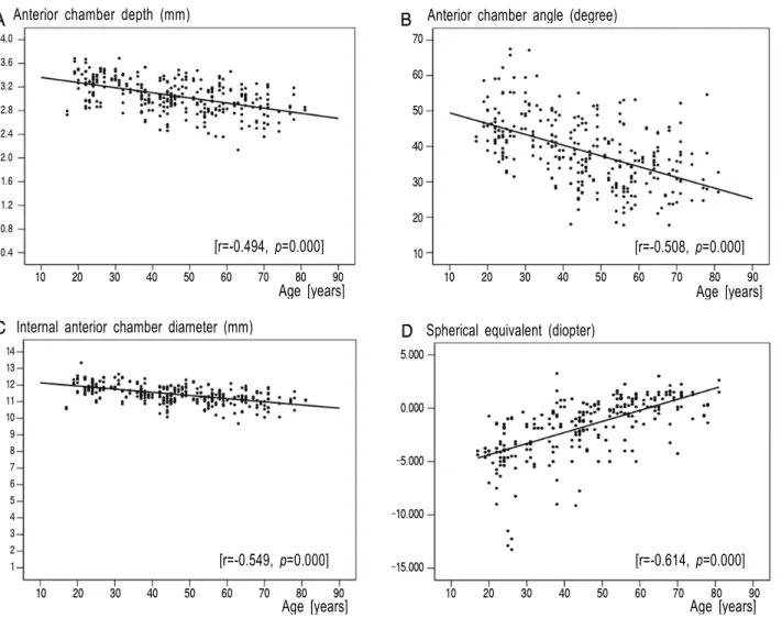
Figure 5.Relationship between several parameters of the anterior segment and age. (A) Anterior chamber depth, (B) anterior chamber angle, and (C) internal anterior chamber diameter show a significant decrease in relation to age. (D) Spherical equivalent shows a significant shift from myopia to hyperopia in relation to age.
깊이가 감소하는 소견을 보였으며 상관계수는 -0.494였다 (p=0.000)(Fig. 5).
전방각(anterior chamber angle)은 전체 연령에서 8방향 의 평균값을 비교하였을 때 남자는 40.1±9.7°, 여자는 37.3±9.4°로 여자가 남자보다 유의하게 전방각이 얕은 것 으로 나타났다(p=0.014). 각 연령대별로 나누어 남녀를 비 교하였을 때도 여자가 남자보다 전방각이 얕은 것이 관찰 되었지만 통계적으로 유의하지는 않았다(Table 3). 여덟 방향에서 측정한 전방각을 살펴보면 비측보다는 이측이, 상 비측보다는 하이측이, 상측보다는 하측이, 상이측보다는 하 비측이 전방각이 얕은 것이 관찰되었으나(Table 4), 비측 과 이측 사이 그리고 상비측과 하이측 사이에서만 통계적 으로 유의한 차이를 보였다(p<0.05). 또한 연령의 증가에 따라 유의하게 전방각이 감소하는 소견을 보였으며 상관계 수는 -0.508이었다(p=0.000)(Fig. 5).
전방각간거리(internal anterior chamber diameter)는 전체 연령의 평균값을 구하였을 때 남자 11.54±0.55 mm, 여자 11.39±0.57 mm로 여자가 남자보다 유의하게 전방각 간거리가 짧은 것으로 나타났다(p=0.017). 각 연령대별로 나누어 남녀를 비교하였을 때에도 전 연령층에서 여자가 남자보다 전방각간거리가 짧은 소견이 관찰되었으나 통계 적으로 유의하지는 않았다(Table 3). 또한 연령의 증가에 따라 유의하게 전방각간거리가 감소하는 소견을 보였으며 상관계수는 -0.549였다(p=0.000)(Fig. 5).
고 찰
고해상도의 전안부 영상을 촬영하고 정확히 계측하는 것 은 안과 환자의 진단이나 추적관찰에 있어서 매우 중요한 가치를 지닌다. 이를 위하여 여러 가지 기기들이 개발되어
Age (years) Demographics Spherical equivalent (mean±SD)
Male Female Male Female
20~29 33 30 -3.84±1.88 -5.15±3.54
30~39 26 26 -2.48±2.37 -2.04±2.11
40~49 29 34 -1.94±1.84 -1.58±2.21
50~59 29 31 -0.35±2.07 -0.60±1.67
60~ 28 32 +0.55±1.74 +0.36±1.22
Total (298) 145 153 -1.67±2.52 -1.75±2.92
Table 1. Demographics & Refractive error of the subjects
Age (years) Visante OCT Ultrasonic pachymeter
Male Female Male Female
20~29 535.2±30.4 522.4±32.4 551.8±24.9 543.6±31.3
30~39 536.5±27.4 525.2±29.2 553.3±26.3 545.3±25.8
40~49 529.1±30.3 517.2±32.3 547.6±30.6 534.4±34.4
50~59 535.0±40.2 511.8±32.5 553.4±38.7 530.5±34.4
60~ 529.1±32.4 518.8±33.7 541.0±29.1 536.8±38.7
Total 533.0±32.2* 518.8±32.0* 549.5±30.2† 537.8±33.5†
*p-value=0.000, comparing male and female. †p-value=0.002, comparing male and female.
Table 2. Central corneal thickness measured by Visante OCT and ultrasonic pachymeter (mean±SD, µm)
왔으며 현재 사용되는 기기로는 초음파생체현미경(UBM), Pentacam, RTVue, Visante OCT를 들 수 있다.
초음파생체현미경은 50 MHz의 탐촉자(transducer)를 사용하여 기존의 안구초음파에 비하여 미세하고 선명한 단 면영상을 제공하는 진단기구로, 2차원 영상 내의 측경 기능 을 이용한 계측이 가능하여 전안부 질환의 진단 및 치료효 과의 정성적, 정량적 평가에 이용할 수 있다. 하지만 환자가 반드시 앙와위를 취해야 하고, 침수 과정이 필요하며, 한 번 에 촬영할 수 있는 영역이 제한되어 있어 전체 전안부를 촬 영하려면 탐촉자를 여러 번 이동하여야 한다. 따라서 환자 에게 불편을 줄 수 있고 시간이 오래 걸리는 단점이 있다.
Pentacam은 475 nm 파장의 Blue LED를 광원으로 하여 전안부를 360도 회전하는 Scheimpflug 카메라로 촬영하는 기기이다. 전안부의 단면영상을 제공할 뿐 아니라 전방 구 조물들의 3D 모델링, 백내장밀도 측정, 각막 전후면의 굴절 력 측정 및 만곡지형도 생성 등의 기능을 제공한다. 하지만 짧은 파장의 빛을 사용함으로써 홍채, 공막 및 각막혼탁을 통과하지 못하고 과다노출을 일으켜서 홍채, 공막, 전방각 의 윤곽이 불분명해지는 단점이 있다.
후안부 OCT인 RTVue (Optovue Inc, Fremont, California, USA)에 CAM (Cornea Anterior Module) lens를 추가로 장 착하면 Visante OCT 보다 해상력이 좋은 각막 및 전안부의 영상을 얻을 수 있다. 하지만 RTVue에 CAM lens를 장착
할 경우 망막에 최적화된 830 nm의 파장을 사용하기 때문 에 전안부 촬영 시 의도하지 않은 에너지가 망막에 노출될 수 있으며, Visante OCT에 비해 투과력이 낮아서 공막 투 과를 잘하지 못하므로 전방각의 구조가 선명하지 않은 단 점이 있다. 또한 한 번에 촬영 가능한 최대구간이 6×2 mm 로 Visante OCT의 16×6 mm보다 좁기 때문에 전방깊이와 전방각간 거리는 측정할 수 없다. 따라서 각막 및 전안부의 국소적인 병변을 미세하게 보고자 할 때는 Visante OCT보 다 유용할 수 있으나 전체적인 전안부의 관찰 및 계측에 사 용되기에는 제한점이 있다.
820 nm의 파장을 가지는 Optical Coherence Tomographer (OCT)는 후안부의 영상을 촬영하는 장치로 개발되었다.6,7 1994년에 Izatt et al8에 의해서 OCT를 이용하여 전안부 영 상을 촬영하는 것이 제안되었고, 2001년에는 1310 nm의 파장을 이용하며 촬영 속도가 향상된 전안부 OCT가 소개 되었다.9현재 사용되고 있는 전안부 OCT (Visante OCT, Carl Zeiss Meditec, Germany)는 1310 nm 파장의 적외선을 이용하여 18 µm의 axial resolution과 60 µm의 transverse resolution을 가지며 high-resolution corneal software를 이용할 경우 axial resolution은 8 µm에 이르게 된다.1 Visante OCT에 의한 영상은 서로 다른 두 조직 간의 후방 산란 대비(back scattering contrast)를 나타내는 것으로 단면에서 후방 산란된 빛의 강도의 차이에 따라 회색음영
Age (years)
Anterior chamber depth (mean±SD, mm)
Anterior chamber angle (mean±SD, degree)
Internal anterior chamber diameter (mean±SD, mm)
Male Female Male Female Male Female
20~29 3.23±0.24 3.31±0.23 46.7±8.1 44.9±7.7 12.02±0.47 11.83±0.48
30~39 3.13±0.27 3.16±0.22 46.9±8.8 41.6±6.6 11.69±0.48 11.67±0.44
40~49 3.03±0.22 3.04±0.26 36.7±7.7 36.5±8.7 11.44±0.43 11.35±0.50
50~59 3.01±0.21 2.90±0.29 34.9±9.5 31.5±7.8 11.35±0.42 11.13±0.44
60~ 2.89±0.23 2.86±0.28 34.8±5.4 33.4±9.1 11.13±0.48 11.02±0.53
Total 3.06±0.26* 3.05±0.30* 40.1±9.7† 37.3±9.4† 11.54±0.55‡ 11.39±0.57‡
*p-value=0.665, comparing male and female; †p-value=0.014, comparing male and female; ‡p-value=0.017, comparing male and female.
Table 3. Anterior chamber depth, anterior chamber angle, and internal anterior chamber diameter
Eight orientations Anterior chamber angle (degree)
Nasal 37.3±10.6*
Superonasal 36.3±10.0†
Superior 37.9±10.3
Superotemporal 38.1±11.2
Temporal 41.2±11.9*
Inferotemporal 41.7±11.6†
Inferior 38.3±9.8
Inferonasal 38.7±11.1
*p-value=0.000, comparing nasal and temporal; †p-value=0.000, comparing superonasal and inferotemporal.
Table 4. Anterior chamber angle at 8 orientations (mean±SD, degree)
(gray scale) 또는 위색(false color)의 2차원 사진으로 보 여지게 된다.10 이렇게 얻어진 영상은 해상도와 선명도가 높으며, 기존의 다른 진단 방법들에 비하여 미세하고 선명 한 영상획득이 가능하고 조직 생검을 한 것과 비슷한 단면 영상을 제공하여, 안구의 전안부 병리 및 형태학적 연구에 유용하다. 또한 2차원 영상내의 측경 기능을 이용한 계측이 가능하여 녹내장환자를 비롯한 전안부 질환을 가진 환자에 서 진단 및 치료효과의 정성적, 정량적 평가에 이용할 수 있다.2-4 하지만 Visante OCT에서 사용하는 빛의 파장은 홍채 색소를 투과하지 못하여 모양체 고랑같은 홍채 뒤쪽 의 구조를 촬영할 수 없다는 단점이 있다.
Baïkoff1는 Visante OCT를 이용하여 유수정체용 안내렌 즈 삽입술을 시행받을 환자들을 대상으로 수술 전 검사로 서 전안부 계측을 시행하였고, Fine et al5은 투명각막절개 를 시행한 환자들을 대상으로 수술 후 절개 부위의 구조 관 찰을 위해 Visante OCT를 촬영하기도 하였다. Dada et al10 은 Visante OCT와 UBM을 이용하여 전안부 구조들을 관찰 하고 두 가지 방법을 이용하여 측정한 전안부 계측치(비측
및 이측 전방각, 중심각막두께, 전방깊이) 사이에 통계적 으로 유의한 차이가 없고 서로 밀접한 상관관계를 보이는 것을 보고하였다.
중심각막두께는 각막의 정상 혹은 병적 상태를 평가하는 중요한 지표로 각막질환이나 레이저 굴절수술 등에 있어 필요한 검사이다. 각막두께와 연령 사이에는 뚜렷한 연관성 이 없는 것으로 알려져 있으며,11-14본 연구에서도 통계적 으로 유의한 상관관계는 보이지 않았다. 각막 두께는 그 측 정 장치에 따라 측정값이 차이가 나는 것으로 알려져 있는데, A-scan 안구초음파가 정확하고 재현성이 높아 여러 연구에 서 기준으로 사용되고 있다. 국내에서는 Park and Cho14가 남자 530.99±33.69 µm, 여자 526.32±32.15 µm로 보고하 였고, 본 연구의 A-scan 안구초음파를 이용한 중심각막두 께는 남자 549.5±30.2 µm, 여자 537.8±33.5 µm로 기존 의 A-scan 안구초음파를 사용한 보고에 비해 약간 두꺼운 것으로 나타났다.
최근 보고들에 의하면 Visante OCT에 의한 중심각막두 께의 측정값은 A-scan 안구초음파에 의한 측정값과 비교 할 때 높은 상관관계를 보이기는 하나 그 절대치는 작게 나오는 것으로 알려져 있고,15,16 본 연구에서도 Visante OCT에 의한 중심각막두께의 측정값은 525.7±32.8 µm, A-scan 안구초음파에 의한 측정값은 543.5±32.4 µm로서 이에 부합하는 결과를 보여주고 있다. 이러한 차이가 발생 하는 이유가 정확히 밝혀져 있지는 않지만 Gao et al17은 정 상안에서 0.5% Tropicamide와 0.5% phenylephrine HCl의 복합점안제 또는 0.9% 생리식염수의 점안 후에 중심각막 두께가 두껍게 측정된다고 보고하였다. 또한 Nam et al18은 0.5% proparacaine 점안제의 사용 후에 중심각막두께가 일 시적으로 증가한다고 보고하였고, 그 기전으로서 점안제에 들어있는 보존제에 의한 각막부종과 눈물막의 불안정성을 제시하였다. 이에 비추어 생각해볼 때 저자들이 A-scan 안구초음파를 위해 사용한 점안마취제가 중심각막두께의
차이를 유발한 원인일 가능성이 있다.
서양인에 대한 보고를 살펴보면 Piñero et al19은 Visante OCT를 이용하여 스페인인의 평균 중심각막두께를 528.00
±20.93 µm로 보고하였고, Fishman et al20은 전안부 OCT 를 이용하여 미국인의 평균 중심각막두께를 533.4±37.8 µm로 보고하였다. Wirbelauer et al21은 전안부 OCT를 이 용하여 독일인의 평균 중심각막두께를 541±43 µm로 보고 하였다. 본 연구에서는 한국인의 중심각막두께가 525.7±
32.8 µm로 서양인보다 약간 얇게 나타났다.
전방깊이와 전방각은 녹내장 발생과 밀접한 관계가 있으 며 수정체의 두께, 홍채의 안구내 상대적 위치, 각막의 두 께, 각막의 직경, 각막의 곡률 반경, 홍채의 산동 정도, 홍채 기시부의 위치에 따라 결정되고 성별, 연령, 약물의 사용과 굴절이상 및 안축장의 길이에 따라서도 달라질 수 있다.22-26 일반적으로 전방깊이와 전방각은 연령이 증가할수록, 남자 에 비해 여자가 얕은 것으로 알려져 있다.26-28본 연구에서 는 전방깊이는 남녀 간에 유의한 차이가 존재하지는 않았 지만 연령의 증가에 따라 얕아졌고, 전방각은 남자에 비해 여자가 유의하게 얕은 것과 연령의 증가에 따라 얕아지는 것이 관찰되어서 이전의 보고들과 비슷한 경향을 보였다.
Lee et al26은 22세부터 63세까지의 정상 성인 64명 126안 을 대상으로 EAS-1000 Scheimpflug camera를 이용하여 측정한 전방깊이를 남자 3.017 mm, 여자 2.809 mm로 보 고하였고, 전방각을 남자 32.719°, 여자 31.661°로 보고하 였다. Oh et al29은 정상 성인 114명 114안을 대상으로 초 음파생체현미경(UBM)을 이용하여 측정한 전방깊이를 남 자 2.997±0.297 mm, 여자 2.763±0.314 mm로, 전방각을 남자 38.86±3.62°, 여자 36.01±3.23°로 보고하였다. 본 연구의 결과에서는 이전의 보고들에 비해 전방깊이가 약간 깊게 나타났는데, 이는 이전의 보고들이 전방깊이를 중심각 막내피의 내측경계면에서 수정체 전낭에 이르는 거리로 측 정한데 비해, 본 연구에서는 중심각막내피의 내측경계면 에서 양측 iris recess를 연결한 선까지의 거리로 측정한 것과 측정 방법이 각각 달랐던 것에서 기인하리라 생각된 다. Nemeth et al30은 전방깊이의 측정에 있어서도 Visante OCT가 초음파 침수 A-scan에 비해 더 뛰어난 반복성과 동등한 수준의 재현성을 가진다고 보고하였고, 두 관찰자가 헝가리인을 대상으로 Visante OCT를 이용하여 측정한 평 균 전방깊이는 각각 3.12±0.33 mm, 3.11±0.33 mm였다.
Baikoff et al31은 프랑스인을 대상으로 전안부 OCT를 이용 하여 측정한 전방깊이를 3.64±0.33 mm로 보고하였고, Piñero et al19은 Visante OCT를 이용하여 스페인인의 평균 전방깊이를 3.16±0.41 mm로 보고하였다. 최근의 연구에 의하면 Müller et al4은 Visante OCT를 이용하여 전방각을
측정시 관찰자내 재현성 및 관찰자간 재현성이 우수한 것 으로 보고하였고, 두 관찰자가 독일인을 대상으로 측정한 평균 전방각은 각각 35.9±5.7°, 36.2±5.7°였다. Piñero et al19은 Visante OCT를 이용하여 스페인인의 평균 전방각을 비측과 이측에서 각각 36.51±10.39°와 36.13±8.60°로 보고하였다. Nolan et al3은 Visante OCT가 폐쇄각 녹내장 발견에 높은 민감도를 보이는 것으로 보고하였다. 본 연구 에서는 서양인에 대한 보고에 비해 전방깊이는 약간 얕게, 전방각은 약간 크게 나타났다.
전방각간거리(internal anterior chamber diameter)는 유수정체용 전방 인공수정체(anterior chamber phakic intraocular lens) 삽입술 시행 시 필요한 중요한 계측치로 서,1,32Baikoff et al33은 7세부터 82세 사이 연령의 프랑스 인 56명 104안을 대상으로 Visante OCT를 이용한 전방각 간거리의 평균이 12.334±0.571 mm이며 연령과의 연관성 이 없다고 보고하였다. Kohnen et al32은 Visante OCT로 측정한 독일인의 평균 전방각간거리는 12.45±0.53 mm 이 고 높은 반복성을 보이며, IOL Master나 Orbscan IIz로 측 정한 수평 각막 직경(horizontal corneal diameter)에 비해 값이 크게 나온다고 보고하였다. 또한 Goldsmith et al34은 전안부 OCT를 이용한 미국인의 평균 전방각간거리를 12.53±
0.47 mm로 보고하였다. Piñero et al19은 Visante OCT를 이용하여 스페인인의 평균 전방각간거리를 12.14±0.52 mm 로 보고하였다. 본 연구에서는 전방각간 거리가 남자 11.54±
0.55 mm, 여자 11.39±0.57mm로 서양인보다 약간 적게 나타났으며 연령의 증가에 따라 유의하게 감소하는 경향을 보였다. 본 연구의 대상자들에서 측정한 굴절 이상 값을 살 펴보면 젊은 연령일수록 유의하게 보다 더 심한 근시를 보이는 것을 관찰할 수 있었는데(r=0.614, p=0.000) (Fig.
5) (Table 1), 이로 인해 연령의 증가에 따라 전방각간거리 (internal anterior chamber diameter)가 감소하는 것으로 나타난 것으로 생각된다.
결론적으로 본 연구에서는 Visante OCT를 이용한 전안 부의 촬영을 통해 한국 성인의 연령 및 성별에 따른 전안부 의 계측치를 도출할 수 있었다. 이는 굴절 교정 수술을 비 롯한 각종 전안부 수술 전의 참고치 및 전안부 질환의 진단 및 추적관찰에 있어서 표준이 될 수 있는 값으로 유용하게 사용될 수 있으리라 생각한다.
참고문헌
1) Baïkoff G. Anterior segment OCT and phakic intraocular lenses:
a perspective. J Cataract Refract Surg 2006;32:1827-35.
2) Memarzadeh F, Li Y, Chopra V, et al. Anterior segment optical coherence tomography for imaging the anterior chamber after
laser peripheral iridotomy. Am J Ophthalmol 2007;143:877-9.
3) Nolan WP, See JL, Chew PT, et al. Detection of primary angle closure using anterior segment optical coherence tomography in Asian eyes. Ophthalmology 2007;114:33-9.
4) Müller M, Dahmen G, Pörksen E, et al. Anterior chamber angle measurement with optical coherence tomography: intraobserver and interobserver variability. J Cataract Refract Surg 2006;32:
1803-8.
5) Fine IH, Hoffman RS, Packer M. Profile of clear corneal cataract incisions demonstrated by ocular coherence tomography. J Cataract Refract Surg 2007;33:94-7.
6) Huang D, Swanson EA, Lin C-P, et al. Optical coherence tomography. Science 1991; 254:1178-81.
7) Puliafito CA, Hee MR, Schuman JS, Fujimoto JG. Optical Coherence Tomography of Ocular Diseases. Thorofare, NJ: Slack, 1996.
8) Izatt JA, Hee MR, Swanson EA, et al. Micrometer-scale resolution imaging of the anterior eye in vivo with optical coherence tomography. Arch Ophthalmol 1994;112:1584-9.
9) Radhakrishnan S, Rollins AM, Roth JE, et al. Real-time optical coherence tomography of the anterior segment at 1310 nm. Arch Ophthalmol 2001;119:1179-85.
10) Dada T, Sihota R, Gadia R, et al. Comparison of anterior segment optical coherence tomography and ultrasound biomicroscopy for assessment of the anterior segment. J Cataract Refract Surg 2007;
33:837-40.
11) Alsbirk PH. Corneal thickness. I. Age variation, sex, difference and oculometric correlations. Acta Ophthalmol (Copenh) 1978;56:
95-104.
12) Cho P, Lam C. Factors affecting the central corneal thickness of Hong-Kong Chinease. Curr Eye Res 1999;18:468-74.
13) Siu A. Herse P. The effect of age on human corneal thickness.
Stastical implications of power analysis. Acta Ophthalmol (Copenh) 1993;71:51-6.
14) Park SO, Cho BJ. Central Corneal Thickness Measured by Ultra- sonic Pachymeter in Normal Koreans. J Korean Ophthalmol Soc 2000; 41:2332-7.
15) Ho T, Cheng AC, Rao SK, et al. Central corneal thickness measurements using Orbscan II, Visante, ultrasound, and Pentacam pachymetry after laser in situ keratomileusis for myopia. J Cataract Refract Surg 2007;33:1177-82.
16) Zhao PS, Wong TY, Wong WL, et al. Comparison of central corneal thickness measurements by visante anterior segment optical coherence tomography with ultrasound pachymetry. Am J Ophthalmol 2007;143:1047-9.
17) Gao L, Fan H, Cheng AC, et al. The effects of eye drops on corneal thickness in adult myopia. Cornea 2006;25:404-7.
18) Nam SM, Lee HK, Kim EK, et al. Comparison of Corneal Thickness After the Instillation of Topical Anesthetics: Propara- caine Versus Oxybuprocaine. Cornea 2006;25:51-4.
19) Piñero DP, Plaza AB, Alió JL. Anterior segment biometry with 2 imaging technologies: very-high-frequency ultrasound scanning versus optical coherence tomography. J Cataract Refract Surg 2008;34:95-102.
20) Fishman GR, Pons ME, Seedor JA, et al. Assessment of central corneal thickness using optical coherence tomography. J Cataract Refract Surg 2005;31:707-11.
21) Wirbelauer C, Scholz C, Hoerauf H, et al. Noncontact corneal pachymetry with slit lamp-adapted optical coherence tomography.
Am J Ophthalmol 2002;133:444-50.
22) Philips GI. Aetiology of angle closure glaucoma. Br J Ophthalmol 1972;56:248-53.
23) Fontana ST, Brubaker RF. Volume and depth of the anterior chamber in the normal aging human eye. Arch Ophthalmol 1981;
98:1803-8.
24) Thomlison A, Leighton DA. Ocular dimensions and the heredity of open angle glaucoma. Br J Ophthalmol 1974;58:68-74.
25) Kim HK, Kim HB, Choi O. The Effects of the Mydriatics on the Anterior Chamber Depth. J Korean Ophthalmol Soc 1984;25:
181-7.
26) Lee JH, Park WC, Rho SH. The Effects of Pilocarpine on the Anterior Chamber Depth and Angle. J Korean Ophthalmol Soc 1994;35:572-9.
27) Choi HH. A Consideration for Corneal Curvature , Its Thickness and Anterior Chamber Depth. J Korean Ophthalmol Soc 1978;
19:417-22.
28) Lee HS, Kwon JY. The Differences of the Anterior Chamber Depth and the Corneal Thickness by the Refractive Error. J Korean Ophthalmol Soc 1982;23:395-400.
29) Oh DE, Jun RM, Choi KR. Quantified Values of Anterior Segment in Normal Adult Koreans Using Ultrasound Biomicro- scopy. J Korean Ophthalmol Soc 2004;45:251-8.
30) Nemeth G, Vajas A, Tsorbatzoglou A. Assessment and reproduci- bility of anterior chamber depth measurement with anterior segment optical coherence tomography compared with immersion ultrasonography. J Cataract Refract Surg 2007;33:443-7.
31) Baikoff G, Jitsuo Jodai H, Bourgeon G. Measurement of the internal diameter and depth of the anterior chamber: IOLMaster versus anterior chamber optical coherence tomographer. J Cataract Refract Surg 2005;31:1722-8.
32) Kohnen T, Thomala MC, Cichocki M, et al. Internal anterior chamber diameter using optical coherence tomography compared with white-to-white distances using automated measurements. J Cataract Refract Surg 2006;32:1809-13.
33) Baikoff G, Lutun E, Ferraz C. Static and dynamic analysis of the anterior segment with optical coherence tomography. J Cataract Refract Surg 2004;30:1843-50.
34) Goldsmith JA, Li Y, Chalita MR, et al. Anterior chamber width measurement by high-speed optical coherence tomography.
Ophthalmology 2005;112:238-44.
=ABSTRACT=
Measurement of Anterior Segment Using Visante OCT in Koreans
Deok Goo Lee, MD, Si Hwan Choi, MD
Department of Ophthalmology, College of Medicine, Chungnam National University, Daejeon, Korea
Purpose: To obtain the average values of anterior segment structures of adult Koreans using Visante OCT in relation to age and sex, and to compare the central corneal thickness values with these measurements using an A-scan ultrasonic pachymeter.
Methods: Anterior segment images were obtained in 185 Korean (298 eyes) using Visante OCT. Four different parameters (central corneal thickness, anterior chamber depth, anterior chamber angle, and internal anterior chamber diameter) were measured at four meridians (vertical, horizontal, 45 degrees, and 135 degrees). Afterwards, the same examiner performed A-scan ultrasonic pachymetry to measure central corneal thickness.
Results: Central corneal thickness values measured using Visante OCT were significantly less than those obtained using an A-scan ultrasonic pachymeter (p=0.000). Central corneal thickness, anterior chamber angle, and internal anterior chamber diameters were all smaller in females than in males (p<0.05). Anterior chamber depth, anterior chamber angle, and internal anterior chamber diameter showed a significant decrease according to increasing age in both sexes (p<0.05).
Conclusions: Average values and changes of anterior segment structures in relation to age and sex were established using Visante OCT in Korean adults. These results may be useful as standard values in anterior segment surgeries as well as in the diagnosis and follow-up of certain anterior segment diseases.
J Korean Ophthalmol Soc 2009;50(4):542-550
Key Words: Anterior segment, Average values, Measurement, Visante OCT
Address reprint requests to Si Hwan Choi, MD
Department of Ophthalmology, Chungnam National University Hospital
#640 Daesa-dong, Jung-gu, Daejeon 301-721, Korea
Tel: 82-42-280-7609, Fax: 82-42-255-3745, E-mail: [email protected]