pISSN: 0378-6471 eISSN: 2092-9374 http://dx.doi.org/10.3341/jkos.2013.54.2.289
= 증례보고 =
한국인의 스펙트럼영역빛간섭단층촬영 변수의 나이에 따른 차이
서지영⋅서홍융⋅노세현 동아대학교 의과대학 안과학교실
목적: 정상 한국인의 시신경유두주위 망막신경섬유층 두께와 시신경유두 변수의 나이에 따른 차이와 상관관계를 알아보고자 하였다.
대상과 방법: 스펙트럼 영역 빛간섭단층촬영술 이용하여 20세부터 70세에 이르는 정상인 128명의 시신경유두주위 망막신경섬유층 두께 및 시신경유두 변수를 검사하였다.
결과: 평균 시신경유두 주위의 망막신경섬유층 두께는 나이에 따라 감소하는 것으로 나타났다. 아래쪽 사분면의 망막신경섬유층이 나 머지 사분면 영역에 비해 유의하게 두꺼웠다. 평균 시신경유두함몰비와 수직 시신경유두함몰비는 모두 나이에 따라 증가하는 경향을 보였으며, 평균 유두함몰비는 r=0.249, 수직 유두함몰비는 r=0.537의 상관계수를 보였다. 그러나 신경테면적, 유두면적, 유두함몰면적 은 연령증가와는 무관한 것으로 나타났다.
결론: 평균 시신경유두 주위의 망막신경섬유층 두께는 나이에 따라 감소하는 경향을, 평균 시신경유두함몰비와 수직 시신경유두함몰비 는 모두 나이에 따라 증가하는 경향을 보였으며, 수직 유두함몰비가 나이증가와 더 강한 상관성을 보이는 것으로 분석되었다.
<대한안과학회지 2013;54(2):289-295>
■ 접 수 일: 2012년 7월 27일 ■ 심사통과일: 2012년 10월 24일
■ 게재허가일: 2013년 1월 2일
■ 책 임 저 자: 노 세 현
부산광역시 서구 대신공원로 26 동아대학교의료원 안과
Tel: 051-240-5227, Fax: 051-254-1987 E-mail: shrho@donga.ac.kr
* 이 논문은 동아대학교 학술 연구비 지원에 의하여 연구되었음.
녹내장에서는, 망막신경절 세포들이 소실되면서 시신경 의 망막신경섬유층이 점진적으로 얇아진다고 알려졌다. 이 는 결국 시신경유두의 생리적 함몰이 커지는 시신경 유두 함몰(optic disc cupping)을 야기하고, 이는 다른 시신경증 (optic neuropathy)에는 존재하지 않는 특징이다.1,2이러한 시신경의 변화는 비가역적이고, 손상이 상당히 진행된 후에 야 시야결손 증상이 나타나기 때문에3,4시신경 유두와 망막 신경섬유층에 대한 검사가 녹내장의 조기진단을 위해 중요 하다. 이러한 검사법에는 동일초점 레이저검안경(confocal scanning ophthalmoscope, CSLO), 주사레이저편광측정기 (scanning laser polarimetry, SLP), 빛간섭단층촬영기(optical coherence tomography, OCT) 등이 있는데,5,6이 중에서도 빛간섭단층촬영기는 다른 기기들과 비교하여 망막신경섬유 층의 결손을 발견하는데 있어 보다 우수하다고 보고된 바 있다.7,8 빛간섭단층촬영기는 비접촉성, 비침습적인 검사방 법으로, 830-850 nm 근적외선 영역 파장이 조직에서 반
사되어 나오는 빛의 강도를 측정하여 영상으로 재구성하여 망막의 단층상을 구현하는 장비로, Hee et al9에 의해 1991 년 소개된 이래, 현재 녹내장 및 망막질환의 진단에 널리 사용되고 있다.10 빛간섭단층촬영기는 초창기 OCT 1과 OCT 2 이후 3세대 시간영역 빛간섭단층촬영기(time do- main OCT)가 개발되어 현재 널리 사용되고 있으며, 최근 에는 4세대에 해당하는 스펙트럼 영역 빛간섭단층촬영기 (spectral domain OCT)가 개발되었다. 시간영역 빛간섭단 층촬영기는 조직과 기준거울(reference mirror)에서 반사 되는 빛의 시간 차이를 간섭계로 측정하여 고해상도의 2차 원적인 망막 단층상을 보여주며, 망막신경섬유층 두께와 시 신경유두 형태를 알 수 있어 녹내장의 진단에 매우 유용하 고 그 객관성과 재현성은 여러 연구에서 입증되었다.10-12최 근에 개발된 스펙트럼영역 빛간섭단층촬영기는 기준거울의 움직임 없이 주파수 스펙트럼(frequency spectrum)에서 간 섭 정보를 획득하며 인접A-라인 데이터를 평균화하여 푸 리에 변환(Fourier transform)을 하는 방식이며 점차 보편 화되고 있다. 스펙트럼영역 빛간섭단층촬영기는 많은 데이 터를 매우 빠르게 처리할 수 있어 시간영역 빛간섭단층촬 영기보다 50배 이상 빠르고, 민감하며, 고해상도의 2차원 및 3차원 망막 영상을 제공한다.13,14
스펙트럼영역 빛간섭단층촬영기는 비교적 최신의 진단 기법으로 정상인의 망막신경섬유층 및 시신경유두 형태에 대한 연구는 많이 이루어져 있지 않으며, 국내에서는 망막
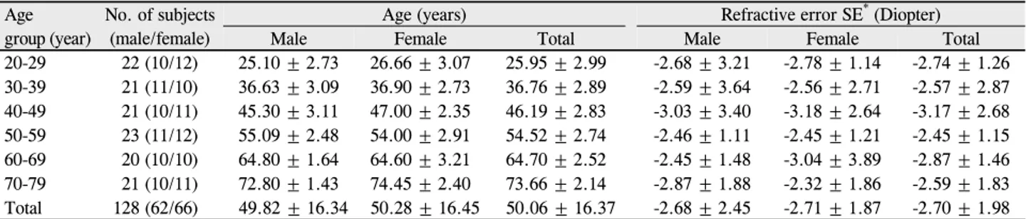
Table 1. Demographic data
Agegroup (year)
No. of subjects (male/female)
Age (years) Refractive error SE* (Diopter)
Male Female Total Male Female Total
20-29 22 (10/12) 25.10 ± 2.73 26.66 ± 3.07 25.95 ± 2.99 -2.68 ± 3.21 -2.78 ± 1.14 -2.74 ± 1.26 30-39 21 (11/10) 36.63 ± 3.09 36.90 ± 2.73 36.76 ± 2.89 -2.59 ± 3.64 -2.56 ± 2.71 -2.57 ± 2.87 40-49 21 (10/11) 45.30 ± 3.11 47.00 ± 2.35 46.19 ± 2.83 -3.03 ± 3.40 -3.18 ± 2.64 -3.17 ± 2.68 50-59 23 (11/12) 55.09 ± 2.48 54.00 ± 2.91 54.52 ± 2.74 -2.46 ± 1.11 -2.45 ± 1.21 -2.45 ± 1.15 60-69 20 (10/10) 64.80 ± 1.64 64.60 ± 3.21 64.70 ± 2.52 -2.45 ± 1.48 -3.04 ± 3.89 -2.87 ± 1.46 70-79 21 (10/11) 72.80 ± 1.43 74.45 ± 2.40 73.66 ± 2.14 -2.87 ± 1.88 -2.32 ± 1.86 -2.59 ± 1.83 Total 128 (62/66) 49.82 ± 16.34 50.28 ± 16.45 50.06 ± 16.37 -2.68 ± 2.45 -2.71 ± 1.87 -2.70 ± 1.98 Values are presented as mean ± SD.
*Spherical equivalent.
신경섬유층 두께에 대한 연구15가 발표된 바 있으나 그 이 외의 시신경유두 파라미터의평균치 연구는 거의 이루어지 지 않은 상태이다. 이에 저자는 스펙트럼영역 빛간섭단층촬 영기를 이용하여 정상인의 망막신경섬유층 두께 및 시신경 유두 형태를 측정하여 정상 한국인의 연령별 기본 자료를 파악하고 그들의 상관관계를 알아보고자 하였다.
대상과 방법
2010년 12월부터 2012년 3월까지 본원에 내원한 환자 들 중 안과검사에서 정상소견을 보인 128명의 양안을 대상 으로 검사를 시행하였다. 환자들은 남녀, 연령별로 20-29 세, 30-39세, 40-49세, 50-59세, 60-69세, 70-79세 총 6군으로 나누어 검사를 시행하였다.
모든 대상자에서 나안시력 및 교정시력 측정, 굴절검사, 세극등현미경을 이용한 전안부 및 앞방각경검사, 검안경을 이용한 시신경유두검사, 골드만압평안압계를 이용한 안압 검사, 험프리 시야계를 이용한 시야검사, 시신경섬유층 사 진촬영, 광간섭단층촬영을 시행하였다.
정상인은 최대교정시력이 0.8 이상, 2회의 안압 측정에 서 21 mmHg 이하, 구면렌즈 대응치(Spherical equivalent) -5.0D에서 +3.0D 사이의 굴절 오차를 보이면서, 앞방각검 사상 개방각, 안저검사상 유두함몰비 0.5 미만 또는 양안의 차이가 0.1 이하, 시신경섬유층 사진촬영, 광간섭단층촬영 상 녹내장성 시신경손상 및 시야검사상 녹내장성 시야결손 이 없는 경우로 정의하였다. 또한 포도막염, 망막질환 등의 다른 안질환이나 눈에 영향을 줄만한 전신질환이 있는 환 자, 안수술이나 외상의 기왕력이 있는 사람들은 연구대상에 서 제외하였다.
광간섭단층촬영은 본원에 있는 Cirrus HD-OCT (Carl Zeiss Meditec, Inc., Dublin, CA, USA)를 이용하였다. Cirrus HD-OCT 내의 내장된 소프트웨어 중 Optic Disc Cube 200 ×200 스캔을 이용하였으며 검사는 숙련된 1명의 숙 련된 검사자에 의해 시행되었다. Signal strength 값이 6 미
만이거나 중심이 이탈되어 있는 경우는 대상에서 제외하였다.
시신경유두 주위 망막신경섬유층은 평균 두께와 4개의 사분면(4-quadrant), 즉 귀쪽(316-45도), 위쪽(46-135 도), 코쪽(136-225도), 아래쪽(226-315도)과 12개의 시 간 영역(12-clock hour)을 측정하였으며, 양안에 측정된 망막신경섬유층은 결과지에서 서로 거울상으로 나타난다.
시신경유두 분석은 신경테면적(rim area), 유두면적(disc area), 평균유두함몰비(average C/D ratio), 수직유두함몰 비(vertical C/D ratio) 그리고 유두함몰면적(cup volume) 을 구하였다.
통계학적인 분석에는 SPSS software version 15.0 for windows (SPSS Inc., Chicago, Illinois, USA)를 사용하였 으며, 각 연령군 간의 차이 비교에는 일원배치 분산분석 후 duncan의 사후검정을 시행하였고, 연령과의 상관관계 분석 에는 피어슨상관계수분석(Pearson's correlation analysis) 을 이용하였다. 통계학적 유의성은 p<0.05 이하일 때로 판 정하였다.
결 과
대상군은 총 128명의 정상인이었으며, 평균 연령은 50.06 ±16.37세였으며, 평균 구면렌즈대응치는 -2.27 ± 2.16 디옵터였다. 대상자는 남자 62명, 여자 66명이었고, 각각의 평균 연령은 49.82 ±16.34세, 50.28 ±16.45세로 남녀간의 유의한 차이는 없었고(p=0.821), 평균 구면렌즈 대응치는 각각 -2.68 ±2.45 디옵터, -2.71 ±1.87 디옵 터였다(Table 1).
시신경유두 주위 시신경섬유층 두께는 평균 100.84 ± 4.21 μm, 위쪽 119.05 ±2.12 μm, 아래쪽 130.31 ±3.33 μm, 코쪽 71.80 ±0.57 μm, 귀쪽 77.72 ±0.16 μm였다.
평균 망막신경섬유층 두께는 20대, 30대, 40대 사이에는 유의한 차이가 없었으나, 40대 이후로는 나이에 따라 유의한 감소경향을 보였다(F=38.548, p<0.05). 사분면별(quadrant scan area) 신경섬유층 두께값 분석에서, 아래쪽 영역이 코
Table 2. Correlation of age with RNFL thickness in quadrant scan area
Age (year) Superior (μm) T† Nasal (μm) T† Inferior (μm) T† Temporal (μm) T† Mean p-value* 20-29 121.86 ± 1.87 a 71.10 ± 0.39 a 135.02 ± 2.98 a 77.68 ± 0.11 a 103.80 ± 1.45 <0.001 30-39 121.08 ± 2.15 a 70.26 ± 0.58 a 133.45 ± 3.03 a 77.71 ± 0.13 a 103.48 ± 1.77 <0.001
40-49 120.03 ± 2.02 a 71.12 ± 0.61 a
b
131.01 ± 3.38 77.94 ± 0.17 b 102.57 ± 2.36 <0.001
50-59 118.24 ± 1.98 71.53 ± 0.56 b 129.54 ± 3.45 77.93 ± 0.21 b 100.61 ± 4.72 <0.001
60-69 117.22 ± 2.35 71.09 ± 0.52 b 127.89 ± 3.12 77.55 ± 0.17 b 98.03 ± 2.05 <0.001
70-79 115.88 ± 2.10 70.41 ± 0.60 125.05 ± 3.41 77.52 ± 0,19 96.33 ± 4.79 <0.001
Mean ± SD 119.05 ± 2.12 71.06 ± 0.57 130.32 ± 3.33 77.72 ± 0.16
Values are presented as mean ± SD.
SD = standard deviation.
*Statistical significances were tested by oneway analysis of variances among groups; †The same letters indicate non-significant difference between groups based on duncan’s multiple comparison test.
Table 3. Correlation of age with RNFL thickness in clock hour scan area
(year)AgeSuperior Nasal Inferior Temporal
p-value* Area
(μm)12 T† Area 1
(μm) T† Area 2
(μm) T† Area 3
(μm) T†Area 4
(μm) T† Area 5
(μm) T† Area 6 (μm) T† Area 7
(μm) T† Area
(μm)8 T† Area
(μm)9 T† Area
(μm)10 T† Area (μm)11 T† 20-29 130.7
± 3.1 a 121.1
± 2.9 a 96.9 ±
1.1 a 70.1 ±
0.9 a 74.4 ±
0.7 a 103.9 ±
2.4 a 141.2
± 3.1 a 141.8
± 3.9 84.9 ±
0.2 a 52.4 ±
0.2 a 95.6 ± 0.7 a 143.8
± 3.9 a <0.001 30-39 127.1
± 2.9 a 119.8
± 3.1 a b 95.1 ±
1.2 a 64.5 ±
0.3 a 76.1 ±
0.5 a 99.8 ±
1.9 a 138.3
± 3.7 a 137.4
± 3.4 83.5 ±
0.4 a 54.6 ± 0.2 a
b 93.2 ± 0.6 a 142.7
± 4.2 a b <0.001 40-49 127.1
± 3.2 a 118.2
± 3.0 b 94.4 ±
1.0 a 66.5 ±
0.5 a 73.4 ±
0.4 a 103.2 ±
2.3 b 135.7
± 3.5 133.1
± 3.1 79.6 ±
0.5 56.8 ±
0.3 b 90.4 ± 0.7 b 135.4
± 4.1 b <0.001 50-59 124.2
± 2.7 116.3
± 3.4 91.3 ±
1.3 65.7 ± 0.4 b 71.5 ±
0.3 b 101.2 ±
2.5 b 136.6
± 2.9 134.1
± 3.3 81.1 ±
0.3 52.4 ±
0.4 b 95.1 ± 1.0 b 137.5
± 3.6 <0.001 60-69 122.4
± 3.2 117.1
± 3.6 88.3 ±
1.0 63.8 ± 0.7 b
c 69.7 ±
0.6 b 92.7 ±
2.0 134.5
± 3.0 132.7
± 2.9 77.7 ±
0.4 48.6 ±
0.2 b 91.8 ± 0.9 c 138.5
± 3.8 <0.001 70-79 119.5
± 2.6 111.7
± 2.8 86.3 ±
0.9 60.4 ± 0.6 c 63.5 ±
0.9 96.4 ±
1.8 131.5
± 3.2 129.4
± 3.1 75.3 ±
0.3 53.4 ±
0.3 88.2 ± 0.8 c 133.1
± 3.6 <0.001 Mean
± SD 125.1
± 3.9 117.3
± 3.2 92.0 ±
4.1 65.1 ± 3.2 71.4 ±
4.4 99.5 ±
4.3 136.3
± 3.30 134.7
± 4.3 80.3 ±
4.9 53.0 ±
2.7 92.4 ± 2.9 138.5
± 4.13 Values are presented as mean ± SD.
SD = standard deviation.
*Statistical significances were tested by oneway analysis of variances among quadrant groups; †The same letters indicate non-significant difference between groups based on duncan’s multiple comparison test.
Table 4. Correlation of age with Optic disc head parameters
Age (year) Rim area (mm2) Disc area (mm2) Cup volume (mm3) Average C/D ratio* Vertical C/D ratio*
20‐29 1.47 ± 0.18 1.70 ± 0.27 0.04 ± 0.009 0.29 ± 0.09 0.26 ± 0.07
30‐39 1.48 ± 0.25 1.78 ± 0.29 0.05 ± 0.010 0.37 ± 0.11 0.28 ± 0.09
40‐49 1.45 ± 0.24 1.79 ± 0.32 0.06 ± 0.011 0.39 ± 0.15 0.33 ± 0.10
50‐59 1.43 ± 0.20 1.83 ± 0.34 0.06 ± 0.013 0.40 ± 0.16 0.37 ± 0.12
60‐69 1.41 ± 0.22 1.73 ± 0.30 0.07 ± 0.012 0.42 ± 0.15 0.41 ± 0.14
70‐79 1.45 ± 0.26 1.74 ± 0.31 0.04 ± 0.008 0.43 ± 0.14 0.44 ± 0.13
Mean ± SD 1.45 ± 0.22 1.76 ± 0.31 0.05 ± 0.01 0.38 ± 0.14 0.35 ± 0.11
p-value† 0.371 0.156 0.083 <0.01 <0.01
Values are presented as mean ± SD.
*Average C/D ratio and vertical C/D ratio were compared by paired t-test and showed statistical difference (p < 0.001); †Statistical significances were tested by one-way analysis of variances among age groups.
쪽 영역, 귀쪽 영역 및 위쪽 영역에 비해 시신경섬유층이 두꺼운 것으로 나타났으며(Table 2), 12개 시간 영역별
(clock hour scan area) 분석에서도 아래쪽 영역에 해당하 는 영역 6, 7, 8이 다른 영역에 비해 유의하게시신경섬유
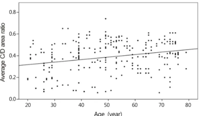
Average C/D area ratio
Age (year)
20 30 40 50 60 70 80 0.8
0.6 0.4 0.2 0.0
Figure 1. The relationship between average cup/disc (C/D)
area ratio and age. Average C/D area ratio increases with age significantly (p < 0.05).0.6 0.5 0.4 0.3 0.2 0.1 0.0
Age (year)
20 30 40 50 60 70 80
Vertical C/D area ratio
Figure 2. The relationship between vertical cup/disc (C/D)
area ratio and age. Vertical C/D ratio increases with age sig- nificantly (p < 0.05).층 두께가 두꺼운 것으로 나타났다(Table 3). 또한 나이에 따른 각 사분면별, 각 12개 시간 영역별 분석에서 위쪽 영 역과 아래쪽 영역, 영역 12, 1, 2(위쪽 영역에 해당하는 영 역)와 6, 7, 8(아래쪽 영역에 해당하는 영역)에서는 유의한 감소가 있었다(Table 2, 3).
시신경유두 분석에서 신경테 면적, 유두 면적, 유두함몰 면적의 평균값은 각각 1.45 ± 0.22 mm3, 1.76 ± 0.31 mm3, 0.05 ±0.06 mm3였다. 평균 유두함몰비와 수직 유두 함몰비는 0.38 ±0.14, 0.35 ±0.11로 나타나 두 수치 사 이에는 통계학적으로 유의한 차이를 보였다(p=0.04). 나이에 따른 비교분석에서, 신경테 면적, 유두 면적, 유두함몰 면적은 모두 나이증가와는 무관한 것으로 나타났으며(Table4), 평 균 유두함몰비와 수직 유두함몰비는 나이가 증가할수록 증 가하는 경향을 보였으며(p<0.01)(Fig. 1, 2), 평균 유두함 몰비는 r=0.249, 수직 유두함몰비는 r=0.537의 상관계수 를 보여 수직 유두함몰비가 나이증가와 더 강한 상관성을 보이는 것으로 분석되었다.
고 찰
녹내장은 시야 변화로 측정되는 기능적 손상 보다 시신 경유두와 망막신경섬유층의 변화가 선행되는 질환이며 망 막신경섬유층의 두께 감소가 시신경유두 변화보다 선행한 다고 알려졌다.3,4,16,17 이러한 망막신경섬유층의 변화는 녹 내장 조기 진단의 지표로서 가치를 가지며 이를 진단하기 위한 장비들이 개발되고 발전되어 왔다.3,16 1973년 Hoyt et al18에 의해 red-free opthalmoscopy로 처음 측정이 시 도되었으며 그 후 stereo photogrammetry, HRT, NFA 등 이 쓰였다. 1993년 Fercher와 Swanson 등에 의해 최초로 빛간섭단층촬영기(Optical coherence tomography)를 이용 한 생체내(in vivo) 논문이 발표된 이후로 빛간섭단층촬영 기는 현재까지 가장 널리 쓰이고 진단법이 되었다.19,20
최근 개발되어 널리 사용되고 있는 스펙트럼영역(spectral domain 혹은 fourier-domain) 빛간섭단층촬영기는 기존의 시간영역 빛간섭단층촬영기(time domain)에 비해 고속으 로 고해상도의 정보를 제공하는 뛰어난 기기이다.13,14스펙 트럼영역 빛간섭단층촬영기의 편위지도는 국소 망막신경섬 유층 결손의 진단에 있어서 다른 시신경섬유층단면 측정값 지도보다도 진단능력이 뛰어난 것으로 보고되었고, 이는 정 상인과 매우 초기의 녹내장 환자를 식별해 내는데 도움이 된다.21,22
본 연구에서 평균 망막신경섬유층 두께는 100.84 ±4.21 μm였다. Hirasawa et al23은 SD-OCT를 이용한 일본인 251예의 정상안 연구에서 평균 102 μm의 두께를 보고하였 고 통상 동양인의 망막신경섬유층 두께는 100 μm 이상으 로 보고되고 있다.24
Quigley and Addicks17는 원숭이의 시신경유두 주위 망 막신경섬유층의 두께를 연구하였고, 조직학적으로 시신경 유두부 주위로 갈수록 두껍고, 위쪽 사분면과 아래쪽 사분 면이 두껍고 코쪽 사분면과 귀쪽 사분면의 순서로 그 두께 가 감소함을 보고하였다. 그 후 Jonas et al25은 정상인에서 의 망막신경섬유층의 형태를 연구하였고, 그 두께가 아래 쪽, 위쪽, 코쪽, 귀쪽 사분면 순서로 얇아진다고 보고하였 다. 이는 ISNT 규칙으로 현재까지 대부분의 검사자들이 시 신경검사에 적용해왔다.23,25저자는 이전 연구에서 위코쪽, 아래귀쪽과 위귀쪽에서 나이가 들어갈수록 유의하게 망막 신경섬유층의 두께가 감소함을 보고하였는데, 이번 연구에 서도 망막신경섬유층의 두께는 아래쪽과 위쪽 사분면에서 두껍고, 코쪽과 귀쪽 사분면은 얇은 double hump형태를 취 하여, 이전에 시행된 연구결과들과 유사한 결과를 보였
다.26,27 하지만 귀쪽 사분면이 코쪽 사분면보다 조금 더 두
꺼운 결과를 보여 정확한 ISNT 규칙을 따르지는 않았다.
다른 정상안에 대한 연구들에서도 ISNT 규칙을 따르는 비 율이 42%, 56%, 79% 등으로 보고되었고, 이는 망막신경 세포의 다양성 때문에 생기는 결과로 정상안에서 꼭 ISNT 규칙에 맞지 않을 수 있음을 시사한다.23,28,29
Schuman et al30은 나이가 들수록 망막신경섬유층의 두 께가 감소함을 보고하였는데, 본 연구에서도 나이가 증가함 에 따라 망막신경섬유층의 두께가 감소하는 결과를 보였다.
정상적으로 망막신경절세포는 70만개에서 140만개 정도이 며 나이에 영향을 받아 사람은 매년 평균 5,000개 가량의 망막신경절세포가 감소한다. 50세 이전에는 매년 약 2,500 개, 50세 이후부터는 매년 약 7,500개 정도로 감소하여 나 이가 들수록 두께감소가 가속화된다.31,32본 연구에서는 10 년간 평균 망막신경섬유층 두께 감소가 대략 1.6-1.7 μm로 나타났다. Spectralis SD-OCT를 사용하여, Alasil et al24은 1.5-1.6 μm/decade의 감소를 보고하였고 Bendschneider et al33은 1.9-2.0 μm/decade의 감소를 보고하였으며, Lee et al15은 Cirrus OCT를 이용한 연구에서 2.1 μm/decade의 감소를 보고하였다.,
최근의 OCT 연구들에서 나이에 의한 망막신경섬유층 두 께 감소는 위쪽과 아래쪽 사분면에서 주로 일어난다고 보 고하고 있으며, Alasil et al24과 Feuer et al34은 위쪽 사분 면의 두께 감소가 가장 많이 보인다고 보고한 바 있다. 이 연구에서도 귀쪽과 코쪽 사분면의 두께 감소는 거의 일어 나지 않았으며, 위쪽과 아래쪽 사분면에서 두께 감소가 관 찰되었고 이는 통계학적으로 유의하였다. 이는 정상안의 노 화에 따른 망막신경섬유층 두께 감소가 모든 사분면에서 똑같은 속도로 일어나는 것이 아니며, 상대적으로 두꺼운 위쪽 사분면과 아래쪽 사분면이 주된 두께 감소의 영역이 라고 할 수 있는 근거가 된다.
평균 시신경유두함몰비와 수직 시신경유두함몰비는 각 각 약 0.55 ±0.16와 0.48 ±0.15을 보였고, 두 수치간에 는 통계학적으로 유의한 차이를 보였다. 정상인에서는 시신 경유두의 수평비가 수직비보다 큰 형태를 지니게 되며 이 연구의 분석에서 시신경유두 함몰비가 나이 증가에 따라 커지는 것을 확인할 수 있었는데, 평균 시신경유두함몰비의 상관계수는 0.249, 수직 시신경유두함몰비의 상관계수는 0.537로, 연령이 증가함에 따라 수직 시신경유두함몰비가 더 뚜렷한 증가를 보임을 알 수 있었다.
이미 Lee et al15이 스펙트럼영역 빛간섭단층촬영기를 이 용한 정상인 연구에서 망막신경섬유층두께가 나이에 따라 감소하며, 특히 위쪽과 아래쪽 사분면에서 나이와의 연관성 이 높음을 보고한 바가 있으나, 국내에서는 이 연구를 제외 하고는 아직 스펙트럼영역 빛간섭단층촬영기를 이용한 시 신경유두촬영과 관련한 연구결과가 발표된 것이 많지 않다.
Cirrus OCT Normative Database Study Group의 연구에 따르면, 연령을 보정하였을 때 인종간에 시신경유두 및 망 막섬유층두께 지표 차이가 유의하게 존재한다.35이렇듯 지 역간, 인종간 차이를 파악하는 것은 역학적인 중요성을 가 지며, 이런 점에서도 정상 한국인을 대상으로 한 이번 연구 는 의미 있을 것으로 생각한다. 이번 연구는 이전에 있었던 정상 한국인 연구의 결과를 재확인하였으며, 또한 망막신경 섬유층두께 뿐만 아니라 다른 변수들의 나이와의 관계를 분석했다는 점에서 의의를 찾을 수 있을 것이다.
이 연구는 대상군의 크기가 비교적 작은 한계점이 있으 며, 그 결과를 일반화하기 위해 향후 보다 큰 환자군을 대 상으로 한 연구가 필요할 것이다. 그러나 정상 한국인의 망 막신경섬유층 두께 및 시신경유두의 형태를 나이별로 정량 적으로 측정하여 각 연령군 간의 차이를 알아봄으로써, 향 후 임상적으로 스펙트럼영역 빛간섭단층촬영술을 이용한 녹내장의 조기 진단과 경과관찰에 유용한 정보를 제공하는 데 이번 연구 결과가 기여할 수 있을 것으로 생각한다.
참고문헌
1) Sugiyama K, Tomita G, Kitazawa Y, et al. The associations of optic disc hemorrhage with retinal nerve fiber layer defect and peripapil- lary atrophy in normal-tension glaucoma. Ophthalmology 1997;
104:1926-33.
2) Tezel G, Trinkaus K, Wax MB. Alterations in the morphology of lamina cribrosa pores in glaucomatous eyes. Br J Ophthalmol 2004;88:251-6.
3) Quigley HA, Katz J, Derick RJ, et al. An evaluation of optic disc and nerve fiber layer examinations in monitoring progression of early glaucoma damage. Ophthalmology 1992;99:19-28.
4) Sommer A, Katz J, Quigley HA, et al. Clinically detectable nerve fiber atrophy precedes the onset of glaucomatous field loss. Arch Ophthalmol 1991;109:77-83.
5) Medeiros FA, Zangwill LM, Bowd C, Weinreb RN. Comparison of the GDx VCC scanning laser polarimeter, HRT II confocal scan- ning laser ophthalmoscope, and stratus OCT optical coherence to- mograph for the detection of glaucoma. Arch Ophthalmol 2004;
122:827-37.
6) Zangwill LM, Bowd C, Berry CC, et al. Discriminating between normal and glaucomatous eyes using the Heidelberg Retina Tomograph, GDx Nerve Fiber Analyzer, and Optical Coherence Tomograph. Arch Ophthalmol 2001;119:985-93.
7) Bowd C, Weinreb RN, Williams JM, Zangwill LM. The retinal nerve fiber layer thickness in ocular hypertensive, normal, and glaucomatous eyes with optical coherence tomography. Arch Ophthalmol 2000;118:22-6.
8) Hoh ST, Greenfield DS, Mistlberger A, et al. Optical coherence to- mography and scanning laser polarimetry in normal, ocular hyper- tensive, and glaucomatous eyes. Am J Ophthalmol 2000;129:
129-35.
9) Hee MR, Izatt JA, Swanson EA, et al. Optical coherence tomog-
raphy of the human retina. Arch Ophthalmol 1995;113:325-32.
10) Blumenthal EZ, Williams JM, Weinreb RN, et al. Reproducibility of nerve fiber layer thickness measurements by use of optical co- herence tomography. Ophthalmology 2000;107:2278-82.
11) Lalezary M, Medeiros FA, Weinreb RN, et al. Baseline optical co- herence tomography predicts the development of glaucomatous change in glaucoma suspects. Am J Ophthalmol 2006;142:576-82.
12) Leung CK, Chan WM, Yung WH, et al. Comparison of macular and peripapillary measurements for the detection of glaucoma: an optical coherence tomography study. Ophthalmology 2005;112:
391-400.
13) Koizumi H, Spaide RF, Fisher YL, et al. Three-dimensional evalu- ation of vitreomacular traction and epiretinal membrane using spectraldomain optical coherence tomography. Am J Ophthalmol 2008;145:509-17.
14) Moon SW, Kim ES, Kim YG, et al. The comparison of macular thickness measurements and repeatabilities between time domain and spectral domain OCT. J Korean Ophthalmol Soc 2009;50:
1050-9.
15) Lee JY, Hwang YH, Lee SM, Kim YY. Age and retinal nerve fiber layer thickness measured by spectral domain optical coherence tomography. Korean J Ophthalmol 2012;26:163-8.
16) Zeyen TG, Caprioli J. Progression of disc and field damage in early glaucoma. Arch Ophthalmol 1993;111:62-5.
17) Quigley HA, Addicks EM. Quantitiative studies of retinal nerve fi- ber layer defects. Arch Ophthalmol 1982;100:807-14.
18) Hoyt WF, Frisén L, Newman NH. Fundoscopy of nerve fiber layer defects in glaucoma. Invest Ophthalmol 1973;12:814-29.
19) Fercher AF, Hitzenberger CK, Drexler W, et al. In vivo optical co- herence tomography. Am J Ophthalmol 1993;116:113-4.
20) Swanson EA, Izatt JA, Hee MR, et al. In vivo retinal imaging by optical coherence tomography. Opt Lett 1993;18:1864-6.
21) Mwanza JC, Oakley JD, Budenz DL, et al. Ability of Cirrus™
HD-OCT optic nerve head parameters to discriminate normal from glaucomatous eyes. Ophthalmology 2011;118:241-8.e1.
22) Kim NR, Lee ES, Seong GJ, et al. Spectral-domain optical coher- ence tomography for detection of localized retinal nerve fiber layer defects in patients with open-angle glaucoma. Arch Ophthalmol 2010;128:1121-8.
23) Hirasawa H, Tomidokoro A, Araie M, et al. Peripapillary retinal nerve fiber layer thickness determined by spectral-domain optical
coherence tomography in ophthalmologically normal eyes. Arch Ophthalmol 2010;128:1420-6.
24) Alasil T, Wang K, Keane PA, et al. Analysis of normal retinal nerve fiber layer thickness by age, sex, and race using spectral domain optical coherence tomography. J Glaucoma 2012.
25) Jonas JB, Gusek GC, Naumann GO. Optic disc, cup and neuroretinal rim size, configuration and correlations in normal eyes. Invest Ophthalmol Vis Sci 1988;29:1151-8.
26) Ha SW, Rho SH. Age-related differences of optical coherence to- mography data in Koreans. J Korean Ophthalmol Soc 2005;46:
2037-44.
27) Caprioli J, Ortiz-Colberg R, Miller JM, Tressler C. Measurement of peripapillary nerve fiber layer contour in glaucoma. Am J Ophthalmol 1989;108:404-13.
28) Harizman N, Oliveira C, Chiang A, et al. The ISNT rule and differ- entiation of normal from glaucomatous eyes. Arch Ophthalmol 2006;124:1579-83.
29) Sihota R, Srinivasan G, Dada T, et al. Is the ISNT rule violated in early primary open-angle glaucoma--a scanning laser tomography study. Eye (Lond) 2008;22:819-24.
30) Schuman JS, Hee MR, Puliafito CA, et al. Quantification of nerve fiber layer thickness in normal and glauomatous eyes using optical coherence tomography. Arch Ophthalmol 1995;113:586-96.
31) Quigley HA, Dunkelberger GR, Green WR. Retinal ganglion cell atrophy correlated with automated perimetry in human eyes with glaucoma. Am J Ophthalmol 1989;107:453-64.
32) Balazsi AG, Rootman J, Drance SM, et al. The effect of age on the nerve fiber population of the human optic nerve. Am J Ophthalmol 1984;97:760-6.
33) Bendschneider D, Tornow RP, Horn FK, et al. Retinal nerve fiber layer thickness in normals measured by spectral domain OCT. J Glaucoma 2010;19:475-82.
34) Feuer WJ, Budenz DL, Anderson DR, et al. Topographic differ- ences in the age-related changes in the retinal nerve fiber layer of normal eyes measured by Stratus optical coherence tomography. J Glaucoma 2011;20:133-8.
35) Knight OJ, Girkin CA, Budenz DL, et al. Cirrus OCT Normative Database Study Group. Effect of race, age, and axial length on op- tic nerve head parameters and retinal nerve fiber layer thickness measured by Cirrus HD-OCT. Arch Ophthalmol 2012;130:312-8.
=ABSTRACT=
Age-Related Differences of Spectral-Domain Optical Coherence Tomography Data in Koreans
Ji Young Suh, MD, Hong Ryung Seo, MD, Sae Heun Rho, MD, PhD
Department of Ophthalmology, Dong-A University College of Medicine, Busan, Korea
Purpose: We evaluated the thickness of RNFL and optic nerve head parameters with age in normal eyes using Spectral-Domain Optical Coherence Tomography (SD-OCT).
Methods: A total of 128 normal Korean volunteers in different age groups were recruited (age range, 20-70 years).
Results: A significant negative correlation in average RNFL thickness with increasing age was found. The inferior areas (130.31 ± 3.33 μm) were significantly thicker than other areas (superior area 119.05 ± 2.12 μm, nasal area 71.80 ± 0.57 μm, temporal area 77.72 ± 0.16 μm). The average C/D ratios (mean 0.38 ± 0.14) and vertical C/D ratios (mean 0.35 ± 0.11) both showed significant increases with age, and the vertical C/D ratio correlation coefficient was higher (average C/D ratio r = 0.249, vertical C/D ratio r = 0.537). However, rim area, disc area, and cup volume were not correlated with age.
Conclusions: From these findings, we conclude that, in normal Koreans, the mean RNFL thickness decreases and the C/D ratio increases with age, with the increase in the vertical C/D ratio being greater.
J Korean Ophthalmol Soc 2013;54(2):289-295
Key Words: Optic disc parameter, Retinal nerve fiber layer thickness, Spectral-domain optical coherence tomography
Address reprint requests to Sae Heun Rho, MD, PhD
Department of Ophthalmology, Dong-A University Medical Center
#26 Daesingongwon-ro, Seo-gu, Busan 602-715, Korea
Tel: 82-51-240-5227, Fax: 82-51-254-1987, E-mail: shrho@donga.ac.kr