□ 증 례 □
Vol. 14, No. 2, November, 20061)
Introduction
Congenital fiber type disproportion (CFTD) is a rare form of congenital myopathy charac- terized by the smallness and marked predo- minance of type 1 fibers and which presents as congenital hypotonia, delayed motor milestones, joint contracture, and skeletal deformities.
Muscle biopsy reveals the type 1 fibers are more than 12% smaller than the type 2 fibers in seize1-3). The term has been used to describe the histological pattern, resulting in confusion about the clinical features of CFTD4, 5). To
책임저자 : 김흥동, 연세대학교 의과대학 소아과학교실 Tel : 02)2228-2061, Fax : 02)393-9118 E-mail : [email protected]
date, our understanding of CFTD has been hampered by the lack of large case series and general consensus on diagnostic criteria. Here we describe a 24-month-old boy who pre- sented with muscle hypotonia, delayed motor milestones, mental retardation with generalized tonic clonic seizure, and pathologic findings of CFTD. Additional findings included left cryp- torchidism, congenital subluxation of hip, con- tracture of ankle joint, diffuse brain atrophy, and optic nerve atrophy.
Case Report
A 2-year-old boy presented with progres- sive muscle weakness, hypotonia, generalized tonic clonic seizure and delayed psychomotor
A Case of Congenital Fiber Type Disproportion with Multiple Anomalies
Su Young Seo, M.D., Young Mock Lee, M.D., Se Hoon Kim, M.D.*
Tai Seung Kim, M.D.*, Joon Soo Lee, M.D. and Heung Dong Kim, M.D.
Departments of Pediatrics, Pathology
*, Institute for Handicapped Children, Severance Children's Hospital, Yonsei University College of Medicine, Seoul, Korea
= 국문 요약 =
복합 기형을 동반한 Congenital Fiber Type Disproportion 1례
연세대학교 의과대학 소아과학교실, 병리학교실
*,장애아동 연구소
서수영·이영목·김세훈
*·김태승
*·이준수·김흥동
Congenital fiber type disproportion (CFTD)은 근긴장도 저하, 발달 지연, 관절 구축, 골 격계 이상 등의 임상 증상을 나타내는 선천성 근육질환으로 근육 조직 소견에서 1형 근섬유 의 크기 감소와 수적 증가를 특징으로 하는데 1형 근섬유가 2형 근섬유보다 지름이 12% 이 상 작아야 한다. 저자들은 근긴장도 저하, 발달 지연, 정신 지체, 전신성 강직 간대 발작, 잠 복고환, 고관절 불완전탈구, 발목관절 구축, 미만성 뇌위축, 시신경 위축 등의 소견을 보이면 서 근육조직 검사에서 확진된 CFTD 1례를 경험하였기에 이를 보고하고자 한다.
Key Words : Congenital fiber type disproprtion (CFTD), Congenital myopathy, Mental
retardation, Seizure
milestones. He was born at 37 weeks with birth weight of 2,460 g due to his mothers pregnancy induced hypertension. The Apgar score was 9 at 1 min and he had no perinatal problem. At the age of 12 months, he was hospitalized due to feeding difficulties and ab- normal movements such like head dropping and arm extension associated with regurgitation of meals at supine position since 5 months of age.
Neither clinical nor electrographic seizure acti- vities are noted on 24 hour video monitoring during the hospital stay. His abnormal move- ment disappeared after introducing H2 ant- agonist and metoclopramide. His head circum- ference and weight were below the 5th per- centile. Height was around the 25th percentile.
He showed no dysmorphic feature such as long face or high arched palate. His deep tendon re- flexes were decreased and he had no Babinski response. Mental milestones were abnormal. He obtained head control at 3 months, creeping at 7 months, sitting with arm support at 10 months, but could not crawl and no sit by himself yet. His verbal speech was limited by two to three words and his eyes could not follow objects. Deterioration of motor and men- tal function was more rapidly progressed after onset of seizure at 13 months. He also showed left cryptorchidism, congenital subluxation of hip, contracture of ankle joint and optic nerve atrophy.
He had a normal male karyotype. Serum CK level, metabolic tests including serum/CSF lactate and pyruvate, urine organic acid and serum aminoacids/long chain fatty acid results were not specific. At the age of 13 months, asymmetric slowing on left side of brain and frequent sharp wave discharges from right frontal area were noted on EEG. Brain MRI
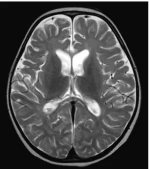
revealed only diffuse atrophy of brain with passive dilatation of ventricles at 18 months of age (Fig. 1). Since he had profound psycho- motor retardation, unexplained diffuse brain atrophy on MRI, and abnormal EEG, we per- formed muscle biopsy to find the cause of his illness.
Muscle tissue was obtained from left rectus femoris muscle at 2 years of age. Sections of the muscle (8 µm thick) were stained with hema- toxylin-eosin (H-E), modified Gomori trichrome technique, succinate dehydronase, adenosine tri- phosphatase (ATPase, at pH 9.4, 4.6, and 4.3), and nicotinamide adenine dinucleotide (NADH)- tetrazolium reductase (TR) statin. There showed selective type 1 fiber atrophy with type 1 fiber predominance of more than 90% of the myofibers (Fig. 2). The mean diameter of type 1 fibers are more than 12% smaller than type 2 fiber. There were no special findings such as central cores,
Fig. 1. Axial T2-weighted brain MRI at 18
months of age. Brain MRI showed diffuse atrophy
of brain with passive dilatation of ventricles.
minicores, central nuclei, nemaline bodies, reducing bodies, target fibers, ragged red fibers, and specific metabolic accumulation.
Discussion
CFTD is a congenital myopathy first de- lineated by Brooke in 19731). The criterion of relative type 1 hypotrophy rather than the mean type 2A or type 2B fiber diameter became the central diagnostic feature of the CFTD. Patients with CFTD shared congenital hypotonia, generalized weakness, failure to thrive, multiple joint contractures, scoliosis, long thin face, and high arched palate. Brooke suggested that CFTD was likely to have a genetic basis. Cases with autosomal dominant or recessive inheri- tance have been reported6). However, nobody in the family had symptoms of myopathy in our case.
Since many neuromuscular disorders share fiber size disproportion (FSD) of 12% or great- er, they should be considered as differential diagnoses to exclude before making diagnosis
of CFTD. It has been shown that while FSD is a feature observed in approximately 7% of pediatric muscle biopsies, only 10 to 20% of those children actually have CFTD7). In addi- tion, CFTD could accompany many central nervous system dysfunction such as perinatal asphyxia, idiopathic mental retardation, cerebral malformations, or Leigh disease. Many myopa- thic processes have FSD as an ancillary fea- ture, too. They cannot be distinguished from CFTD by clinical features alone. Diagnosis of these disorders needs their own histological findings. Other authors have used the term secondary fiber size disproportion when FSD occurs in this context8).
In our case, he was manifested as a floppy infant and demonstrated unexplained progres- sing hypotonia, and proximal muscle weakness with pathological criteria of FSD>12% and type I fiber predominance >90% in muscle biopsy in the absence of other specific histolo- gical abnormalities. These features met the criteria for CFTD by Brooke1). His clinical fea- tures including congenital subluxation of the hip, ankle joint contracture, and foot defor- mities are the same as other previous cases of CFTD8). Contractures are usually evident at birth, but in some patients with marked weak- ness and severely impaired mobility, they can develop in later period. The most commonly affected joints are described to be the ankles8). Cryptorchidism, short stature and poor weight gain have frequently been reported as clinical characteristics of CFTD9). These features are similar to clinical characteristics of our case.
The mental milestones of our patient were markedly retarded than those of any previously reported case. To the best of our knowledge, his generalized tonic clonic seizure is rarely
Fig. 2. Pathologic examination of left rectus
femoris muscle at 2 years of age. Muscle biopsy
shows selective type 1 fiber atrophy with type 1
fiber predominance of more than 90% of the
myofibers (NADH-TR stain, ×400)
found in CFTD cases. In addition, his brain MRI showed evident cerebral atrophy in the absence of other causes like metabolic or peri- natal asphyxia. Though the findings of the case were also reported in congenital muscular dystrophy in terms of structural changes of the central nervous system10), the characteristic histological features such as many degenera- ting or regenerating fibers, or increased con- nective tissue of congenital muscular dystrophy were absent in the case8). Nevertheless, mental function in CFTD is usually preserved and our literature search also revealed only a few re- ports of mental retardation in CFTD cases.
Clarke et al reported only two patients (3%) with intellectual disability after reviewing 117 cases of CFTD8).
Shibita et al11) reported a boy with feeding difficulties from early infancy, who required artificial ventilation suffering mental retarda- tion. His first muscle biopsy done at the age of 1 month showed CFTD, but the histologic findings significantly improved on the second biopsy at 1 year of age showing type 1 fiber predominance and characteristic morphologic findings such as the formation of nemalines and cores. Quazi et al12) also reported three unrelated boys with marked, central, nonpro- gressive hypotonia, severe psychomotor retar- dation, seizures, delayed and disharmonic skeletal maturation, and CFTD.
It still remains unclear whether the seizures affected his mental milestones or not and what the main cause was for his seizure. He may be a case of CFTD with type I predominance associated with seizure, mental retardation, dif- fuse brain atrophy, left cryptorchidism, optic nerve atrophy, congenital subluxation of hip, and contracure of ankle joint.
Unlike other previous cases, he did not make a significant improvement in both motor and mental development. It needs to be clari- fied why he did not. Many children with CFTD reported later generally had good prog- nosis. Apparently the disproportion of diameter is not a permanent feature during the course of CFTD. It has been established that a strik- ing correlation between changes in the degree of FSD over time and changes in disease se- verity in some cases is likely to exist8). These findings suggest that for an individual patient, the clinical course may in part depend on whether type 1 fiber hypotrophy resolves or progresses over time.
In conclusion, the frequent association of CFTD with other diseases suggests that CFTD may not be a specific myopathy but a histo- logical abnormality due to different pathogenic insults which interfere with the normal growth and maturation of the muscle fibers. CFTD is a congenital myopathy whose diagnosis can be made only by muscle biopsy, rather than a distinct syndrome whose diagnosis can be as- sumed on the basis of clinical characteristics alone. We believe its pathological pattern could be shared with other conditions. Moreover, we should consider CFTD as the differential diag- nosis of early onset myopathies.
Abstract
Congenital fiber type disproportion (CFTD) is a rare form of congenital myopathy charac- terized by the smallness and the marked pre- dominance of type 1 fibers, which presents congenital hypotonia, delayed motor milestones, joint contractures, and skeletal deformities. The muscle biopsy reveals the type 1 fibers are
more than 12% smaller than the type 2 fibers in size. We describe a 24-month-old boy who presented muscle hypotonia, delayed motor milestones, mental retardation with generalized tonic clonic seizures, and the muscle pathologic findings of CFTD. Additional findings included left cryptorchidism, congenital subluxation of the hip, contractures of ankle joints, diffuse brain atrophy, and optic nerve atrophy. We should consider CFTD as a differential diagno- sis of early onset myopathy.