Vol. 17, No. 2, November, 2009 □ 증 례 □
- 253 -
1)
서 론
(midbrain), (pons), (medulla oblongata)
(spinal cord) .
(mesence-
phalitis) (rhom-
bencephalitis)
1)
.
,
7 7
1 .
증 례
: 2009 8 19 , : 2009 10 16
: ,
Tel : 02)2228-2063, Fax : 02)393-3080 E-mail : [email protected]
환 아 : ○○ , , 7 7
주 소 : ,
출생력 : 3.0 kg .
가족력 : 2 , ,
. 과거력 :
현병력 : 5 ,
, .
.
진찰 소견 : 37.3 , ℃
90 / , 20 / , 105/60 mmHg
.
, ,
.
. .
두통과 복시를 주소로 내원한 뇌간 뇌염 1례
권혜은 박은정 이윤진 이준수․ ․ ․
= Abstract =
A Case of Brainstem Encephalitis Presented with Diplopia and Headache
Hye Eun Kwon, M.D., Eun Jung Park, M.D.
Yun Jin Lee, M.D. and Joon Soo Lee, M.D., Ph.D.
Department of Pediatrics, Pediatric Epilepsy Clinics, Severance Childrens Hospital Brain Reserch Institute, Yonsei University College of Medicine, Seoul, Korea
Brainstem encephalitis is a rare disease and patients typically present with symptoms of areflexia, ataxia, and ophthalmoplegia. We experienced a case of brainstem encephalitis in a 7 years old girl, who presented with diplopia and headache. It can be reliably confirmed by magnetic resonance imaging(MRI) and CSF analysis. We report a case of brainstem encephalitis in a child, with a brief review of literature.
Key Words : Brainstem encephalitis, Diplopia, Headache
― 3 : 1 ―
- 254 - .
. .
. ,
.
.
검사 소견 : 6,460/
mm
3, 13.3 g/dL, 37.4%,
280,000/mm
3. .
90/µL 99% ,
0/µL, 25 mg/dL, 75 mg/dL , .
. 1:320 3
.
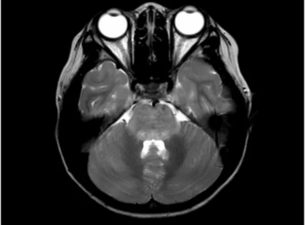
방사선학적 소견 :
. T2
(left rectus gyrus), ,
(Fig. 1A, 1B).
뇌파 검사 :
. 치료 및 경과 :
,
.
,
(vancomycin 60 mg/kg/day, ceftriaxone 100 mg/kg/day)
(acyclovir 30 mg/kg/day)
. 2, 3 1 g/kg
2 . 3
6
. 14
(Fig. 2A, 2B).
.
.
Fig. 1A. Axial T2-weighted brain MRI showed increased signal intensity with edema in pons.
Fig. 1B. Axial T2-weighted brain MRI showed increased signal intensity with edema in left rec- tus gyrus and midbrain.
― : 17 2 2009 ―
- 255 -
고 찰
(mesen-
cephalitis) (rhom-
bencephalitis) 1951 Bickerstaff Cloak
2)
. (areflexia), (ataxia), (ophthalomoplegia)
, , .
.
1, 3, 4)
, Listeria monocytogenes
3, 5, 6)
. Myco -
plasma pneumoniae , influenza A, adenovirus, enterovirus 71, rubella, Epstein-Barr virus
7-12)
.
.
1-6, 11)
,
, ,
6)
.
. 1:
320
.
2, 13, 14)
.
11, 14)
15)
.
15)
.
.
T2 (midbrain), (pons),
(medulla)
1, 11). T2
, , , ,
, , ,
,
15)
.
,
1, 3-6)Fig. 2A. Two weeks later, follow up brain MRI did not show the increased signal intensity or parenchymal swelling in T2 weighted image com- pared to previous images.
Fig. 2B. Two weeks later, follow up brain MRI did not show the increased signal intensity or parenchymal swelling in T2 weighted image com- pared to previous images.
― 3 : 1 ―
- 256 -
16)
. Enterovirus
,
16)
.
L. monocytogenes
17)
. M. pneumoniae
7)
.
.
요 약
, 7
7
1 .
References
1) Soo MS, Tien RD, Gray L, Andrews PI, Fri- edman H. Mesenrhombencephalitis: MR fin- dings in nine patients. Am J Roentgenol 1993;
160:1089-93.
2) Bickerstaff ER, Cloake PC. Mesencephalitis and rhombencephalitis. Br Med J 1951;2:77-81.
3) Hall WA. Infectious lesions of the brain stem.
Neurosurg Clin N Am 1993;4:543-51.
4) Tien RD, Dillon WP. Herpes trigeminal neu- ritis and rhombencephalitis on Gd-DTPA-en- hanced MR imaging. Am J Neuroradiol 1990;
11:413-4.
5) Kohler J, Winkler T, Wakhloo AK. Listeria brainstem encephalitis: two own cases and literature review. Infection 1991;19:36-40.
6) Alper G, Knepper L, Kanal E. MR findings in listerial rhombencephalitis. Am J Neuroradiol 1996;17:593-6.
7) Sakoulas G. Brainstem and striatal encephalitis complicating Mycoplasma pneumoniae pneumo- nia: possible benefit of intravenous immuno- globulin. Pediatr Infect Dis J 2001;20:543-5.
8) Zagardo MT, Shanholtz CB, Zoarski GH, Rothman MI. Rhombencephalitis caused by adenovirus: MR imaging appearance. Am J Neuroradiol 1998;19:1901-3.
9) Shen WC, Chiu HH, Chow KC, Tsai CH. MR imaging findings of enteroviral encephaloyme- litis: an outbreak in Taiwan. Am J Neurora- diol 1999;20:1889-95.
10) Senol U, Haspolat S, Cevikol C, Saatci I. Sub- acute sclerosing panencephalitis: brain stem involvement in a peculiar pattern. Neuroradio- logy 2000;42:913-6.
11) Yaqub BA, al-Deeb SM, Daif AK, Sharif HS, Shamena AR, al-Jaberi M, et al. Bickerstaff brainstem encephalitis. A grave non-demyeli- nating disease with benign prognosis. J Neurol Sci 1990;96:29-40.
12) Shian WJ, Chi CS. Fatal brainstem encephalitis caused by Epstein-Barr virus. Pediatr Radiol 1994;24:596-7.
13) Protheroe SM, Mellor DH. Imaging in influen- za A encephalitis. Arch Dis Child 1991;66:
702-5.
14) Wali GM. Bickerstaff's brainstem encephalitis associated with typhoid fever. Postgrad Med J 1991;67:1011-2.
15) Wasenko JJ, Park BJ, Jubelt B, Lieberman KA, Swarnkar A, Joy SE, et al. Magnetic re- sonance imaging of mesenrhombencephalitis.
Clin Imaging 2002;26:237-42.
16) Rumboldt Z. Imaging of topographic viral CNS infections. Neuroimaging Clin N Am 2008;18:
85-92.
17) Armstrong RW, Fung PC. Brainstem enceph- alitis (rhombencephalitis) due to Listeria mono- cytogenes: case report and review. Clin Infect Dis 1993;16:689-702.