Received January 2, 2019; revised February 18, 2019; accepted February 25, 2019.
Corresponding author: In-Uk Song, Department of Neurology, Incheon St. Mary’s Hospital, College of Medicine, The Catholic University of Korea, 56 Dongsu-ro, Bupyeong-gu, Incheon 21431, Korea. E-mail: siuy@catholic.ac.kr; or Hyeonseok Jeong, Department of Radiology, Incheon St. Mary’s Hospital, College of Medicine, The Catholic University of Korea, 56 Dongsu-ro, Bupyeong-gu, Incheon 21431, Korea. E-mail: hsjeong@catholic.ac.kr
Copyright Ⓒ 2019 The Korean Academy of Clinical GeriatricsThis is an open access article distributed under the term s of the Creative Com m ons Attribution Non-Com m ercial License (http://creativecom m ons.org/licenses/by-nc/4.0) which perm its unrestricted non-com m ercial use, distribution, and reproduction in any m edium , provided the original work is properly cited.
혼합치매와 알츠하이머병의 국소뇌혈류 차이
이명아
1
, 정현석2
, 송인욱1
1가톨릭대학교 인천성모병원 신경과, 2가톨릭대학교 인천성모병원 영상의학과
Differences in Regional Cerebral Blood Flow between Mixed Dementia and Alzheimer’s Disease
Myeong-A Lee
1, Hyeonseok Jeong
2, In-Uk Song
11
Department of Neurology, Incheon St. Mary’s Hospital, College of Medicine, The Catholic University of Korea, Seoul, Korea;
2
Department of Radiology, Incheon St. Mary’s Hospital, College of Medicine, The Catholic University of Korea, Seoul, Korea
Background: Previous studies have suggested that mixed dementia (MD) has distinct characteristics of brain structure and function compared to other types of dementia such as Alzheimer’s disease (AD). However, the patterns of altered regional cerebral blood flow (rCBF) in MD remain elucidated. This study aimed to investigate the differences in rCBF between MD and AD patients.
Methods: Twent-seven MD patients and 27 AD patients in their early stages underwent brain technetium-99m hexamethylpropy- lene amine oxime single-photon emission computed tomography scans. Voxel-wise differences in rCBF between the two groups were examined using Statistical Parametric Mapping.
Results: MD patients presented lower rCBF in the superior/inferior frontal and lateral orbital gyri compared to AD patients (P<0.005). On the other hand, AD patients demonstrated lower rCBF in the amygdala and hippocampus compared to their counterparts (P<0.005).
Conclusion: The distinct characteristics of rCBF vary by underlying dementia pathology, MD and AD.
Key Words: Alzheimer disease, Mixed dementia, Regional cerebral blood flow, Single-photon emission computed tomography
서 론
혼합치매(mixed dementia)는 치매의 주요 원인으로 알 려진 알츠하이머병(Alzheimer’s disease)과 혈관치매(vascular dementia)가 공존하는 경우로 정의된다[1]. 신경병리학적 연구들에 따르면 치매 환자 중 혼합치매의 비율은 약 22%로서 비교적 흔하게 관찰되는 것으로 알려져 있다 [2]. 세포 외 아밀로이드판(amyloid plaque)과 세포 내 신
경원섬유매듭(neurofibrillary tangles) 등의 알츠하이머병 관련 뇌 병변 및 허혈뇌경색증, 다발성 열공뇌경색증, 뇌 실주변백질병터 등의 혈관치매 뇌 병터가 함께 발생하 는 경우, 이들 간의 상승작용에 의해 인지기능의 저하가 발생할 위험이 유의하게 높아질 수 있다[3,4].
혼합치매의 진단 및 치료법 개발을 위해서는 다른 형 태의 치매와의 비교연구가 중요하고, 실제로 이에 대한 다양한 임상 및 뇌영상 연구들이 보고되고 있다. 신경심
리학적 특성을 비교한 연구들에서는 알츠하이머병 환자 군에 비하여 혼합치매 환자군의 주의집중 및 실행기능 등이 유의하게 낮은 것으로 나타났다[5,6]. 또한 치매에 흔하게 동반될 수 있는 신경정신증상들의 양상도 알츠 하이머병, 혈관치매, 혼합치매 환자군 사이에서 차이가 있는 것으로 알려졌다[7]. 자기공명영상(magnetic resonance imaging, MRI) 등을 이용한 뇌영상 연구에서는 혼합치매 군에서 전두엽-측두엽-두정엽의 피질 두께 감소 및 백질 의 미세구조 손상[8], 알츠하이머병 및 혈관치매군에 비하 여 default mode network 및 중앙집행기능 네트워크(central executive network)에서 휴지기 기능적연결성(resting-state func- tional connectivity)의 저하[9], 혈관치매 및 정상 대조군에 비하여 후두엽에서 높은 myoinositol/creatine ratio [10] 등 이 확인되었다. 이와 같은 결과들은 혼합치매의 임상양상 과 뇌의 구조 및 기능이 다른 형태의 치매와 차이를 보인 다는 점을 시사한다. 그러나 혼합치매에 대한 뇌영상 연 구는 다른 형태의 치매에 비해 상대적으로 부족하여 일관 된 결론을 얻기 위해서는 추가적인 연구가 필요하다. 특 히 알츠하이머병에 비해 혈관치매에서 이마관자엽, 마루 엽, 시상에 비대칭적으로 산재된 국소뇌혈류(regional cere- bral blood flow) 저하가 관찰된 것과는 달리, 혼합치매의 국소뇌혈류 양상에 대해서는 잘 알려지지 않았다.
본 연구는 단일광자방출컴퓨터단층촬영(single-photon emission computed tomography, SPECT)을 이용하여 알츠 하이머병 환자군과 혼합치매 환자군 간 국소뇌혈류를 비교분석하였다. 혼합치매군에서 실행기능과 전두엽의 휴지기 기능적연결성이 저하되어 있다고 보고한 선행연 구에 근거하여[6,9], 알츠하이머병군에 비하여 혼합치매 군에서 전두엽의 국소뇌혈류가 유의하게 낮을 것으로 가정하였다.
대상 및 방법
1. 연구 대상자
가톨릭대학교 인천성모병원에서 모집한 초기 알츠하 이머병 및 혼합치매 환자를 대상으로 하였으며, 진단은 Diagnostic and Statistical Manual of Mental Disorders, Fourth edition [11] 및 National Institute of Neurological and Communicative Disorders and Stroke and the Alzheimer disease and Related Disorders Association 기준에[12] 근거 하였다. 임상치매척도(Clinical Dementia Rating, CDR) [13]
점수는 0.5인 경우로 하였다. 두부외상, 뇌경색증, 뇌전 증, 다른 신경과적 또는 정신과적 질환의 과거력이 있는 환자는 연구에서 제외되었다. 본 연구는 인천성모병원 임상연구심사위원회의 승인을 받았으며, 모든 연구참여 자로부터 서면동의를 받았다.
2. 임상적 평가
신경과 전문의가 병력청취, 신체검사, 실험실적 검사 등의 임상적 진찰을 실시하였다. 인지기능 및 신경정신 증상은 각각 간이정신상태검사(Mini-Mental State Examination, MMSE) [14]와 Neuropsychiatric Inventory (NPI)로[15] 평 가하였다. 치매의 전반적인 심각도는 CDR [13], CDR-Sum of Boxes (CDR-SOB) 및 치매단계평가척도(Global Deterio- ration Scale, GDS) [16]로 평가하였다.
3. 뇌영상 획득 및 분석
대상자에게 550 MBq의 technetium-99m hexamethylpro- pylene amine oxime을 정맥주사하고 약 20분 후에 저에너 지부채꼴빔조준기가 탑재된 dual-head 감마카메라(Discovery NM630, GE Healthcare, Milwaukee, WI, USA)를 이용하 여 SPECT 뇌 영상을 획득하였다. 영상은 6o 간격으로 총 720o를 회전하는 카메라에 의해 획득되었으며, filtered back projection 기술을 이용하여, 128×128 matrix 및 1.95×1.95 mm의 픽셀(pixel) 크기(field of view=250 mm, slice thickness=2.08 mm, 20% symmetric energy window at 140 keV)로 재구성되었다.
영상의 전처리와 통계분석에는 Statistical Parametric Mapping 12 (SPM; Wellcome Department of Cognitive Neurology, Institute of Neurology, London, UK)가 사용되 었다. 전처리를 위하여 각 영상을 SPM SPECT template 에 공간정규화(spatial normalization)시킨 다음, 2.0×2.0×
2.0 mm3의 복셀(voxel) 크기로 reslicing하였고, 12 mm full- width half-maximum Gaussian kernel을 이용하여 smoothing 을 적용하였다. 각 복셀의 국소뇌혈류 값은 proportional scaling에 의해 뇌 전체의 평균 혈류를 기준으로 정규화 되었다.
4. 통계 분석
군간 연속변수의 비교를 위하여 독립 t 검정이 이용되 었다. 통계적 유의성은 P<0.05 (양측검정)을 기준으로 하였으며, 통계분석에는 Stata 13.1 (StataCorp., College
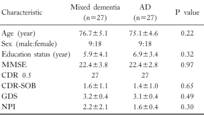
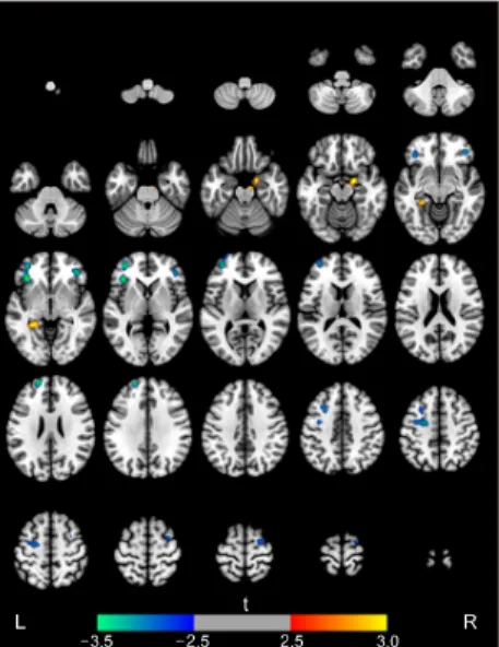
Table 2. Differences in regional cerebral blood flow between patients with mixed dementia and Alzheimer disease
Brain region t P value Cluster size (voxels) Coordinates*
Mixed dementia<Alzheimer disease
Left superior frontal gyrus 3.99 <0.001 197 −20, 58, 30
Left inferior frontal gyrus 3.73 <0.001 595 −38, 30, 0
Right lateral orbital gyrus 3.35 0.001 208 40, 40, −6
Left superior frontal gyrus 3.16 0.001 258 −22, −10, 52
Right superior frontal gyrus 3.01 0.002 131 24, −8, 70
Mixed dementia>Alzheimer disease
Right amygdala 3.54 <0.001 294 18, −4, −18
Left hippocampus 3.31 0.001 274 −26, −42, −4
*The coordinates refer to the Montreal Neurological Institute coordinate system.
Table 1. Demographic and clinical characteristics of the study participants
Characteristic Mixed dementia (n=27)
AD
(n=27) P value
Age (year) 76.7±5.1 75.1±4.6 0.22
Sex (male:female) 9:18 9:18
Education status (year) 5.9±4.1 6.9±3.4 0.32
MMSE 22.4±3.8 22.4±2.8 0.97
CDR 0.5 27 27
CDR-SOB 1.6±1.1 1.4±1.0 0.65
GDS 3.2±0.4 3.1±0.4 0.49
NPI 2.2±2.1 1.6±0.4 0.30
AD, Alzheimer’s disease; CDR, Clinical Dementia Rating; CDR- SOB, Clinical Dementia Rating-Sum of Boxes; GDS, Global Deterioration Scale; MMSE, Mini-Mental State Examination;
NPI, Neuropsychiatric Inventory.
Station, TX, USA)이 사용되었다.
알츠하이머병군과 혼합치매군의 국소뇌혈류 차이를 분석하기 위하여 SPM에서 각 복셀 단위로 두표본 t 검 정(two-sample t-test)이 실시되었으며, 나이, 성별, MMSE 총점이 공변량으로 포함되었다. 각 복셀의 height thresh- old는 P<0.005로, extent threshold는 100 복셀 이상으로 설정하였다.
결 과
총 27명의 알츠하이머병 환자와 27명의 혼합치매 환 자가 분석에 포함되었으며, 이들의 인구학적 및 임상적 특성이 Table 1에 정리되었다. 나이는 알츠하이머병군이 75.1±4.6세, 혼합치매군이 76.7±5.1세로, 두 집단 간 유 의한 차이가 없었으며(P=0.22), 성별은 양군 모두 남성
9명과 여성 18명으로 동일하였다. 그 밖에 교육정도(P=
0.32), MMSE (P=0.97), CDR (P=1.00), CDR-SOB (P=
0.65), GDS (P=0.49), NPI (P=0.30)에도 유의한 차이는 없었다.
두 집단 간 국소뇌혈류 비교 결과는 Table 2와 Figure 1에 제시되었다. 알츠하이머병군에 비하여 혼합치매군에 서 국소뇌혈류가 유의하게 낮았던 영역은 왼쪽 위이마 이랑, 왼쪽 아래이마이랑, 오른쪽 가쪽눈확이랑, 왼쪽 위 이마이랑, 오른쪽 위이마이랑으로 나타났다. 혼합치매군 에 비하여 알츠하이머병군에서 국소뇌혈류가 유의하게 낮았던 뇌영역은 오른쪽 편도 및 왼쪽 해마였다.
고 찰
본 연구는 SPECT 뇌영상을 이용하여 혼합치매와 알 츠하이머병 환자군의 국소뇌혈류 차이를 비교 분석하였 다. 그 결과, 인구학적 특성, 전반적 인지기능, 치매의 심 각도, 신경정신증상 등에는 군간 차이가 유의하지 않은 가운데, 혼합치매 환자군은 위/아래이마이랑 및 가쪽눈 확이랑에서 상대적으로 국소뇌혈류가 낮았고, 알츠하이 머병 환자군은 편도와 해마에서 낮았다. 이와 같은 결과 는 혼합치매와 알츠하이머병의 기저 병태생리의 차이에 따라 국소뇌혈류의 분포가 다를 수 있음을 시사한다.
혼합치매군에서는 전두엽 여러 영역에서 국소뇌혈류 가 상대적으로 낮게 나타났다. 이와 유사하게, 휴지기 기 능적 MRI를 이용한 선행연구에서도 혈관치매 및 알츠 하이머병군에 비하여 혼합치매군에서 아래이마이랑을 중심으로 중앙집행기능 네트워크 내부의 기능적연결성 이 감소한 것으로 나타났다[9]. 또한 혈관치매 중 가장 흔한 피질하혈관치매(subcortical vascular dementia) 환자군
Figure 1. Brain regions with increases (red-yellow) or decreases (blue-green) in regional cerebral blood flow in patients with mixed dementia compared to those with Alzheimer’s disease. The height threshold was P<0.005 and extent threshold was 100 or more contiguous voxels. Images are shown in neurological con- vention. Color bar represents voxel-level t-values. L, left; R, right.
에서도 알츠하이머병 환자군에 비하여 전두엽의 인지기 능 및 국소뇌혈류 저하가 보고되었다[17]. 피질하혈관치 매 환자군에서는 T2-강조영상에서 피질하열공경색과 백 질병터(white matter hyperintensity)가 관찰되는 경우가 많 은데, 이러한 허혈병터는 전두엽의 피질과 피질하부위를 이어주는 신경회로에 손상을 야기하고[18], 수행기능에 장애를 초래할 수 있는 것으로 알려졌다[19]. 또다른 연 구에서는 백질병터가 전두엽에 더 많이 발견되며, 이는 전두엽의 포도당 대사 및 수행기능 저하와 연관되는 것 으로 보고되었다[20]. 본 연구에서 나타난 혼합치매 환 자군의 전두엽 국소뇌혈류 저하는 허혈성 병태생리가 반영된 것으로 생각되며, 혼합치매의 특징적인 전두엽 기능 저하[6]와 연관되었을 가능성을 시사한다.
알츠하이머병군에서 편도 및 해마의 국소뇌혈류 저하 는 기존의 병리학 및 뇌영상 연구 결과들과 잘 부합한 다. 알츠하이머병에 대한 병리학적 연구들에서는 내측두 엽, 편도, 해마, 내후각피질, 해마곁이랑 등의 영역에서 가장 먼저 손상이 시작되는 것으로 알려져 있다[21]. 특 히 편도의 덧기저편도핵(accessory basal nucleus), 피질핵 (cortical nucleus)와 cortical transition area 등 해마 투영 (hippocampal projection)과 연결된 핵 중심으로 신경원섬 유매듭과 아밀로이드판이 관찰되었고[22], 해마의 CA1
영역에서도 신경손상이 보고된 바 있다[23]. 관류강조 (perfusion-weighted) MRI를 이용한 연구에서는 정상 대조 군과 비교하여 알츠하이머병군에서 편도와 해마의 국소 뇌혈류가 저하된 것으로 나타났다[24]. 본 연구에 포함 된 환자들은 초기 단계의 치매였기 때문에, 측두정엽 등 의 영역에서는 차이가 유의하지 않았던 것으로 생각된 다.
본 연구의 제한점은 다음과 같다. 본 연구에는 정상 대조군이 포함되지 않아 알츠하이머병과 혼합치매 간 국소뇌혈류의 상대적인 차이만을 비교할 수 있었다. 국 소뇌혈류의 정상범위와 비교한 방향성을 파악하기 위하 여 향후 정상 대조군과의 비교분석이 필요하고, 추가적 으로 혈관치매와 혼합치매 간의 비교도 이루어진다면 혈관치매와는 다른 혼합치매의 특이적인 국소뇌혈류 양 상을 규명하는데 도움이 될 것이다. 두 번째, 종합적인 신경심리학적 검사가 수행되지 않아 알츠하이머병과 혼 합치매 간 구체적인 인지기능의 차이 및 국소뇌혈류와 인지기능 사이의 연관성을 확인하지 못하였다. 추후 혼 합치매군에서 전두엽의 국소뇌혈류 저하가 실행기능 등 의 인지기능 손상과 연관관계를 보이는지에 대한 연구 가 더 필요할 것이다.
결론적으로 본 연구는 알츠하이머병과 혼합치매 환자 군 사이의 SPECT 뇌영상을 비교분석해 국소뇌혈류 양 상의 차이를 확인하고자 하였다. 그 결과 혼합치매에서 는 전두엽의 국소뇌혈류가 상대적으로 낮았고, 알츠하이 머병군에서는 피질하영역 및 내측두엽에서 국소뇌혈류 가 낮았다. 앞서 언급한 제한점들을 고려하여, 향후 정상 대조군 및 혈관치매 환자군까지 포함하고 뇌영상과 신 경심리학적 검사 결과를 추적하여 연계 분석한다면, 혼 합치매의 감별진단 및 예후 예측 등에도 도움이 될 것으 로 기대된다.
CONFLICTS OF INTEREST
No potential conflict of interest relevant to this article was reported.
ACKNOWLEDGEMENT
This research was supported by Global Frontier Program through the National Research Foundation of Korea (NRF)
funded by the Ministry of Science and ICT (2018M3 A6A3058651).
REFERENCES
1. Langa KM, Foster NL, Larson EB. Mixed dementia: emerg- ing concepts and therapeutic implications. JAMA. 2004;
292:2901-8.
2. Custodio N, Montesinos R, Lira D, Herrera-Perez E, Bardales Y, Valeriano-Lorenzo L. Mixed dementia: A review of the evidence. Dement Neuropsychol 2017;11:364-70.
3. Snowdon DA, Greiner LH, Mortimer JA, Riley KP, Greiner PA, Markesbery WR. Brain infarction and the clinical ex- pression of Alzheimer disease. The Nun Study. JAMA 1997;
277:813-7.
4. de la Torre JC. Vascular basis of Alzheimer's pathogenesis.
Ann N Y Acad Sci 2002;977:196-215.
5. Dong Y, Gan DZ, Tay SZ, Koay WI, Collinson SL, Hilal S, et al. Patterns of neuropsychological impairment in Alzheimer disease and mixed dementia. J Neurol Sci 2013;333:5-8.
6. Kang HS, Kwon JH, Kim S, Na DL, Kim SY, Lee JH, et al. Comparison of neuropsychological profiles in patients with Alzheimer disease and mixed dementia. J Neurol Sci 2016;
369:134-8.
7. Anor CJ, O'Connor S, Saund A, Tang-Wai DF, Keren R, Tartaglia MC. Neuropsychiatric symptoms in Alzheimer dis- ease, vascular dementia, and mixed dementia. Neurodegener Dis 2017;17:127-34.
8. Jang H, Kwon H, Yang JJ, Hong J, Kim Y, Kim KW, et al. Correlations between gray matter and white matter degener- ation in pure Alzheimer disease, pure subcortical vascular de- mentia, and mixed dementia. Sci Rep 2017;7:9541.
9. Kim HJ, Cha J, Lee JM, Shin JS, Jung NY, Kim YJ, et al.
Distinctive resting state network disruptions among Alzheimer disease, subcortical vascular dementia, and mixed dementia patients. J Alzheimers Dis 2016;50:709-18.
10. Waldman AD, Rai GS, McConnell JR, Chaudry M, Grant D. Clinical brain proton magnetic resonance spectroscopy for management of Alzheimer's and sub-cortical ischemic vascular dementia in older people. Arch Gerontol Geriatr 2002;35:
137-42.
11. American Psychiatric Association. Diagnostic and Statistical Manual of Mental Disorders. 4th ed. Text Revision. Washington, DC: American Psychiatric Association; 2000.
12. McKhann GM, Knopman DS, Chertkow H, Hyman BT, Jack Jr CR, Kawas CH, et al. The diagnosis of dementia due
to Alzheimer disease: Recommendations from the National Institute on Aging-Alzheimer’s Association workgroups on di- agnostic guidelines for Alzheimer disease. Alzheimers Dement 2011;7:263-9.
13. Morris JC, Heyman A, Mohs RC, Hughes JP, van Belle G, Fillenbaum G, et al. The Consortium to Establish a Registry for Alzheimer disease (CERAD). Part I. Clinical and neuro- psychological assessment of Alzheimer disease. Neurology 1989;39:1159-65.
14. Kang Y, Na DL, Hahn S. A validity study on the Korean Mini-Mental State Examination (K-MMSE) in dementia patients. J Korean Neurol Assoc 1997;15:300-8.
15. Choi SH, Na DL, Kwon HM, Yoon SJ, Jeong JH, Ha CK.
The Korean version of the neuropsychiatric inventory: a scor- ing tool for neuropsychiatric disturbance in dementia patients.
J Korean Med Sci 2000;15:609-15.
16. Reisberg B, Ferris SH, de Leon MJ, Crook T. Global Deterioration Scale (GDS). Psychopharmacol Bull 1988;24:
661-3.
17. Starkstein SE, Sabe L, Vazquez S, Teson A, Petracca G, Chemerinski E, et al. Neuropsychological, psychiatric, and cer- ebral blood flow findings in vascular dementia and Alzheimer disease. Stroke 1996;27:408-14.
18. Roman GC, Erkinjuntti T, Wallin A, Pantoni L, Chui HC.
Subcortical ischaemic vascular dementia. Lancet Neurol 2002;1:426-36.
19. O'Brien JT, Wiseman R, Burton EJ, Barber B, Wesnes K, Saxby B, et al. Cognitive associations of subcortical white mat- ter lesions in older people. Ann N Y Acad Sci 2002;977:
436-44.
20. Tullberg M, Fletcher E, DeCarli C, Mungas D, Reed BR, Harvey DJ, et al. White matter lesions impair frontal lobe function regardless of their location. Neurology 2004;63:246- 53.
21. Braak H, Braak E. Neuropathological stageing of Alzheimer- related changes. Acta Neuropathol 1991;82:239-59.
22. Kromer Vogt LJ, Hyman BT, Van Hoesen GW, Damasio AR. Pathological alterations in the amygdala in Alzheimer disease. Neuroscience 1990;37:377-85.
23. West MJ, Coleman PD, Flood DG, Troncoso JC. Differences in the pattern of hippocampal neuronal loss in normal ageing and Alzheimer disease. Lancet 1994;344:769-72.
24. Luckhaus C, Fluss MO, Wittsack HJ, Grass-Kapanke B, Janner M, Khalili-Amiri R, et al. Detection of changed region- al cerebral blood flow in mild cognitive impairment and early Alzheimer’s dementia by perfusion-weighted magnetic reso- nance imaging. Neuroimage 2008;40:495-503.