서 론
연골점액유사 섬유종은 드문 종양으로 대개 장골의 골간단에 발생하며, 일반적으로 경골근위부와 대퇴골 원위부에 호발하는 것으로 알려져 있으나 드물게 5.4%
정도로 두개골 및 안면골 등의 두경부에 발생하기도 한 다. 보통 이 종양은 하악과 상악골에 존재하고 부비강 으로 침범하는 경우는 10예 정도만 외국 문헌에 알려질
정도로 흔하지 않다. 또한 국내외적으로 아직까지 비강 및 하비갑개에 발생이 보고된 예는 없다. 치료는 근치적 절제술이며 재발율은 15% 정도로 보고되며 예후는 양 호하다. 조직학적 특징은 풍부한 점액성 또는 연골성 기 질에 세포형태는 방추상형 또는 성상형의 형태를 취한 다. 본 증례는 46세 여자 환자로 양측 비폐색과 비루를 주소로 내원하여 하비갑개에서 발생한 혈관종으로 오 인된 연골점액유사 섬유종에 대해 문헌 고찰과 함께 보 고하는 바이다.
증 례
46세 여자 환자가 수개월전부터 발생한 양측 비폐색, 양측 비루를 주소로 내원하였다. 환자는 5년 전 잦은 비 폐색, 비루 및 출혈을 주소로 개인이비인후과 내원하여 논문접수일 :2018년 2월 23일
논문수정일 :2018년 4월 11일 심사완료일 :2018년 5월 21일
교신저자 :최지윤, 61452 광주광역시 동구 필문대로 365 조선대학교 의학전문대학원 이비인후과학교실
전화 :(062) 220-3200・전송 :(062) 225-2702 E-mail:[email protected]
J Clinical Otolaryngol 2018;28:99-103 증 례
하비갑개에서 기원한 연골점액유사 섬유종 1예
조선대학교 의학전문대학원 이비인후과학교실,1 코웰의원2
한지혜1
·
오정현1·
김성현2·
최지윤1Chondromyxoid Fibroma Originated from Inferior turbinate : A Case Report
Ji Hye Han, MD1, Jeong Hyun Oh, MD1, Sung Hyun Kim, MD, PhD2 and Ji Yun Choi, MD, PhD1
1Department of Otorhinolaryngology-Head and Neck Surgery, School of Medicine, Chosun University, Gwangju; and 2Cowell Clinic, Gwangju Korea
- ABSTRACT -
Chondromyxoid fibroma is a rare benign tumor that usually occurs in the long bones. A 46-year-old female pa- tient presented with masses in her nasal cavities the masses were subsequently diagnosed as a chondromyxoid fibroma. The masses originated from the inferior turbinate. Wide mass excision that included the turbinate and surrounding tissues was performed. No recurrence or new lesion was observed during the one-year follow-up.
A diagnosis of chondromyxoid fibroma, that involves the head and neck is extremely rare. There are no reports of such masses arising from the nasal cavities of patients in our country. Furthermore, available literature does not discuss such masses originating from the inferior turbinate. In this paper, we report on a rare case of a very large-sized chondromyxoid fibroma within the nasal cavity of a patient; we also review the relevant literature. (J Clinical Otolaryngol 2018;29:99-103)
KEY WORDS:Chondromyxoid fibromaㆍNasal cavityㆍInferior turbinate.
비강 내 종양 소견 관찰되어 부분마취 하에 내시경적 절제술을 시행받았으며 수술 후 시행한 조직학적 검사 상 혈관종으로 진단되었다. 수술 후 별다른 합병증 없 이 잘 치료되고 증상호전 보였으며 별다른 문제없이 지 내던 중 수개월 전부터 양측 비폐색이 다시 발생하여 이비인후과 방문하여 시행한 이학적검사상 비강내 같 은 부위에 종양의 재발소견 보여 본원으로 전원되었다.
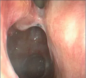
환자는 이전 수술 기왕력 외에 과거력상 특이소견은 없 었다. 본원 내원 당일 시행한 부비동 내시경 검사상 우 측 비강내 종괴소견 관찰되었으며 종괴는 4 cm 정도로 비교적 크기가 크고 하비갑개에서 기시하여 비강의 후 방을 가득 채우고 상악동의 입구와 후비공 및 비인강까 지 확장된 소견을 보였다. 종괴는 표면이 비교적 매끈 하고 단단하였으며 출혈소견은 보이지 않았다(Fig. 1).
수술 전 시행한 전산화단층촬영상 비교적 경계가 뚜 렷한 4×4 cm 크기의 종괴가 우측 하비갑개 후방부터 상악동 내벽을 부분적으로 파괴하고 후방으로 날개돌 기공간(Pterygoid fossa)과 몸체(Pterygoid Body)의 일 부를 파괴하고 후비강 및 비인두까지 이어지는 것을 확 인하였다. 종괴는 비중격을 반대쪽으로 밀고 있었으며 위로는 접형동의 바닥까지 닿아있는 양상이었다. 종괴 는 근육과 비교하여 동등한 세포 밀도를 보여주었으나 조영된 전산화단층촬영 영상에서 종괴 변연부 주변으 로 불균질한 조영증강소견이 관찰되었다. 부비동내에 이차적인 부비동염 소견은 관찰되지 않았다(Fig. 2). 또 한 수술 전 시행한 자기공명영상에서는 T1 강조 영상상 비교적 균일한 음영의 저신호 강도 및 T2 강조 영상상 균일한 음영의 고신호 강도를 보이는 경계가 비교적 좋 은 종괴가 확인 되었다(Fig. 3). 과거력, 내시경 소견과 영상의학적 검사를 토대로 기존의 혈관종의 재발로 의 Fig. 1. Preoperative finding of right nasal cavity shows a
solid and smooth surface soft tissue masses originated from inferior turbinate (arrow).
Fig. 2. Preoperative contrast enhanced paranasal sinus computer tomogram shows a 4 cm sized expansile heterog- enous soft tissue mass originated from inferior tubinate extend to medial wall of maxillary sinus, septum, pterygoid plate and nasopharynx. A : Axial view. B : Coronal view.
A B
심하였다. 그러나 나이, 성별, 위치 그리고 CT와 MRI 소 견을 고려하여 볼 때 다른 종양의 가능성을 염두에 두 고 수술을 계획하였다. 수술 전 혈관색전술은 시행하지 않았으며 전신 마취 하에 내시경을 이용한 광범위 절제 술을 계획하였다. 수술 시 종괴가 하비갑개 후방에서 시
작하여 상악동 자연공 주변, 비중격후방 및 후비공 상부 까지 유착된 소견을 보여 주변조직을 포함한 광범위한 절제술을 계획하였다. 수술시야를 확보하기 위해 우선 우측 하비갑개의 전절제를 시행 후 충분한 절제연을 확 보하고 종괴를 주변조직과 함께 근치적절제술을 시행하
T1 weighted image T2 weighted image
Fig. 3. Preoperative paranasal sinus Magnetic resonance imaging. T1 weighted image (A. axial view C. coronal view) shows well marginated hypotensive soft tissue mass originated from inferior turbinate. T2 weighted image (B.
axial view D. coronal view) shows well marginated highertensive soft tissue mass with heterogenous enhancement extend to maxillary sinus, septum, pterygoid plate and nasopharynx.
T1 weighted image T2 weighted image
A
C
B
D
였다. 수술 중 시행한 신속조직절편검사상 악성세포는 관찰되지 않았으며 수술 중 출혈은 심하지 않았다. 제 거된 종괴는 표면이 비교적 매끈하고 단단한 4×4 cm 정도의 원형의 고형 종괴 형태였다. 절제생검상 H-E 염색 후 저배율에서 종괴는 섬유질, 점액질 기질과 연골 성 요소로 구성되어 있었으며 고배율상에서는 교원성 물질이 섬유 조직과 섞여 있고 엽상 구조물의 주변부로 세포 밀도가 비교적 높게 나타나고 엽상 구조물 중앙부 위는 방추세포와 성상세포로 구성된 섬유아세포가 관 찰되어 연골점액유사 섬유종으로 진단되었다. 면역염 색검사상 S-100 단백에 음성반응을 보였고 CD34, Desmin, Actin에서도 음성소견이었다(Fig. 4). 외래에 서 시행한 1년 이상의 추적관찰에서 재발소견은 보이지 않고 있다(Fig. 5).
고 찰
연골점액유사 섬유종은 국소적으로 분엽화된 섬유성, 점액성, 연골성의 특징을 가지는 양성종양으로, 1948년
Jaffe와 Lichtenstein에 의해 처음 보고되었다.1) 이 종양 은 주로 10대나 20대의 청소년에 호발하는 질환으로 주 로 장골의 골간단에 발생한다. 이 양성종양은 연골육종
Fig. 5. Postoperative endoscopic finding. The operated site was healed well without recurrence on 1 year after surgery.
Fig. 4. Histopathologic findings of chondromyxoid fibroma. Higher magnifications show stellate and spindle-shaped cells, as well as chondroid, myxoid and fibrous components (left) (H&E ×400). The tumor consists of fibrous myxoid matrix and chondroid elements (right) (H&E ×100).
과 감별이 필요한 질환으로 슬관절 주변으로 대게 경골 근위부와 대퇴골원위부의 골간단 부분에 호발하고 25% 정도에서는 장골과 같은 편평한 골에서 발견되기 도 한다.2) 그 외에도 종종 연골점액유사 섬유종이 발생 하기도 하는데 수부,3-6) 족부,7) 척추골,8) 늑골9) 순서로 발생빈도를 보인다. 두경부 침범은 드물지만 두개골 기 저부, 하악골 및 상악골에서 보고 되기도 한다. 또한 전 두동, 사골동, 접협동 등의 부비동에서 발생한 예도 이 미 보고 되었으나 아직까지 하비갑개에 발생한 예는 보 고된 바가 없다. 방사선 영상에서 골간단부 가까이에 편심성의 골용해 소견을 보이고 주변은 골경화 소견 및 스캘럽트모양(scalloped shape)의 경계부가 관찰되어지 는 특징을 보인다. Soder 등은 연골점액유사 섬유종에 서 기질 조성과 유전자 발현 양상을 분석하여 연골모세 포종, 골연골종, 연골종 및 연골육종을 비롯한 다른 중 간 엽의 신생물에서는 발견되지 않는 연골점액유사 섬 유종의 특정 기질 조성을 입증했다.10) 점액질과 섬유질 성분의 양에 변화가 있을 수 있지만, 전형적인 성상 세 포 및 방추 모양의 세포는 전형적으로 교원성 물질 및 점액성 기질에 놓여 있으며, 이형성은 거의 없다. 보고 된 몇몇 사례는 소엽의 말단에서 다핵 거대 세포를 보 여주기도 하고 본 예에서는 존재하지 않지만 드물게 석 회화를 동반하는 경우도 나타내는 것으로 알려져 있다.11) S-100 단백질은 몇몇 경우에서 확인되었지만 S-100 염 색은 연골모세포종 및 연골점액유사 섬유종을 감별진 단하는데 있어 신뢰할 수 없는 표지 인 것으로 나타났
다.12,13) Steiner는 연골점액유사 섬유종과 다른 양성 연
골성 종양의 미세 구조를 비교한 결과 감별할 수 있는 특징을 제공하지 못한다고 결론지었다.14)
치료는 가능하다면, 병소 및 주변부위를 포함하는 근 치적절제술이 최상의 치료법이다.2,15) 소파술은 일반적 으로 성공하지만 재발의 위험성이 높다.15) 방사선 요법 은 외과적 접근이 불가능한 경우를 제외하곤 적응증이 되지 않는다. 본 증례는 5년전 동일한 부위에 발생한 종 괴에 관해 혈관종으로 진단되었으나 이 후 재발된 종괴 에서는 연골점액유사 섬유종으로 진단된 경우로 비강 내 연골점액유사 섬유종이 드물고 조직학적 진단이 까 다로워 이전의 조직학적 진단에 오류가 있었을 것으로
사료된다.
중심 단어:연골점액유사 섬유종・비강・하비갑개.
This study was supported by research funds from Chosun University Hospital 2016.
REFERENCES
1) Jaffe HL, Lichtenstein L. Chondromyxoid fibroma of bone:
a distinctive benign tumor likely to be mistaken especial- ly for chondrosarcoma. Arch Pathol 1948;45:541-51.
2) Unni KK, Inwards CY, Bridge JA, Kindblom L-G, Wold LE. Cartilaginous lesions. In: AFIP atlas of human pa- thology, 4th Series, Fascicle 2. Tumors of bones and joints.
Washington, DC: American Registry of Pathology;2005.
p.67-73.
3) Hau MA, Fox EJ, Rosenberg AE, Mankin HJ. Chondro- myxoid fibroma of the metacarpal. Skeletal Radiol 2001;
30:719-21.
4) Nalbantoglu U, Aktas S, Peker KR. Chondromyxoid fi- broma involving the entire metacarpal: a case report. J Hand Surg Am 2005;5:1083-6.
5) Strauch RJ, Kleinman WB. Chondromyxoid fibroma of a metacarpal: a case report and review of the literature. J Hand Surg 1996;21:293-5.
6) Yamamoto T, Mizuno K. Chondromyxoid fibroma of the finger. Kobe J Med Sci 2000;46:29-32.
7) O’Connor PJ, Gibbon WW, Hardy G, Butt WP. Chondro- myxoid fibroma of the foot. Skeletal Radiol 1996;25:143-8.
8) Kikuchi F, Dorfman HD, Kane PB. Recurrent chondro- myxoid fibroma of the thoracic spine 30 years after prima- ry excision: case report and review of the literature. Int J Surg Pathol 2001;9:323-9.
9) Lawson JP, Barwick KW. Case report 209: chondromyx- oid fibroma of left first rib. Skeletal Radiol 1982;9:53-5.
10) Soder S, Inwards C, Muller S, Kirchner T, Aigner T. Cell biology and matrix biochemistry of chondromyxoid fibro- ma. Am J Clin Pathol 2001;116:271-7.
11) Noh H, Park YJ. A Neurofibroma Arising from the Nasal Septum: A Case Report. J Clinical Otolaryngol 2006;17:
261-5.
12) Bleiweiss IJ, Klein MJ. Chondromyxoid fibroma: a re- port of six cases with immunohistochemical studies. Mod Pathol 1990;3:664-6.
13) Desai SS, Jambhekar NA, Samanthray S, Merchant NH, Puri A, Agarwal M. Chondromyxoid fibromas: a study of 10 cases. J Surg Oncol 2005;89:28-31.
14) Steiner GC. Ultrastructure of benign cartilaginous tumors of intraosseous origin. Hum Pathol 1979;10:71-86.
15) Rahinmi A, Beabout JW, Ivins KL, Dahlin DC. Chondro- myxoid fibroma: a clinicopathologic study of 76 cases.
Cancer 1972;30:726-36.