J. of Korean Orthopaedic Research Society V o l u m e 8 , N u m b e r 1 , A p r i l , 2 0 0 5
기니아 피그의 골절 치유에 대한 저출력 레이저의 효과
단국대학교 의과대학 정형외과학교실, 단국대학교 의과대학 이비인후과학교실*, 서울대학교 의과대학 해부학교실†
김명호・박희곤・이정구*・박경한†・박홍근・김영재
= Abstract =
The Effect of Low Level Laser on the Fracture Healing in Guinea Pig Femur
Myung-Ho Kim, M.D., Hee-Gon Park, M.D., Chung-Ku Rhee,M.D.*, Kyung-Han Park, M.D.†, Hong-Keun Park, M.D., Young-Jae Kim, M.D.
Department of Orthopaedic Surgery, Dankook University College of Medicine,
Department of Otolaryngology-Head and Neck Surgery, Dankook Univerisity College of Medicine Department fo anatomy, Seoul national university college fo Medicine†
Purpose: This study was conducted to evaluate the effectiveness of the low level laser on the fracture-heal- ing of the guinea pig femurs.
Materials and Methods : This study was performed on 36 guinea pigs(17 in the experimental group and 19 in the control group). After anesthesia of the guinea pigs, the shaft of femur was fractured with Gigli saw under aseptic conditions, and then the intramedullary nailing of K-wire was performed. The application of low level laser on the fracture sites in the experimental group began 2 days after the operation under anesthesia, for 30 minutes every 2 days. The laser we used was a CW-type microchip laser with output wave length of 890 nm and intensity of 13 mWatts, measuring upto 4.68 J/cm2per each application. In order to compare the femur fracture healing process of both groups; gross findings, simple radiologic findings, and histologic findings of fracture site stability were evaluated. The simple radiologic and histologic findings were assessed by the scor- ing system invented by Zorlu and his colleagues. The statistical evaluation was done by using repeative mea- sured ANOVA test.
R e s u l t s: In both experimental and control groups, the gross findings progressed favorably. Although the improvement in radiologic findings were found in both groups with the passage of time, the experimental group showed a greater rate of callus formation with statistical significance(p<0.0) compared to the control group.
The result of histological findings showed that a increase of osteoblastic proliferation was also greater in the
※ 통신저자: 박 희 곤
충청남도 천안시 안서동 1 6 - 5 단국대학교 의과대학 정형외과학교실
TEL: 041) 550-3950 FAX: 041) 556-3238 E-mail: heegon@chol.net
✽ 본 논문은 2 0 0 4년도 단국대학교 의학레이저 연구센터 지원을 받아 이루어졌음.
서 론
골절의 치유는 장기간의 시간을 필요로 하고 불 유합, 지연유합 등 다양한 합병증의 가능성이 있 다. 최근에는 골절 치유를 촉진시키는 여러 방법 에 대한 다양한 연구가 보고 되고 있다. 적당양의 초음파, 전기 자극, 미세 운동 등이 골절의 치유 과정을 단축시키는 방법으로 알려져 있다
1 , 2 , 8 , 1 3 , 2 1 , 2 2 ). 최근 레이저 역시 초음파나 전기자극등
과 유사한 효과가 있다는 보고가 있다3 , 4 , 5 , 2 3 )
. 과거 정형외과나 구강외과 등에서 초음파와 레이저의 동시 조사에 대한 실험이 부분적으로 시행된 적
있었으나3 , 5 , 2 0 , 2 3 )
, 레이저 단독 조사에 대한 연구는 현재까지 많은 논란이 있다. 레이저는 일종의 광 파 혹은 전자파로서, 보통 우리가 일상적으로 접 할 수 있는 광선과는 몇 가지 다른 특성을 가지고 있다. 레이저는 일종의 특이한 빛으로 파장이 비 교적 짧은 전자기파의 일종으로 빛의 속도로 전파 된 전기장(electrical wave)과 자기장( m a g n e t i c w a v e )으로 구성되어 있다. 저출력 레이저 광선 을 흡수한 생체 세포들은 광 에너지를 세포의 손 상을 치유할 수 있는 화학적인 에너지로 전환시 켜, 이를 손상된 부위의 치유 (healing) 및 고통 완화 (pain relief)에 이용하게 되는데, 이러한 현상을 총체적으로 생체 촉진 효과 ( b i o - s t i m u- lation effect)라고 한다. 또한 저출력 레이저를 이용한 광 조사( p h o t o - i r r a d i a t i o n )는 다양한 효 소나 세포, 조직, 기관들에서 생화학적, 생리적 효과를 유발할 수 있다. 저출력 레이저 경우 Chen 등6 , 7 , 9 , 1 7 )
에 의해 1 9 8 1년 콜라겐 합성의 촉 진, 상처 치료 촉진 효과 등 일부 연부 조직에 대 한 효과는 이미 밝혀졌고, 세포에서 성장 인자의 발현을 증가시킬 수 도 있다고 알려져 있다. 이에 저자는 저출력 레이저의 현재까지 밝혀진 생역학 적 특성과 생화학적인 특성에 따른 혈액 순환의 증가, 세포 감수 분열의 시간 단축, 그리고 A T P
합성을 통한 세포의 포텐셜 에너지의 증가, 광 에 너지 효과 등이 골절 치유를 촉진시킬 수 있을 것 이라는 가정 하에 기니아 피그를 대상으로 실험을 시행하였다.
대상 및 방법 1. 실험 대상
본 실험은 생후 9주된 잉글리시( E n g l i s h )종 H a r t l e y계 암컷 기니아 피그를 대상으로 하였다.
체중은 최저 552 gm에서 635 gm으로 평균 6 1 0
±56 gm이었다. 42마리를 두군으로 나누어 실험 군은 우측 대퇴골을, 대조군은 좌측 대퇴골을 골 절시켜 실험 하였다. 3주와 5주째 사망한 3마리 와 수술 부위에 감염소견이 관찰되었던 3마리는 제외하고, 최종 실험까지 가능하였던 기니아 피그 는 실험군이 1 7마리 대조군이 1 9마리였다.
2. 실험 방법
1) 수술
5% Ketamin(Yuhan. corp. Korea)을 그램 ( g m )당 0.5 cc을 둔부에 근육 주사하여 마취시 키고 무균 조작 하에서 대퇴의 외측을 종으로 절 개를 하였다. 그 후 근육을 박리하여 대퇴간부을 노출시키고 소독된 Gigli saw을 이용하여 가능한 횡형 골절을 발생시키고자 하였다. 하지만 골절 시키는 과정에서 다양한 형태의 골절이 발생되었 다. 그 후 소독된 드릴을 이용하여 1.4 mm K- 강선의 골수강내 고정술를 시행하였다. 내고정술 은 대퇴부의 전장을 고정시켰으나, 추시 과정 중 기니아 피그의 움직임에 의하여 강선의 위치 변화 가 관찰되기도 하였다. 내고정후 절개창은 나일론 으로 봉합하였다. 술 후 7일까지 양군 모두에게 항생제( C e f a z e d o nⓇ Kukje Phar. Korea)를 체 experimental group(p<0.01).
Conclusion: In this study, the low level laser was revealed to be effective on acceleration of femur fracture healing in guinea pigs.
Key Words: Guinea Pig, Fracture healing, Low level laser
중 100 gm당 0.05 gm씩 근육 주사하였다.
2) 레이저 조사
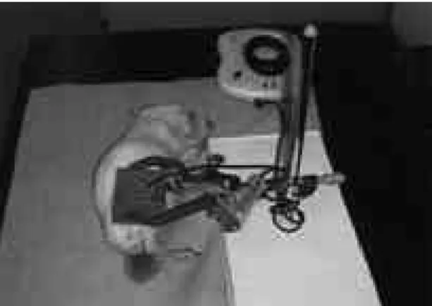
술 후 2일 째부터 6주째 까지 실험 군에게 마취 하에 저출력 레이저를 3 0분씩 2일에 한번씩 조사 하였다(Fig. 1). 레이저는 C W형 반도체 레이저 (L-Dr. 890, Pros International, 2002, K o r e a )로 출력 파장 890 nm, 강도 1 3 m W a t t s로 한번 조사 때마다 4.68 J/cm2를 조 사하였다. 조사 거리는 피부에서 1 c m의 거리에서 실험을 진행하였다.
3) 조직 표본 작성
수술 후 2 주 째에 다량의 K e t a m i n을 심장 내 주사하여 군당 무작위로 추출된 6마리씩의 기니아 피그를 희생시킨 후 즉시 양군의 대퇴골을 적출하 여 4% paraformaldehyde로 고정하였다. 이어 서 탈회용액에 1 0일간 넣어 골을 탈회 시킨 후 5% sodium sulfate solution에 2 4시간 동안 담 가 두었다. 이후 일반적인 표본 제작 과정을 거쳐 hematoxyline-eosin 염색 표본 슬라이드를 제 작하였다. 또한 수술 후 4주, 6주째에도 위의 과 정을 반복하여 역시 총 3 6마리의 기니아 피그로 부터 조직을 채취하였다.
4) 방사선 검사
방사선 검사는 수술 후 2주째, 4주째, 그리고 6 주째 사망 직후 시행하였다. 방사선 검사 조건은 43 Kv, 3.2 mAs로 대퇴의 측면 사진을 촬영하 였다.
5) 분석 방법
실험 결과 분석은 육안적 소견, 단순 방사선 검 사, 조직학적 검사를 이용하여 양군의 대퇴부의 골유합 진행 정도를 비교 분석하였으며, Zorlu
등1 8 )의 scoring system을 사용하였다(Table 1).
또한 Scion ImageⓇ (Scion version 16, USA) 프로그램을 이용하여 가골 면적을 정량적으로 분 Fig. 1. Application of low level laser under anesthesia is
shown.
Table 1. Modified Zorlu Scoring System
Score Findings
1 Mobile, easy to manipulate
Gross findings 2 Elastic, angulation by manipulation
3 Solid, stable fracture site
0 No visible callus
1 Beginning stage of periosteal callus
Radiologic findings
2 Mature periosteal callus
3 Finishing stage of pariosteal callus
0 Nonunion
1 Fibrous union
Histologic findings 2 Osteochondral union
3 Bone union
4 Complete reorganization
U. Zorlu et al (Am. J. of Physical Medicine & Rehabilitation, 1998)
석하여 통계 처리하였다. 육안적 소견은 3인의 정 형외과 전문의가 기니아 피그를 희생시킨 직후 직 접 골절 부위를 손으로 조작하여 강도 및 탄력 정 도를 점수화 하여 평균 처리하였다. 방사선 검사 항목에 대한 평가는 2인의 방사선과 전문의와 1 인의 정형외과 전문의가 평가하여 각각 점수화 한 후 그 평균을 이용하였으며, 조직학적 평가는 2인 의 해부 병리과 전문의가 각각 점수화 하여 그 평 균을 이용하였다. 통계학적 분석은 SPSS 12Ⓡ (Sigma, USA) 프로그램을 이용하였고 각 주에 따라 대조군과 실험군 사이와 각 군내의 차이를
분 석 하 기 위 하 여 repeative measured ANOVA test를 사용하였다.
결 과 1. 육안적 소견
실험군의 경우 술 후 2주째 육안적 소견은 1 . 1 3±0 . 5 2점, 4주째는 2 . 1 7±0 . 7 5점, 6주째는 3 . 0점으로 통계학적으로 의미 있게 증가되었으며 (p<0.01), 대조군의 경우에도 2주째 1 . 0 1점, 4주 째 1 . 5 0±0 . 5 5점, 6주째 2 . 1 7±0 . 4 1점으로 증가 되어(p<0.01) 두 군 모두 시간이 경과함에 따라 회복되었다(Fig. 2).
2주와 6주에서는 통계학적으로 양군의 의미 있 는 차이는 없었으나, 4주에서는 실험군이 대조군 보다 더 빠르게 골절이 회복되는 양상을 보였으며 통계학적으로도 유의성이 있었다( p < 0 . 0 1 ) .
2. 단순 방사선 검사
실험군에서는 술 후 2주째 0 . 8 3±0 . 4 1점, 4주 째는 1 . 8 3±0 . 4 2점, 6주째는 2 . 6 7±0 . 5 2점으로 증가되는 소견을 보였다(p<0.01). 대조군의 경우 에도 2주째 0 . 3 3±0 . 5 2점, 4주째 1 . 3 3±0 . 5 7점, 6주째 2 . 3 3±0 . 5 4점으로 증가되어(p<0.01) 실험 Fig. 2. The results of gross findings of experimental and
control groups are shown. There was no statisti- cally significant difference between the two groups at 2 and 6 weeks after fracture. However, at 4 weeks, the difference was statistically signif- icant
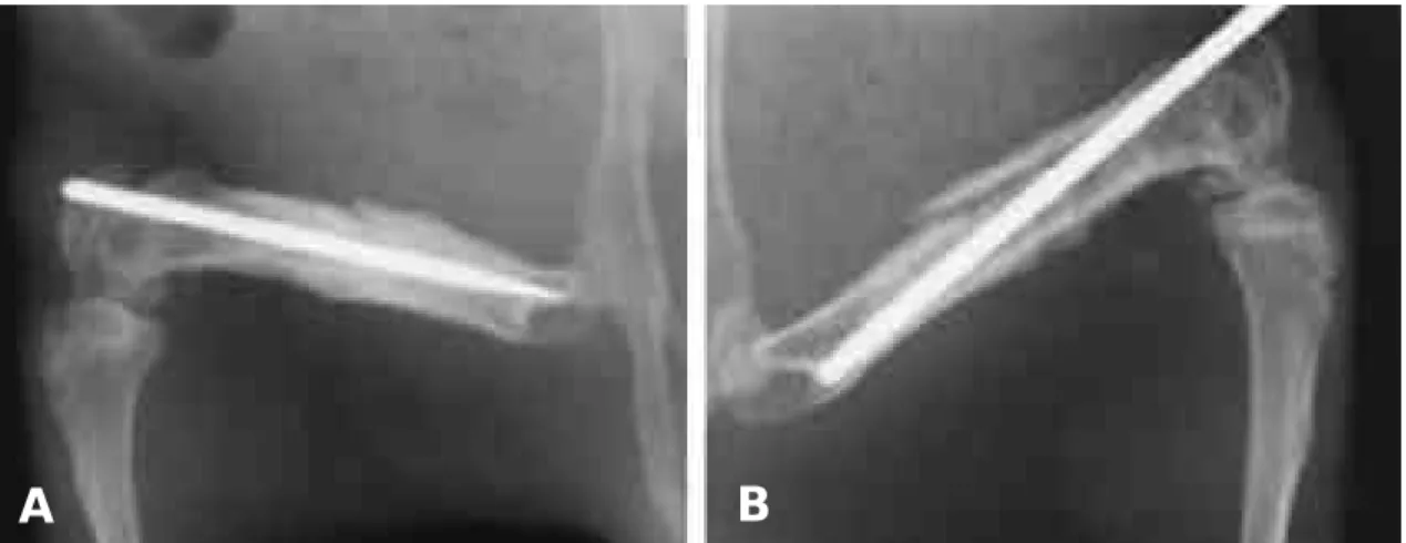
Fig. 3. Radiographs show intramedullary nailing state in both group at postoperative 2weeks.
The bone union was faster in the experimental group(A) than in the control group(B).
A B
군과 마찬가지로 시간이 경과함에 따라 골유합이 진행되고 있었으나, 2주째 이미 실험군이 대조군 보다 더 빠르고 성숙된 가골 형성이 이루어지는 것으로 관찰되었다(Fig. 3-A,B. Fig. 4-A,B.
Fig. 5-A,B.). 통계적으로도 유의한 차이를 보 였다(p<0.01, Fig. 6).
3. 조직학적 검사
실험군에서 술 후 2주째 1 . 3 3±0 . 5 2점, 4주째 는 3 . 0 0±0 . 8 9점, 6주째는 3 . 8 3±0 . 4 1점으로 시
간이 경과함에 따라 가골의 양이 증가되었다 (p<0.01). 대조군의 경우에도 2주째 0 . 5 0±0 . 5 5 점, 4주째 1 . 3 3±0 . 5 2점, 6주째 3 . 0 0±0 . 8 9점으 로 증가되어(p<0.01), 두 군 모두 시간이 경과함 에 따라 의미 있는 조직학적 변화가 관찰되었다 (Fig. 7-A,B,C,D. Fig. 8-A,B,C,D. Fig. 9- A,B,C,D). 마찬가지로 2주부터 실험군이 대조 군보다 더 빠른 조직학적 골절 치유의 양상을 보 였다. 통계적으로도 의미 있는 변화를 나타내었다 (p<0.01, Fig. 10).
Fig. 4. Radiographs taken postoperative 4 weeks show more marked callus formation in the experimental group (A) than in the control group(B).
A B
Fig. 5. Radiographs taken postoperative 6weeks show more complete bone union in the experimental group(A) than in the control group(B).
A B
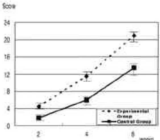
4. Zorlu 등의 scoring system 총 점수
Zorlu 등의 scoring system에 따라 육안적 소 견, 단순 방사선 검사, 조직학적 검사 점수를 통 합 했을 때, 실험군은 술 후 2주째 4 . 5±0 . 5 5점,
4주째 1 1 . 5±1 . 6 4점, 6주째 2 1 . 0±1 . 2 6점으로 점차 증가되었다(p<0.01). 대조군에서도 2주째 1 . 8 3±0 . 7 5점, 4주째 6 . 0 2±1 . 6 7점, 6주째 1 3 . 5 0±1 . 8 7점으로 증가되었으나(p<0.01), 위의 모든 결과와 마찬가지로 실험군에서 대조군에 비 해 빠른 회복정도를 나타내었다(Fig. 11). 역시 통계학적으로도 의의가 있는 차이가 나타났다 ( p < 0 . 0 1 ) .
고 찰
골절의 치유는 호르몬, 비타민과 전신적 상태뿐 아니라, 국소 혈류량, 체중 부하, 골절부의 상태 등의 국소적 인자 등 다양한 요인에 영향을 받는 다. 골재생이라는 과정을 통하여 이루어는 골절 의 치유 과정은 다음과 같은 3가지 방법으로 골절 의 유합을 이루게 된다. 첫째, 직접 골치유 (direct bone healing) 둘째, 신연골 생성( d i s- traction osteogenesis), 셋째로 가골로 골절부 가 일차로 고정된 후 재형성 과정을 거쳐 골절이 치유되는 간접 골치유(Indirect bone healing) Fig. 6. The results of radiologic findings of experimental
group and control group are shown.
There was significant difference between the experimental and control groups at postoperative 2, 4 and 6 weeks.
Fig. 7. Histologic findings of both groups 2weeks after operation are shown. The experimental group(A , B) shows more callus than the control group(C,D). (H-E, × 40)
*F: Fracture site, †H: Hard callus, ‡Fr: Cartilage
A B
C D
Fig. 8. Histological findings 4weeks after operation show more marked new bone formation in the experimental group(A,B) than in the control group(C,D). (H-E, × 40)
*F: Fracture site, †H: Hard callus, ‡Fr: Cartilage
A B
C D
Fig. 9. Histologic findings 6weeks after operation show more complete bone union in the experimental group(A,B) than in the control group(C,D). (H-E, × 40)
*F: Fracture site, †H: Hard callus, ‡Fr: Cartilage
A
C D
B
가 있다. 이러한 간접 골치유에서 외부 가골의 형 성은 골절부 안정성에 크게 기여하며, 생물학적 그리고 물리학적 자극에 반응하여 다양한 변화가 발생하기에 흔히 골절 치유 연구에 활용된다.
최근 간접 골절 치유 과정을 분자생물학적으로 접근하여, 경가골과 연가골을 각각 분리하여 각 치유단계별로 골 특수 기질 단백 (bone specific matrix protein)과 연골 특수 기질 단백( c a r t i- lage specific matrix protein)의 유전자 발현 을 Northern hybridization 기법으로 연구하 여, 각 치유과정과 시기별로 기질 단백의 형성이 일정하고, 이는 유전자 전사 과정에서 조절되는 자체 조절기전에 의할 것으로 추측되어지고 있다
1 9 , 2 6 , 3 0 ). 이러한 정상적인 치유 과정은 1 8 9 2년
W o l f f4 4 )가 물리적인 자극이 근골격계에 구조적, 또는 기능적으로 영향을 줄 수 있다는 발표 후 growth factor나 cytokine 등의 생물학적 자극 뿐만 아니라 물리적인 자극에도 매우 민감하여 5 - 20 μA의 낮은 전류, 30~50 mW/cm2의 낮은 초 음파에너지 영역 또는 특수한 파장의 전자장에 의 해 골형성이 촉진되며, 골절부의 미세한 움직임 역시 골절부의 가골 형성을 촉진시키는 것으로 알 려지고 있다1 , 2 , 8 , 1 3 , 2 1 , 2 2 ).
T. Yaakobi 등2 3 )은 저출력 He-Ne 레이저를
사용하여 토끼의 경골을 대상으로 한 실험에서 실 험군이 대조군에 비해 골절부위에서 알카린 인산 효소(alkaline phosphatase)와 칼슘의 발현시기 가 1주일 정도 빠르다고 하였고, 골생성 세포의 양도 최소 2배 이상 차이가 난다고 보고하였다.
저자들의 실험에서는 육안적 소견에서 2주와 6주 에는 통계적으로 의미 있는 차이가 발견하지 못하 였으나, 4주에서는 육안적 소견에서 의미 있는 차 이가 나타났다. 그 이유는 2주에서 시행한 육안적 소견에서는 양군 모두에서 육안적 차이를 나타낼 수 있는 정도의 골절 유합이 진행되지 않았으며, 6주에는 저출력 레이저 조사와 관계없이 골절 유 합이 이미 이루어져 양군에서 통계적 차이가 나타 나지 않았던 것으로 생각된다. 육안적 소견을 제 외한 방사선학적, 조직학적 소견과 정량적인 가골 의 면적의 비교에서 모든 주에서 실험군이 대조군 에 비해 의미 있는 통계적 차이가 발견되었다. 방 사선학적 분석과 조직학적 분석에서는 2주째 이미 통계적으로 큰 차이가 나타나 저출력 레이저가 2 주전부터 골절 유합 촉진에 영향을 주는 양상을 보였다.
본 논문의 문제점으로는 골절의 양상이 일정치 않고, 골유합 평가 방법에 객관성이 떨어지며, 일 정한 양상으로 K -강선 내고정이 동일한 조건이
Fig. 10. The result of histologic findings of experimental and control groups are shown. There was signif- icant difference in the union of fracture histo- logically between the experimental and control groups at postoperative 2, 4 and 6 weeks.
*F: Fracture site, †H: Hard callus, ‡Fr: Cartilage
Fig. 11. The results of score of Zorlu scoring system are shown. Zorlus score was significantly higher in the experimental group than in the control group at postoperative 2, 4 and 6 weeks.
아니라는 점이 가장 큰 문제점 이였다. 또한 본 실험에서는 레이저의 사용이 골절 치유에 정확하 게 어떠한 작용 기전이 있는지에 대한 연구는 시 행하지 못하였고, 조사 시간과 조사량에 따른 골 절 치유 정도 차이에 대하여도 실험하지 않아 추 후 이 부분에 대한 연구와 골절 부위에 기계적 강 도나 신생 혈관의 연구 및 동물이나 인체에 미치 는 영향에 대하여 다양한 강도, 주파수, 조사시간 등을 바꾸어 효과를 검증해 볼 필요가 있을 것으 로 생각된다.
앞으로 임상 의학 분야에서 레이저는 많은 양 의 에너지를 순간적으로 목표물에 집중 조사할 수 있어 파괴력이 크고 주변조직에 미치는 손상이 적 기 때문에 환부에 대해서만 집중적인 투여가 가능 하다는 장점을 있고, 저출력 레이저의 여러 물리 학적, 생화학적인 활성들이 확인됨에 따라 전에는 치료하지 못했던 질환들에 적용되어질 수 있을 것 으로 예상된다.
결 론
평균 6 1 0±56 gm, 9주짜리 기니아 피그 3 6마 리의 대퇴골을 골절 시킨 후 내고정술을 시행하 여, 저출력 레이저인 출력 파장 890 nm, 강도 13 mWatts의 C W형 반도체 레이저( L - D r . 890, Pros International, 2002, Korea)로 이 틀에 한번씩 3 0분간, 1회 조사 때마다 4 . 6 8 J / c m 2를 6주간 조사한 결과, 저출력 레이저 조 사군의 골절 치유가 촉진된 양상을 관찰 하였다.
R E F E R E N C E S
01) Buckley, M.J. et al: Osteoblasts increase their rate of division and align in response to cyclic, mechanical tension in vitro. Bone and Min., 4:
225-236, 1988.
02) Cochran G.V.B., et al.: Piezoelectric internal fixation devices: A new approach to electrical augmentation of osteogenesis. J Orthop. Res. 3:
508-513, 1985.
03) David R., Nissan M., Cohen I., Soudry M. : Effect of low-power He-Ne laser on fracture
healing in rats: Laser Surg. Med. 1996; 19(40):
458-64.
04) Garavello-Freitas I., Baranauskas V., Joazeiro P.P., Padovani C.R., Dal Pal-Silva M., da Cruz-Hoflin M.A.: Low-power laser irradiation improves histomorphometrical parameters and bone matrix organization during tibia wound healing in rats. J Photochem Photobiol B.2003 May-Jun; 70(20):81-9.
05) Guzzardella G.A., Torricelli P., Nioli Aldini N., Giardino R.: Laser technology in orthope- dics: preliminary study on low power laser ther- apy to improve the bone-biomaterial interface.
Int J Artif Organs. 2001 Dec; 24(12): 898-902.
06) Hall, R, R.: The healing of tissure incised by a carbon dioxade laser. Brit. J. Surg. 58, 222-225, 1971.
07) Horoszowski H., Farine I. & Engel J.: The laser in orthopaedic surgery. Proceedings of the 1st International Symposium on Laser Surgery.
1975, Jerusalem Academic Press.
08) Heckman, J.D. et at : Acceleration of tibial frac- ture-healing by non-invasive, low-intensity pulsed ultrasound. J. Bone joint Surg. 76-A: 26- 34, Jan. 1994.
09) Luger E.J., Rochkind S., Wolliam Y., Kogan G .: Effect of low-power laser irradiation on the mechanical properities of bone fracture healing in rats.:Laser Surg Med. 1998; 22(20:97-102).
10) Mester E., Mester A.F.: The biomedical effects of laser aplication. laser Surg. Med 5;31-39 11) Ninomiya T., Miyamoto Y., Yamashita A.,
Wakita M.: High-intensity pulsed laser irradia- tion accelerate bone formation in metaphyseal trabecular bone in rat femur. J Bone Miner Meta., 2003;21(2):67-73.
12) Pai-Silva M., da Cruz-Hofling M.A. : L o w - power laser irradiation improves histomorpho- metrical parameters and bone matrix organiza- tion during tibia wound healing in rats. J Pho- tochem Photobiol B. 2003 M a y - J u n; 70(20:81- 9).
13) Rubin J.: Pressure regulates osteoclast formation and MCSF expression in marrow culture. J. Cell Physiol., 170 : 81-87, 1997.
14) Tadashi Ninomiya, Yuuichi Miyamoto, Taku Ito, Atsushi Yamashita, Masayoshi Wakita:
High intensity pulse laser irradiation accelerates bone formation in metaphyseal trabecular bone in rat femur: J bone Miner Metab( 2 0 0 3 ) 2 1 : 6 7 - 73.
15) Tang X.M., Chai B.P.: Effect of CO2 laser irra- diation on experimental fracture healing: a trans- mission electron microscopic study.: Laser Surg.
Med.1986;6(3):346-52.
16) Taubr C., Farine I., Horoszoowski H., Gassner S. : Fracture healing in rabbits after osteotomy using CO2 laser. Acta Orthop.
17) Trelles M.A., Mayayo E. : Bone fracture consol - idates faster With low-power laser. Lasers Surg Med; 7 ;36-45. 1987.
18) Umran Zorlu, et al: Comparative study of the
effect of ultrasound and eletrostimulation on bone healing in rats. American Journal of Physi - cal Medicine& Rehabilitation, No.15: 427-432, 1998.
19) Wolff J.: The law of bone remodelling, translat- ed by P. Maquet and R. Furlong. New York, Springer, 1986.
20) Yang K.B. et at: Low intensity ultrasound stim- ulates fracture healing in rat model: biomechani- cal and gene expression analysis. Trans. Orthop.
Res. Soc., 19 :519, 1994.
21) Yasuda: Electrical Callus and Callus Formation by Electricity. Clin. Orthop., 124: 53, 1977.
22) Ziskin M.C.: Applications of ultrasound in med- icine-comparison with other modalities. U l t r a - sound. 49-59, 1987.
23) Yaakobi T., Maltz L., Oron U.; Promotion of bone repair in the cortical bone of the tibia in rats by low energy laser(He-Ne) irradiation : Calcif.
Tissue Int. 1996 Oct;59(4):297-300.