Introduction
Temporomandibular joint (TMJ) disorders are common problems among children and adolescents. Their incidence has been reported as being between 6% and 68% in pre- vious studies.1-5 Radiographic examination may serve to provide additional information along with clinical findings.
Despite the high incidence of TMJ dysfunction in children and adolescents, radiographic studies have rarely been carried out. It has been reported that osteoarthritis of the TMJ is an age-related change and is more frequent in older persons than in younger persons.6,7However, a study re-
ported that the pediatric age group with TMJ pain and dys- function showed evidence of degenerative arthritis in 37%
of cases.8Osteoarthritis (OA) of the TMJ in children might potentially have an effect on mandibular growth and lead to an altered skeletal structure.9Therefore, attention should be paid to any osteoarthritic changes in the TMJ in child- ren.
The clinical significance of condylar positioning in the glenoid fossa is controversial. It seems that TMJ disorder (TMD) patients are more likely to have posterior condylar positioning.10-13However, some investigators have suggest- ed no significant association between condylar positioning and clinical or radiographic findings.14-18
This study was performed to assess the prevalence of osteoarthritic changes and the parasagittal condylar posi- tioning of TMJ in children and adolescents with or with- out TMJ symptoms.
Osteoarthritic changes and condylar positioning of the temporomandibular joint in Korean children and adolescents
Bong-Hae Cho, Yun-Hoa Jung
Department of Oral and Maxillofacial Radiology, School of Dentistry, Pusan National University, Yangsan, Korea ABSTRACT
Purpose: To investigate the prevalence of osteoarthritic changes and condylar positioning of the temporomandibular joint (TMJ) in Korean children and adolescents with or without temporomandibular disorders (TMDs).
Materials and Methods: A total of 101 asymptomatic and 181 symptomatic children and adolescents aged 10 to 18 years old were included in the study. Osteoarthritic changes such as flattening, sclerosis, osteophytes, or erosion, and the parasagittal positioning of the condyle were assessed using cone-beam computed tomography (CBCT) images.
Results: The overall prevalence of osteoarthritic changes was higher in symptomatic (26.8%) than in asymptomatic adolescents (9.9%) (p⁄0.05). In the symptomatic group, the frequency was higher in males (33.3%) than in females (23.0%) (p⁄0.05). Erosion was the most common change for the symptomatic group (15.6%), whereas sclerosis was the most common change for the asymptomatic group (5.4%). Posterior condylar position was more frequently ob- served in the symptomatic group (p⁄0.05). Erosion was more common in the samples with TMJ pain or mouth opening limitations as compared to those without them (p⁄0.05).
Conclusion: This study showed that osteoarthritic changes in TMJ were common in children and adolescents, with a much higher prevalence in symptomatic patients. (Imaging Sci Dent 2012; 42 : 169-74)
KEY WORDS: Adolescent; Temporomandibular Joint; Cone-Beam Computed Tomography
*This study was supported by a grant of Pusan National University Hospital.
Received May 16, 2012; Revised June 7, 2012; Accepted June 14, 2012 Correspondence to : Dr. Yun-Hoa Jung
Department of Oral and Maxillofacial Radiology, School of Dentistry, Pusan National University, Beomeo-ri, Mulgeum-eup, Yangsan, Gyeongsangnam-do 626- 870, Korea
Tel) 82-55-360-5255, Fax) 82-55-360-5029, E-mail) [email protected]
http://dx.doi.org/10.5624/isd.2012.42.3.169
Copyright ⓒ 2012 by Korean Academy of Oral and Maxillofacial Radiology
This is an Open Access article distributed under the terms of the Creative Commons Attribution Non-Commercial License (http://creativecommons.org/licenses/by-nc/3.0) which permits unrestricted non-commercial use, distribution, and reproduction in any medium, provided the original work is properly cited.
Imaging Science in Dentistry∙pISSN 2233-7822 eISSN 2233-7830
Materials and Methods
Subjects
We retrospectively reviewed the data of 10- to 18-year- old patients who underwent cone beam computed tomo- graphy (CBCT) images in our hospital between October 2009 and March 2011, and collected 181 symptomatic and 101 asymptomatic patients (Table 1). The symptomatic group consisted of 66 males and 115 females who present- ed for the treatment of TMDs. A clinical examination was performed to assess the presence of mouth opening limi- tation, pain, and TMJ noise in each patient. A maximum opening under 40 mm was considered to be limited mouth opening. TMJ pain was assessed by asking the patient if he or she felt joint or muscle pain during mandibular func- tion. TMJ noise was based on either crepitation or a click- ing sound.
The asymptomatic group was composed of 40 males and 61 females who had no TMJ signs or symptoms. All of them had undergone CBCT scans for the thorough examination of tooth impaction or an orthodontic evalua- tion.
In both groups, the patients who had any conditions that could affect TMJ components such as skeletal abnormali- ties, TMJ tumors, or other infectious disease were exclud- ed.
Methods
CBCT scans were obtained with PaX-Zenith3D (Va- tech, Kihung, Korea) in the maximal intercuspal position, with a rotation of 360 degrees for data acquisition. The exposure factors were a 24×19 cm field of view, 120 kVp, 5-6 mA, and a 24 seconds exposure time. Images were re- constructed using a high spatial frequency reconstruction algorithm immediately after acquisition, and the acquired image data consisted of a 14-bit scale with a 0.3 mm3voxel size. For interpretation, real-time reconstruction was per- formed using an Ez3D 2009 3D image viewer (Vatech, Kihung, Korea), and the axial, coronal, and sagittal two- dimensional (2D) multi-planar reformatted slices were pro- vided.
Two experienced oral and maxillofacial radiologists in- dependently assessed the images on Coronis 5MP moni- tors (Barco, Brussels, Belgium 2,048×2,560 image matric- es, 10-bit viewable gray scale, and 145.9-ft-lambert lumine- scence). Interpretation was made using a combination of all three plane images via an interactive multi-planar dis- play. They were allowed to adjust the window width, win-
dow level, and magnification. No demographic data or cli- nical features were available to them.
Radiographic osteoarthritic changes of flattening, sclero- sis, osteophytes, and erosion for both the temporal part and condyle of the TMJ were evaluated.
The criteria were as follows: flattening-a loss of the round contour of the condyle at the load bearing area, sclerosis-an increased density of the cortical lining or the subchondral bone, osteophyte-a marginal bone outgrowth, and erosion-an interruption or absence of the cortical lining.
Parasagittal condylar positioning was assessed in accor- dance with the method of Pullinger and Hollender,19sug- gesting linear measurement of the subjective closest anteri- or and posterior interarticular space (Fig. 1). Parasagittal plane perpendicular to the long axis of the condyle on axial plane was created and the image passing through the center was used to evaluate condylar positioning. The values of the narrowest anterior (A) and posterior (P) in- terarticular space were transferred to the following for- mula from Pullinger et al.10
(P-A) / (P++A)×100
The position was expressed as a percentage of the anteri-
Table 1.Gender and age distribution of patients included in the study
Male Female Total Mean age
Asymptomatic 40 61 101 15.2
Symptomatic 66 115 181 15.6
Total 106 176 282 15.5
Fig. 1.Linear measurement of anterior (A) and posterior (P) sub- jective closest joint spaces.
or or posterior displacement from absolute concentricity (zero). The range of ±12% was considered to be concent- ric. Values smaller than -12 indicated that the condyle was in a posterior position, and values greater than ++12 indicated that the condyle was in an anterior position.
Statistical analysis
First, we calculated the interobserver agreement by using Cohen’s kappa coefficient,20 which showed substantial agreement (k==0.67). Then, the two observers discussed the disagreed-upon cases together and reached an agree- ment. We used these results for further statistical analysis.
A comparison was carried out to evaluate differences between the asymptomatic and the symptomatic groups.
Additionally, gender and age differences were analyzed within each group. For the symptomatic group, differ- ences in the clinical variables of mouth opening limitation, TMJ pain, and noise were assessed. The statistical analysis was performed using χ2test and the Fisher exact test. The level of statistical significance was set at .05. The calcula- tions were performed with PASW Statistics 18 (SPSS Inc., Chicago, IL, USA).
Results
The occurrence of osteoarthritic change was much high- er in the symptomatic group (26.8%) than in the asympto- matic group (9.9%), showing a significantly higher preval- ence of sclerosis, osteophytes, and erosion in the sympto- matic group (p⁄0.05) (Fig. 2). The concentric condylar position was most common in the asymptomatic group (72.3%), while posterior positioning of the condyle was dominant in the symptomatic group (52.8%) (p⁄0.05) (Fig. 2).
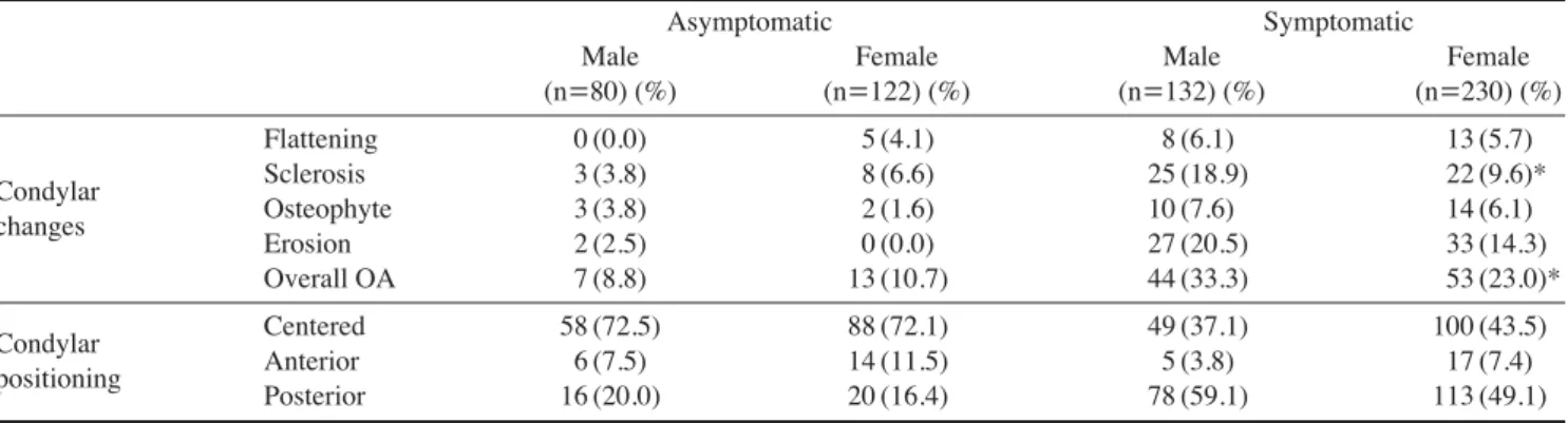
Gender differences were found in the symptomatic group;
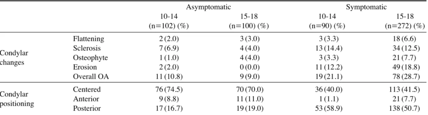
sclerosis and overall osteoarthritic changes were more frequently observed in males than in females (p⁄0.05) (Table 2). In both the asymptomatic and symptomatic groups, there were no significant differences between the two age groups of 10 to 14 and 15 to 18 (Table 3).
In relation to the clinical findings, the patients with mouth opening limitation or TMJ pain showed more ero- sion than those without them (p⁄0.05). TMJ noise show- ed a statistically significant difference in terms of condy- lar positioning; posterior positioning of the condyle was observed more often in cases of TMJ noise (p⁄0.05) (Ta- ble 4).
Fig. 2.Comparison of osteoarthritic changes of TMJs and parasa- gittal positioning of the condyles between the asymptomatic and the symptomatic groups.
*statistically significant difference between asymptomatic and sym- ptomatic groups (p⁄0.05)
OA: one or more osteoarthritic changes of flattening, sclerosis, ost- eophyte, or erosion
80 70 60 50 40 30 20 10 0
Flattening Sclerosis Osteophyte
Erosion OA
Centered Anterior Posterior Condylar bony changes Condylar positioning 2.55.8 5.4
13.0*
Asymptomatic (n==202) Symptomatic (n==362)
2.5 6.6*
1 15.6*
26.8*
72.3
41.2
9.9 6.1
17.8 52.8*
9.9
Table 2.Prevalence of osteoarthritic changes of TMJs and parasagittal positioning of the condyles by gender
Asymptomatic Symptomatic
Male Female Male Female
(n==80) (%) (n==122) (%) (n==132) (%) (n==230) (%)
Flattening 0 (0.0) 5 (4.1) 8 (6.1) 13 (5.7)
Condylar Sclerosis 3 (3.8) 8 (6.6) 25 (18.9) 22 (9.6)*
changes Osteophyte 3 (3.8) 2 (1.6) 10 (7.6) 14 (6.1)
Erosion 2 (2.5) 0 (0.0) 27 (20.5) 33 (14.3)
Overall OA 7 (8.8) 13 (10.7) 44 (33.3) 53 (23.0)*
Condylar Centered 58 (72.5) 88 (72.1) 49 (37.1) 100 (43.5)
positioning Anterior 6 (7.5) 14 (11.5) 5 (3.8) 17 (7.4)
Posterior 16 (20.0) 20 (16.4) 78 (59.1) 113 (49.1)
*statistically significant difference between two gender groups (p⁄0.05)
Overall OA: one or more osteoarthritic changes of flattening, sclerosis, osteophyte, or erosion
Discussion
CBCT is an excellent imaging modality for the assess- ment of bony TMJ components.21,22It provides 2D multi- planar reformatted images with submillimeter spatial resolution and allows the observer to customize the slices interactively by using a volumetric data set. Honey et al23 reported that CBCT images gave superior reliability and great accuracy in evaluating the TMJ, and when the ob- servers interpreted the images through an interactive dis- play, the diagnostic accuracy was much higher than that achieved through static captured images. In this study, the observers were allowed to access the volumetric data and evaluate osteoarthritic changes by scrolling the multi-pla- nar images interactively.
TMJ osteoarthritis is very common in adults. The radio- graphic changes corresponding to osteoarthritis can be observed in 12-44% of the general adult population.1,24 However, aging is not the crucial factor in the pathogene- sis of osteoarthritis.25Our results show that TMJ osteoar-
thritis is also common among children and adolescents. In the asymptomatic group, the prevalence was 9.9%, com- parable to that of Petrikowski and Grace’s study.26 They reported that the overall occurrence of TMJ radiographic abnormalities in preorthodontic patients was 6.9% in the 9 to 11 age group and 7.5% in the 12 to 15 age group.
In the symptomatic group, the prevalence of TMJ osteo- arthritis was 26.8%, being significantly higher than that of the asymptomatic group. Sanchez-Woodworth et al8studi- ed pediatric patients (7 to 16 years) with TMJ pain and dysfunction and indicated that 37% of the patients showed evidence of degenerative arthritis on one or both sides.
Zhao et al25reported a lower prevalence of TMJ osteoar- thritis. In their study using conventional radiography, the frequency was 13.3% in girls and 8.0% in boys over the range of 11 to 14 years, and it was 17.5% in girls and 10.7% in boys over the range of 15 to 19 years. We attri- buted the variation in prevalence mainly to the difference in diagnostic criteria, population sampling, and radiogra- phic examination method used. However, all those studies showed that TMJ osteoarthritis was a common finding in
Table 3.Prevalence of osteoarthritic changes of TMJs and parasagittal positioning of the condyles by age group
Asymptomatic Symptomatic
10-14 15-18 10-14 15-18
(n==102) (%) (n==100) (%) (n==90) (%) (n==272) (%)
Flattening 2 (2.0) 3 (3.0) 3 (3.3) 18 (6.6)
Condylar Sclerosis 7 (6.9) 4 (4.0) 13 (14.4) 34 (12.5)
changes Osteophyte 1 (1.0) 4 (4.0) 3 (3.3) 21 (7.7)
Erosion 2 (2.0) 0 (0.0) 11 (12.2) 49 (18.8)
Overall OA 11 (10.8) 9 (9.0) 19 (21.1) 78 (28.7)
Condylar Centered 76 (74.5) 70 (70.0) 36 (40.0) 113 (41.5)
positioning Anterior 9 (8.8) 11 (11.0) 1 (1.1) 21 (7.7)
Posterior 17 (16.7) 19 (19.0) 53 (58.9) 138 (50.7)
Overall OA: one or more osteoarthritic changes of flattening, sclerosis, osteophyte, or erosion
Table 4.Prevalence of osteoarthritic changes of TMJs and parasagittal positioning of the condyles according to clinical features
Mouth opening limitation Pain Noise
Negative Positive Negative Positive Negative Positive
(n==174) (%) (n==188) (%) (n==46) (%) (n==316) (%) (n==218) (%) (n==144) (%)
Flattening 8 (5.0) 13 (6.9) 0 (0.0) 21 (6.6) 11 (5.0) 10 (6.9)
Osseous Sclerosis 17 (9.8) 30 (16.0) 3 (6.5) 44 (13.9) 30 (13.8) 17 (11.8)
changes Osteophyte 9 (5.2) 15 (8.0) 4 (8.7) 20 (6.3) 15 (6.9) 9 (6.3)
Erosion 20 (11.5) 40 (21.3)* 3 (6.5) 57 (18.0)* 34 (15.6) 26 (18.1)
Overall OA 39 (22.4) 58 (30.9) 6 (13.0) 91 (28.8)* 59 (27.1) 38 (26.4)
Condylar Centered 72 (41.4) 78 (41.5) 18 (39.1) 131 (41.5) 99 (45.4) 50 (34.7)
positioning Anterior 9 (5.2) 13 (6.9) 5 (10.9) 17 (5.4) 16 (7.3) 6 (4.2)
Posterior 93 (53.4) 97 (51.6) 23 (50.0) 168 (53.2) 103 (47.2) 88 (61.1)*
*statistically significant difference between negative and positive groups (p⁄0.05)
Overall OA: one or more osteoarthritic changes of flattening, sclerosis, osteophyte, or erosion
children as well.
A number of studies1,7,14,21have found that degenerative arthritis of TMJ increased with age. However, that would not always be the case for young people. In this study, there was no significant difference between the two age groups (11-14 and 15-18), although there was a tendency toward a higher prevalence of osteoarthritic changes in the older age group. This finding was in agreement with pre- vious studies that sampled similar age groups.25,26
It is widely accepted that TMD is more common in fe- males. Many studies also claimed that the frequency of osteoarthritic changes was higher in females.14,25,26Some investigators, however, found no gender differences.7 In our study, the asymptomatic group showed no significant gender differences, however in the symptomatic group, the prevalence of osteoarthritic changes was higher in males than in females. We speculated that the results arose from gender differences in the willingness to seek help. Boys might be less likely to seek treatment than girls and only visit a hospital in a more advanced stage of the disease.
There has been controversy regarding whether clinical signs and symptoms have effects on radiographic findings.
There were several previous studies7,27-29that indicated a poor correlation between TMJ osteoarthritis and the signs and symptoms of TMD. Wiese et al14 reported that pain was not associated with the increased risk of degenerative findings in TMJ tomograms. On the other hand, Kurita et al30 reported a significant relationship between the pre- sence of TMJ pain upon mandibular function and osteo- arthritic changes at the articular surface. Among other de- generative findings, erosion seems to be associated with subjective symptoms. In this study, erosion was the most common finding in the symptomatic cases, which was consistent with a previous study.31Zhao et al25suggested that the presence of TMJ pain was associated with erosive changes, and Yamada et al9found that erosive bone change was usually accompanied by pain and difficulty in mouth opening. In accordance with these studies, our study de- monstrated that erosion was more frequently observed in the subjects with pain or limited mouth opening. Another controversy existed over the clinical significance of con- dylar positioning in the glenoid fossa. There have been many studies showing no significant association between condylar positioning and clinical and radiographic manife- stations.14-18On the other hand, several studies found that TMD patients appeared to have posterior condyle posi- tioning.10-13For the asymptomatic patients, there have been also conflicting reports concerning condylar positioning.
One study indicated that asymptomatic volunteers showed almost randomly distributed condylar positions in the gle- noid fossa,32 while another claimed that the majority of joints had condyles centered in the glenoid fossa. Our study showed a significantly different distribution between the asymptomatic and the symptomatic groups; a concentric position was more common in the asymptomatic group, and a retruded position was more common in the sympto- matic group. It is usually accepted that a retruded condyle is not always associated with TMD. Nonetheless, the poste- rior positioning of the condyle is commonly observed in TMD patients.
In conclusion, our retrospective study demonstrated that TMJ osteoarthritis was not unusual in children and adole- scents, whether they were complaining of TMD symptoms or not, and considering their age, dentists should pay more attention to monitoring the progression of degenerative changes of TMJ until the stability of mandibular growth would be established.
References
1. Poveda Roda R, Bagan JV, Díaz Fernández JM, Hernández Bazán S, Jiménez Soriano Y. Review of temporomandibular joint pathology. Part I: classification, epidemiology and risk factors. Med Oral Patol Oral Cir Bucal 2007; 12 : E292-8.
2. Dibbets JM, van der Weele LT. Prevalence of structural bony change in the mandibular condyle. J Craniomandib Disord 1992; 6 : 254-9.
3. Vanderas AP. Prevalence of craniomandibular dysfunction in children and adolescents: a review. Pediatr Dent 1987; 9 : 312- 6.
4. Magnusson T, Egermark-Eriksson I, Carlsson GE. Four-year longitudinal study of mandibular dysfunction in children. Com- munity Dent Oral Epidemiol 1985; 13 : 117-20.
5. Gazit E, Lieberman M, Eini R, Hirsch N, Serfaty V, Fuchs C, et al. Prevalence of mandibular dysfunction in 10-18 year old Israeli schoolchildren. J Oral Rehabil 1984; 11 : 307-17.
6. Ishibashi H, Takenoshita Y, Ishibashi K, Oka M. Age-related changes in the human mandibular condyle: a morphologic, ra- diologic, and histologic study. J Oral Maxillofac Surg 1995;
53 : 1016-24.
7. Widmalm SE, Westesson PL, Kim IK, Pereira FJ Jr, Lundh H, Tasaki MM. Temporomandibular joint pathosis related to sex, age, and dentition in autopsy material. Oral Surg Oral Med Oral Pathol 1994; 78 : 416-25.
8. Sanchez-Woodworth RE, Katzberg RW, Tallents RH, Guay JA. Radiographic assessment of temporomandibular joint pain and dysfunction in the pediatric age-group. ASDC J Dent Child 1988; 55 : 278-81.
9. Yamada K, Saito I, Hanada K, Hayashi T. Observation of three cases of temporomandibular joint osteoarthritis and mandibular morphology during adolescence using helical CT. J Oral Reha-
bil 2004; 31 : 298-305.
10. Pullinger AG, Solberg WK, Hollender L, Guichet D. Tomo- graphic analysis of mandibular condyle position in diagnostic subgroups of temporomandibular disorders. J Prosthet Dent 1986; 55 : 723-9.
11. Pereira LJ, Gavi~ao MB, Bonjardim LR, Castelo PM. Ultra- sound and tomographic evaluation of temporomandibular joints in adolescents with and without signs and symptoms of tempo- romandibular disorders: a pilot study. Dentomaxillofac Radiol 2007; 36 : 402-8.
12. Gateno J, Anderson PB, Xia JJ, Horng JC, Teichgraeber JF, Liebschner MA. A comparative assessment of mandibular condylar position in patients with anterior disc displacement of the temporomandibular joint. J Oral Maxillofac Surg 2004;
62 : 39-43.
13. Bonilla-Aragon H, Tallents RH, Katzberg RW, Kyrkanides S, Moss ME. Condyle position as a predictor of temporomandi- bular joint internal derangement. J Prosthet Dent 1999; 82 : 205-8.
14. Wiese M, Svensson P, Bakke M, List T, Hintze H, Petersson A, et al. Association between temporomandibular joint symp- toms, signs, and clinical diagnosis using the RDC/TMD and radiographic findings in temporomandibular joint tomograms.
J Orofac Pain 2008; 22 : 239-51.
15. Abdel-Fattah RA. Optimum temporomandibular joint (TMJ) condylar position. Todays FDA 1989; 1 : 1C-3C.
16. Robinson de Senna B, Marques LS, França JP, Ramos-Jorge ML, Pereira LJ. Condyle-disk-fossa position and relationship to clinical signs and symptoms of temporomandibular disorders in women. Oral Surg Oral Med Oral Pathol Oral Radiol En- dod 2009; 108 : e117-24.
17. Vasconcelos Filho JO, Menezes AV, Freitas DQ, Manzi FR, Bóscolo FN, de Almeida SM. Condylar and disk position and signs and symptoms of temporomandibular disorders in stress- free subjects. J Am Dent Assoc 2007; 138 : 1251-5.
18. Katzberg RW, Keith DA, Ten Eick WR, Guralnick WC. Inter- nal derangements of the temporomandibular joint: an assess- ment of condylar position in centric occlusion. J Prosthet Dent 1983; 49 : 250-4.
19. Pullinger A, Hollender L. Variation in condyle-fossa relation- ships according to different methods of evaluation in tomo- grams. Oral Surg Oral Med Oral Pathol 1986; 62 : 719-27.
20. Cohen J. A coefficient of agreement for nominal scales. Educ Psychol Meas 1960; 20 : 37-46.
21. Alexiou K, Stamatakis H, Tsiklakis K. Evaluation of the seve- rity of temporomandibular joint osteoarthritic changes related to age using cone beam computed tomography. Dentomaxil- lofac Radiol 2009; 38 : 141-7.
22. Tsiklakis K, Syriopoulos K, Stamatakis HC. Radiographic
examination of the temporomandibular joint using cone beam computed tomography. Dentomaxillofac Radiol 2004; 33 : 196-201.
23. Honey OB, Scarfe WC, Hilgers MJ, Klueber K, Silveira AM, Haskell BS, et al. Accuracy of cone-beam computed tomo- graphy imaging of the temporomandibular joint: comparisons with panoramic radiology and linear tomography. Am J Orthod Dentofacial Orthop 2007; 132 : 429-38.
24. Takayama Y, Miura E, Yuasa M, Kobayashi K, Hosoi T. Com- parison of occlusal condition and prevalence of bone change in the condyle of patients with and without temporomandibular disorders. Oral Surg Oral Med Oral Pathol Oral Radiol Endod 2008; 105 : 104-12.
25. Zhao YP, Zhang ZY, Wu YT, Zhang WL, Ma XC. Inve- stigation of the clinical and radiographic features of osteoar- throsis of the temporomandibular joints in adolescents and young adults. Oral Surg Oral Med Oral Pathol Oral Radiol Endod 2011; 111 : e27-34.
26. Petrikowski CG, Grace MG. Age and gender differences in temporomandibular joint radiographic findings before ortho- dontic treatment in adolescents. Oral Surg Oral Med Oral Pathol Oral Radiol Endod 1999; 87 : 380-5.
27. Wiese M, Wenzel A, Hintze H, Petersson A, Knutsson K, Bak- ke M, et al. Osseous changes and condyle position in TMJ tomograms: impact of RDC/TMD clinical diagnoses on agree- ment between expected and actual findings. Oral Surg Oral Med Oral Pathol Oral Radiol Endod 2008; 106 : e52-63.
28. Wiberg B, Wänman A. Signs of osteoarthrosis of the tempo- romandibular joints in young patients: a clinical and radiogra- phic study. Oral Surg Oral Med Oral Pathol Oral Radiol En- dod 1998; 86 : 158-64.
29. Hiltunen K, Peltola JS, Vehkalahti MM, Närhi T, Ainamo A. A 5-year follow-up of signs and symptoms of TMD and radio- graphic findings in the elderly. Int J Prosthodont 2003; 16 : 631-4.
30. Kurita H, Kojima Y, Nakatsuka A, Koike T, Kobayashi H, Kurashina K. Relationship between temporomandibular joint (TMJ)-related pain and morphological changes of the TMJ con- dyle in patients with temporomandibular disorders. Dentomaxil- lofac Radiol 2004; 33 : 329-33.
31. Lee JU, Kim HS, Song JS, Kim KA, Koh KJ. Bone change of mandibular condyle using cone beam computed tomography.
Korean J Oral Maxillofac Radiol 2007; 37 : 139-47.
32. Ren YF, Isberg A, Westesson PL. Condyle position in the temporomandibular joint. Comparison between asymptomatic volunteers with normal disk position and patients with disk displacement. Oral Surg Oral Med Oral Pathol Oral Radiol Endod 1995; 80 : 101-7.