43
IntroductIon
Pituitary apoplexy is hemorrhage or infarction of a pitu- itary gland tumor presented as acute onset of headache, oph- thalmoplegia, and visual impairment. Pituitary apoplexy is an endocrine and surgical emergency that requires prompt treat- ment with glucocorticoid replacement and surgical decom- pression, especially when accompanied by visual symptoms or mental status changes. Known precipitating factors of pitu- itary apoplexy include surgery, anticoagulation therapy, and post-partum hemorrhage [1]. Additionally, bromocriptine, cabergoline, and other pituitary-stimulating treatments, such as gonadotropin-releasing hormone (GnRH) agonist or lu- teinizing hormone-releasing hormone (LHRH) agonist, have been reported to cause apoplexy [2,3]. Most cases of pituitary apoplexy occur in the absence of any precipitating factor. Cy- totoxic chemotherapy for systemic cancer has not been con- sidered as a cause of apoplexy; however, there have been two previously reported cases of pituitary apoplexy following sys-
Extensive Pituitary Apoplexy after Chemotherapy in a Patient with Metastatic Breast Cancer
Je-Hun Jang1, Young San Ko1, Eun Kyeong Hong2, Ho-Shin Gwak3
1Department of Neurosurgery, Seoul National University College of Medicine, Seoul, Korea
Departments of 2Pathology, 3Cancer Control, National Cancer Center, Graduate School of Cancer Science and Policy, Goyang, Korea
Received September 8, 2017 Revised February 6, 2018 Accepted February 7, 2018 Correspondence
Ho-Shin Gwak Neuro-Oncology Clinic, National Cancer Center, 323 Ilsan-ro, Ilsandong-gu, Goyang 10408, Korea Tel: +82-31-920-1666 Fax: +82-31-920-2798 E-mail: [email protected]
Surgery, anticoagulation therapy, pregnancy, and hormone treatments, such as bromocriptine, are well-characterized precipitating factors for pituitary apoplexy. However, whether cytotoxic chemother- apy for systemic cancer could cause pituitary apoplexy has not been investigated. Here, we present a case of a 41-year-old woman who developed a severe headache with decreased visual acuity after intravenous cytotoxic chemotherapy to treat metastatic breast cancer. Preoperative neuroimaging re- vealed pituitary adenoma with necrosis. Operative findings and pathologic examination concluded extensive necrosis with a small intratumoral hemorrhage in a pre-existing pituitary adenoma. We re- viewed two additional previously published cases of pituitary apoplexy after systemic chemotherapy and suggest that cytotoxic chemotherapy may induce pituitary apoplexy.
Key Words Breast cancer; Chemotherapy; Pituitary adenoma; Pituitary apoplexy; Necrosis.
temic chemotherapy [4,5].
cASE rEPort
A 41-year-old woman diagnosed with metastatic breast cancer presented to an emergency room with headache and vomiting a few hours after her third cycle of intravenous cyto- toxic chemotherapy with doxorubicin and cyclophospha- mide. Her breast cancer was confirmed following modified radical mastectomy of her right breast 10 weeks prior. Patho- logic examination revealed stage Ib infiltrative ductal carcino- ma. She was prescribed intravenous treatment with doxorubi- cin and cyclophosphamide every three weeks. Her past medical history included menstrual irregularity, but she did not con- sider it unusual and no work up was performed. After neuro- logical examination in the emergency room, she was prescribed analgesics and anti-emetics and sent home.
The following day, she woke up with diplopia and visual dis- turbances. She returned to the emergency room after having a brain computed tomography (CT) scan at a nearby hospital to rule out intracranial hemorrhage. The CT scan revealed an enlarged sella containing a slightly low-density, round mass 2.8 cm in diameter (Fig. 1A). Upon arrival at the emergency room, she was determined to have left sixth-cranial nerve palsy CASE REPORT Brain Tumor Res Treat 2018;6(1):43-46 / pISSN 2288-2405 / eISSN 2288-2413
https://doi.org/10.14791/btrt.2018.6.e7
This is an Open Access article distributed under the terms of the Creative Commons Attribution Non-Commercial License (http://creativecommons.org/licenses/by-nc/4.0) which permits unrestricted non-commercial use, distribution, and reproduction in any medium, provided the original work is properly cited.
Copyright © 2018 The Korean Brain Tumor Society, The Korean Society for Neuro- Oncology, and The Korean Society for Pediatric Neuro-Oncology
44 Brain Tumor Res Treat 2018;6(1):43-46 Pituitary Apoplexy after Chemotherapy
following a hand motion in the left-eye visual acuity test and temporal hemianopsia in the right-eye visual field test (Fig.
2A). Her initial sodium concentration was 138 mmol/L and there was no definite sign of adrenal insufficiency. Basal hor- mone test results were as follows: growth hormone, 19.32 ng/
mL (normal: 0.38–12.00); cortisol, 66.5 µg/dL (normal: 5.3–
22.5); T3, 0.78 ng/mL (normal: 0.60–1.81); free T4, 1.23 ng/
dL (normal: 0.89–1.76); thyroid stimulating hormone, 0.70 µIU/mL (normal: 0.55–4.78); luteinizing hormone, 0.8 mIU/
mL (normal: 1.0–95.6); follicle stimulating hormone, 11.4 mIU/mL (normal: 1.7–21.5); and estrogen, 30.3 pg/mL (12.4–
341). Magnetic resonance imaging (MRI) confirmed a 2.8-cm mass with solid and cystic portions with spotty intratumoral hemorrhage (Fig. 1B) extending from an enlarged pituitary fossa to suprasellar cistern and left cavernous sinus (Fig. 1C).
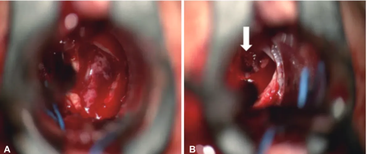
A transsphenoidal approach (TSA) and tumor removal was performed the following day. After dura incision, necrotic ma- terial flowed out and a mass with a blood clot was found upon
microsurgical viewing (Fig. 3). Pathology of the specimen re- vealed pituitary adenoma with extensive mixed-stage (recent and remote) necrosis (Fig. 4). At the end of decompression, a frozen biopsy confirmed a flesh-firm normal gland at the bot- tom of the surgical field. A post-operative MRI, taken 30 hours after surgery, demonstrated that gross total resection was achieved while saving the normal gland as a posteriorly linear enhancing portion in the sagittal view.
The patient felt improved visual acuity immediately postop- erative and developed transient diabetes insipidus on the first postoperative day that was treated with desmopressin and flu- id replacement. Three days postoperative, her left sixth-cranial nerve palsy recovered. One week later, she was discharged fol- lowing tamoxifen chemotherapy. Three days after discharge, she was again admitted to the emergency department due to cere- brospinal fluid rhinorrhea and severe headache. A brain CT scan performed at an outside hospital showed pneumocephalus throughout the brain. After three days of lumbar drainage, the
Fig. 1. Preoperative neuro-images. (A) Pre-operative brain computed tomography scan demonstrating a lobulated, contoured, low-density lesion on the widened sella. There was no evidence of either intracranial or intratumoral hemorrhage. Pre-operative magnetic resonance imaging of T2 axial (B) and T1 weighted (C), gadolinium-enhancement, coronal views showing lobulated, contoured, large, mixed solid and necrotic masses with spotty intratumoral hemorrhage (white arrow) and suprasellar extension.
Fig. 2. Pre- and post-operative visual field examinations. A: Visual field tests were not possible on the left eye. Lateral and inferior medial three-quadrant anopsia was found preoperatively. B: Postoperative 2-month visual field test revealed nearly complete recovery of the right eye and left-eye temporal hemianopsia.
A
A
B
B
C
JH Jang et al.
45 tinoma and prostate cancer, respectively [2,3]. However, most pituitary apoplexy cases lack any known predisposing condi- tion.
Semple et al. [1] analyzed consecutive pituitary apoplexy cases from a single institute; among 38 pituitary apoplexy cas- es, only nine cases (24%) had a precipitating factor(s). In the group with a precipitating factor, a larger portion had an im- paired level of consciousness at the time of presentation than in the group without any precipitating factor. Most patients with a precipitating factor were also more likely to have a hem- orrhage or hemorrhagic infarction than patients without a pre- cipitating factor.
Biousse et al. [6] similarly analyzed 30 consecutive cases of pituitary apoplexy from a single institution. Potential risk fac- tors, including anticoagulation, surgery, post-partum, and bro- mocriptine therapy, were identified in nine cases (30%). Fur- thermore, these patients with associated conditions had a patient continued to suffer from posterior nasal drip. Thus,
TSA revision was performed and the sella floor was enforced with bi-layered lateral oblique muscle fascia. Three months after the initial surgery, the patient was doing well. Ophthal- mologic examination at the 3-month follow-up revealed im- proved visual acuity of the left eye up to 0.2 and normaliza- tion of the right-eye visual field defect (Fig. 2B).
dIScuSSIon
There are several predisposing conditions for pituitary ade- noma, such as coronary artery surgery, pregnancy, head trau- ma, and gamma knife irradiation [1,6]. Hormonally active tu- mors, such as acromegaly and Cushing’s disease, or large non- functioning tumors are vulnerable to apoplexy. Bromocriptine/
cabergoline and GnRH agonist/LHRH agonist are well-char- acterized precipitating treatments that are used to treat prolac-
Fig. 3. Intra-operative picture showing necrotic material flowing out after dura incision (A) and a mass with blood clot (white arrow) (B).
Fig. 4. Pituitary adenoma showing varying degree of necrosis (hematoxylin and eosin staining, ×400). Some areas retain papillary tumor configuration which is readily discernable as pituitary adenoma, although the tumor shows pyknotic nuclei and acidophilic cytoplasm (A) and complete necrosis of tumor cells in this area, showing ghosty cells (B).
A B
A B
46 Brain Tumor Res Treat 2018;6(1):43-46 Pituitary Apoplexy after Chemotherapy
higher frequency of altered mental status at the time of presen- tation and more neuro-ophthalmic sequelae after treatment than the patients without risk factors [6].
To our knowledge, only two cases of pituitary apoplexy re- lated to cytotoxic chemotherapy have been reported [4,5]. Pi- tuitary apoplexy was reported in the second cycle of cisplatin, methotrexate, and vinblastine chemotherapy in a 70-year-old man with penile squamous cell carcinoma [5]. The patient had sudden, severe headache and diplopia due to left third-nerve palsy. He made good progress upon conservative management with steroids alone and the third-nerve palsy gradually re- solved. The other patient, a 55-year-old man, presented with a severe headache, photophobia, and nausea on day 6 following induction chemotherapy for acute myeloid leukemia (AML) [4]. The patient had thrombocytopenia and panhypopituita- rism. The headache resolved after replacement therapy with hydrocortisone and testosterone.
In our case, pituitary apoplexy occurred after the third cycle of doxorubicin and cyclophosphamide in a 41-year-old wom- an with metastatic breast cancer. She presented with severe headache, nausea, diplopia, left sixth-cranial nerve palsy, and decreased visual acuity, which necessitated emergency de- compression via TSA. Thus, our case is the only pathological- ly proven pituitary apoplexy among those related to systemic chemotherapy.
The timing of pituitary apoplexy and administration of sys- temic chemotherapy was not consistent between the three pa- tients. In the AML case, the symptoms arose during the first cycle of chemotherapy (day 6 of induction chemotherapy). In
the penile cancer case, the patient suffered a headache after the second cycle. In our case, the patient had sudden visual symp- toms 1 day after the third cycle of chemotherapy, and we could find multi-stage necrosis in the surgical specimen of the pituitary adenoma. Based on hormone study, all three cases of pituitary apoplexy following cytotoxic chemotherapy were non-functioning pituitary adenomas.
We suggest that when an incidental large pituitary adeno- ma is identified in patients with systemic cancer who are to be treated with cytotoxic chemotherapy, prophylactic pituitary adenoma removal should be considered to prevent pituitary apoplexy.
Conflicts of Interest
The authors have no financial conflicts of interest.
REFERENCES
1. Semple PL, Jane JA Jr, Laws ER Jr. Clinical relevance of precipitating factors in pituitary apoplexy. Neurosurgery 2007;61:956-61.
2. Guerrero-Pérez F, Marengo AP, Planas-Vilaseca A, Flores-Escobar V, Villabona-Artero C. Pituitary apoplexy induced by triptorelin in pa- tient with prostate cancer. Endocrinol Nutr 2015;62:411-2.
3. Chng E, Dalan R. Pituitary apoplexy associated with cabergoline ther- apy. J Clin Neurosci 2013;20:1637-43.
4. Silberstein L, Johnston C, Bhagat A, Tibi L, Harrison J. Pituitary apo- plexy during induction chemotherapy for acute myeloid leukaemia. Br J Haematol 2008;143:151.
5. Davies JS, Rees DA, Evans LM, Scanlon MF. Pituitary apoplexy follow- ing combination chemotherapy-a case report. Endocr Relat Cancer 1998;5:151-3.
6. Biousse V, Newman NJ, Oyesiku NM. Precipitating factors in pituitary apoplexy. J Neurol Neurosurg Psychiatry 2001;71:542-5.