527
Peyronie’s Disease: Current Medical Treatment and Future Perspectives
Ji-Kan Ryu, Jun-Kyu Suh
From the Department of Urology and National Research Laboratory of Regenera- tive Sexual Medicine, Inha University School of Medicine, Incheon, Korea
Purpose: Because of our incomplete understanding of the pathogenesis of Peyronie’s disease (PD), management of PD remains a therapeutic dilemma in the field of sexual medicine. Most currently available medical treatments have not demonstrated conclusive effects. The present review addresses the current status of nonsurgical treatment and emerging new therapeutic targets for PD.
Materials and Methods: A systematic review of clinical or preclinical results of nonsurgical treatment for PD published as original articles in peer-reviewed journals is provided.
Results: Although many studies regarding nonsurgical treatment of PD showed positive outcomes, the majority of these studies were not placebo-controlled approaches. Currently available randomized controlled trials on the use of oral, intralesional injection, and topical agents have not showed conclusive effects, with minor or little effect. However, the outcomes of recent preclinical studies targeting the TGF-β pathway or NO-cGMP pathway are promising.
Conclusions: There is no viable therapeutic option for PD between watchful waiting and surgical manipulation. With further research into the pathologic cascade of cellular and molecular events and an increase in our understanding of the pathophysiology of PD using animal models, the development of novel and effective medical therapies will become a realistic objective. (Korean J Urol 2009;50:527-533)
Key Words: Penile induration, Fibrosis, Therapy, Transforming growth factor beta, TGF-beta type I receptor
Korean Journal of Urology Vol. 50 No. 6: 527-533, June 2009
DOI: 10.4111/kju.2009.50.6.527
Correspondence to: Jun-Kyu Suh Professor of Urology and National Research Laboratory of
Regenerative Sexual Medicine, Inha University School of Medicine, 7-206, 3rd ST, Shinheung-dong, Jung-gu, Incheon 400-711, Korea TEL: 032-890-3441 FAX: 032-890-3097 E-mail: jksuh@inha.ac.kr This study was supported by grant no. R01-2007-000-10075-0 (Ji-Kan Ryu) from the Basic Research Program of the Korea Science and Engineering Foundation.
Ⓒ The Korean Urological Association, 2009
INTRODUCTION
Peyronie’s disease (PD), which affects 3.2-8.9% of the male population, is characterized by localized fibrotic plaques in the tunica albuginea.1,2 These plaques impede the expansion of the tunica albuginea during erection, which results in penile bending and often pain. The etiology of PD is not fully delineated, but an inflammatory process and subsequent aberrant wound healing triggered by repetitive trauma to the penis during intercourse has gained widespread acceptance.3,4 However, management of PD remains a therapeutic dilemma in the field of sexual medicine. Due to our incomplete under- standing of the pathogenesis of PD, the nonsurgical or medical
treatment of this entity has progressed little and has not kept pace with the surgical treatment of this condition. This dis- crepancy has led to tremendous frustration among patients who do not wish to proceed with surgical correction but for whom the pain, disfigurement, and sexual dysfunction are a constant source of anxiety and distress.
The present review addresses the current status of non- surgical treatment and recent investigations into basic science and therapeutic advances in the management of PD.
CURRENT MEDICAL TREATMENT
Multiple nonsurgical remedies including a variety of oral, injectable, and topical agents, as well as noninvasive techniques
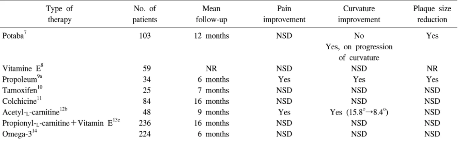
Table 1. Oral therapy for Peyronie’s disease: placebo-controlled, double-blind trials
Type of No. of Mean Pain Curvature Plaque size
therapy patients follow-up improvement improvement reduction
Potaba7 103 12 months NSD No Yes
Yes, on progression of curvature
Vitamine E8 59 NR NSD NSD NR
Propoleum9a 34 6 months Yes Yes Yes
Tamoxifen10 25 7 months NSD NSD NSD
Colchicine11 84 16 months NSD NSD NSD
Acetyl-L-carnitine12b 48 9 months Yes Yes (15.8o→8.4o) NSD
Propionyl-L-carnitine+Vitamin E13c 236 16 months NSD NSD NSD
Omega-314 224 6 months NSD NSD NSD
NSD: no significant difference, NR: not recorded, a: not clinically relevant because available only in Cuba, b: tamoxifen-controlled study,
c: propionyl-L-carnitine and vitamin E were given separately or in combination
Table 2. Intralesional injection therapy for Peyronie’s disease: placebo-controlled trials
Type of No. of Mean Pain Curvature Plaque size
therapy patients follow-up improvement improvement reduction
Collagenase15 49 3 months NR Yesa NR
Verapamil16 14 3 months NR NSD Yes
Interferone α-2b17 117 At least 4 weeks Yes Yes (49.9o→36.4o) Yes
Verapamil20 80 24 weeks NSD NSD NSD
NR: not recorded, NSD: no significant difference, a: thirty-six percent of the patients receiving collagenase expericenced a positive response (curvature improvement, 15o to 20o)
such as extracorporeal shock wave therapy have been attempted to alter the clinical course of PD. The indication for nonsurgical therapy is in the early painful and progressive stage of the disease. However, no conservative treatment is currently avail- able that eventually results in complete relief of all symptoms including pain, plaque formation, and penile curvature. Further- more, most of the oral, injectable, and topical agents either have not been tested in randomized controlled studies or have been shown to be ineffective in placebo-controlled trials. In fact, most studies on the medical treatment of PD are characterized by the absence of a placebo group, small numbers of patients, short follow-up periods, and lack of objective criteria to determine treatment outcomes.5,6
1. Oral therapy
During recent years, several double-blind, placebo-controlled trials on the use of potassium para-aminobenzoate (Potaba), vitamin E, propoleum, tamoxifen, colchicine, acetyl-L-carnitine, propionyl-L-carnitine, and omega-3 for the treatment of PD have been performed, usually with minor or little proven effect
(Table 1).7-14
2. Intralesional injection therapy
In a recent review by Russel et al,5 90% of the studies regarding intralesional injection therapy for PD showed positive outcomes. Unfortunately, most of those studies did not offer convincing evidence-based data, with only 4 placebo-controlled trials being available (Table 2).15-18 However, the power of the former 2 studies was hampered by their small patient popula- tions (27 in the control and 22 in the treatment arm for the collagenase study; 7 in the control and 7 in the treatment arm for the verapamil study).15,16 Hellstrom et al17 present the results of a multicenter, placebo-controlled, single-blind trial of intralesional injection of interferon α-2b for the treatment of PD. A total of 103 subjects completed the study (53 in the saline injection group and 50 in the interferon injection group).
The results of this study were encouraging in that subjects receiving interferon injections experienced improvement in penile curvature, decreased plaque size and density, and reduced pain compared with those who received intralesional
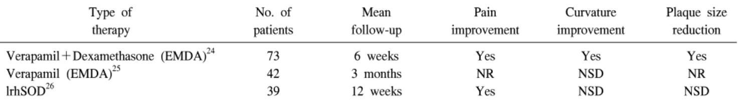
Table 3. Topical or transdermal therapy for Peyronie’s disease: placebo-controlled trials
Type of No. of Mean Pain Curvature Plaque size
therapy patients follow-up improvement improvement reduction
Verapamil+Dexamethasone (EMDA)24 73 6 weeks Yes Yes Yes
Verapamil (EMDA)25 42 3 months NR NSD NR
lrhSOD26 39 12 weeks Yes NSD NSD
NR: not recorded, NSD: no significant difference, EMDA: electromotive drug administration, lrhSOD: liposomally encapsulated recombinant human superoxide dismutase
saline injection. However, repeated dosing of this expensive drug had to be performed, making it less attractive for general use.
Previously, Levine et al18,19 showed that intralesional therapy with verapamil had a positive effect for PD. Unfortunately, these studies did not use a placebo-controlled approach. In a recent placebo-controlled, single-blind trial of intralesional verapamil injection (40 in the saline injection group and 40 in the verapamil injection group), however, no significant improvements were seen in penile curvature, plaque size, and penile pain.20
Recently, Jordan21 reported the result of a prospective, single-center, non-placebo-controlled study with intralesional injection of clostridial collagenase subtypes (Biospecifics Technologies, Lynbrook, USA). In 25 patients with PD, penile curvature and plaque size were significantly improved after treatment. Self-assessment of sexual functioning, such as enjoyment of intercourse, satisfaction with sexual relationships, and freedom from pain during erection, showed significant improvements from baseline.21 However, a double-blind, placebo-controlled multicenter study is required for further validation.
3. Topical or transdermal therapy
A previous study reported that the transdermal application of verapamil gel to the penile shaft fails to infiltrate the tunica albuginea.22 To enhance transdermal drug transport, new tech- niques have been introduced, such as iontophoresis or electro- motive drug administration (EMDA) using electrokinetic transport of charged (ionic) molecules for enhancement of transdermal drug transport into diseased tissue.23 In a randomized controlled study with 5 mg verapamil and 8 mg dexamethasone, a response was observed by a reduction of plaque size in 100%, a response on curvature in 57%, and a response on pain in 76%, which were statistically significant
compared with the control group.24 However, a recent double-blind, placebo-controlled trial to determine the effec- tiveness of verapamil delivered through EMDA failed to show improvement in penile curvature.25 Further study is needed to determine whether EMDA may play a role in the treatment of PD and whether verapamil or other agents delivered via EMDA may exert a therapeutic effect.
A randomized, placebo-controlled series using liposomal recombinant human superoxide dismutase revealed a significant decrease in penile pain compared with the placebo group.26 However, no significant difference in penile curvature or plaque size was noted between groups.26 The result of placebo- controlled trials on the use of topical or transdermal therapy is summarized in Table 3.
4. Extracorporeal shock wave therapy
The data published so far do not justify considering extra- corporeal shock wave therapy as an evidence-based standard procedure for the treatment of PD. It may be beneficial only to achieve freedom from pain within a short time.6,27
Most of the currently available nonsurgical treatment moda- lities so far have not conclusively demonstrated effects. Future randomized controlled studies are needed to investigate whether early conservative intervention can reduce penile deformity or preserve sexual function in men with PD.
FUTURE PERSPECTIVES
Currently, surgical intervention is the only efficacious treatment for PD. Despite the relatively high prevalence of PD, very little scientific information is known about this condition, which is reflected by the lack of any medical treatment to definitively alter the progression of the disorder. Therefore, novel forms of treatment for PD are needed and may emerge
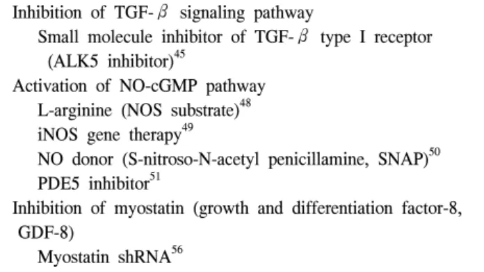
Table 4. Future therapeutic targets for Peyronie’s disease: precli- nical studies
Inhibition of TGF-β signaling pathway
Small molecule inhibitor of TGF-β type I receptor (ALK5 inhibitor)45
Activation of NO-cGMP pathway L-arginine (NOS substrate)48 iNOS gene therapy49
NO donor (S-nitroso-N-acetyl penicillamine, SNAP)50 PDE5 inhibitor51
Inhibition of myostatin (growth and differentiation factor-8, GDF-8)
Myostatin shRNA56
TGF-β: transforming growth factor-β, ALK5: activin receptor- like kinase 5, NO: nitric oxide, cGMP: cyclic 3’,5’-guanosine monophosphate, iNOS: inducible nitric oxide synthase, PDE5: type 5 phosphodiesterase, shRNA: short hairpin RNA
from an understanding of the pathophysiology of this disorder at the molecular level. Recently, several therapeutic targets have emerged in preclinical studies (Table 4).
1. Inhibition of transforming growth factor-β signaling
Transforming growth factor-β1 (TGF-β1) has been iden- tified as the most relevant fibrogenic cytokine and is known to be upregulated in both human PD plaque28 and in the PD-like plaque of the rat model.29 Moreover, genetic variations in the coding region of the TGF-β1 gene have been documented in patients with PD,30 and the expression and activity of the Smad transcription factors of the TGF-β pathway are increased in fibroblasts of patients with PD31 and in the penis of diabetic rats.32
An exciting development has been the recent introduction of animal models for PD to investigate the pathophysiologic mechanisms in vivo and drug development. With the aim of establishing an animal model of PD, several investigators have shown that injection of cytomodulin, a synthetic heptapeptide with TGF-β-like activity; recombinant TGF-β1 protein; or fibrin into the tunica albuginea of rats results in histologic and biochemical changes in the tunica that are similar to those found in human PD plaques.29,33,34 However, the main feature that differentiates these animal models from the human PD condition is the lack of penile curvature. Recently, we for the first time established a PD animal model with penile curvature by using adenoviruses expressing TGF-β1 (ad-TGF-β1).35 Repeated intratunical injections (days 0, 3, and 6) of low-dose
ad-TGF-β1 (1x10¹⁰ particles/0.1 ml) not only induced fibrous scarring in the tunica, but also resulted in significant penile curvature in the artificial erection test 45 days after injection.
However, a single intratunical injection of high-dose ad-TGF- β1 (3x10¹⁰ particles/0.1 ml) did not produce penile curvature.35 This result suggests that repetitive insult to the tunica albuginea plays an important role in the development of penile curvature and agrees with previous suggestions that repetitive trauma to the tunica albuginea is an important etiologic factor in the pathogenesis of PD in humans.3,4 The most striking histologic feature of rats showing penile curvature was a trapping of inflammatory cells at the area of injection in the tunica albuginea and the complete loss of elastin fibers within the area of cartilage formation. In previous studies, ectopic calcification and bone or cartilage formation were observed in human PD plaque.36-38 It is likely that cartilage formation with loss of elastin fibers and a subsequent marked decrease in the expandability of the tunica albuginea in our animal model may be highly responsible for penile bending during erection.35 This finding further supports an etiologic role of TGF-β1 in the pathogenesis of PD. Therefore, therapies aimed at blocking the TGF-β signaling pathway might be efficacious in the treatment or prevention of PD.
Antagonizing TGF-β signaling through the use of neu- tralizing antibodies, soluble type II receptors (TGFβRII), or antisense oligonucleotides has been reported to inhibit various types of TGF-β-mediated fibrosis.39-41 However, these thera- pies can induce unwanted side effects as the result of the multifunctional nature of TGF-β.39 Furthermore, the fusion of the extracellular domain of TGFβRII with the Fc portion of immunoglobulin might create immunogenic epitopes. Activin receptor-like kinase 5 (ALK5) is a type I receptor of TGF-β.
ALK5 inhibition has been reported to attenuate the tissue fibrosis in kidney, lung, and liver.42-44
Recently, we further elucidated the molecular mechanism of PD and have uncovered potential targets for molecular-based therapies.45 In that study, we determined the effectiveness of IN-1130, a novel small molecule inhibitor of ALK5, in promoting the regression of fibrotic scarring and correcting penile curvature in a rat model of PD induced by repeated intratunical injections of ad-TGF-β1. The rats were randomly assigned into 5 groups: control, control with IN-1130 injections, PD group without treatment, PD with saline injections, and PD with IN-1130 injections. IN-1130 (5 mg/kg in 0.1 ml saline)
or saline was given twice (days 30 and 37). The PD rats treated with IN-1130 showed significant regression of fibrotic plaque and improvement in penile curvature compared with those receiving no injections or saline injections. The PD group treated with IN-1130 had a posttreatment curvature of 9.1o versus 23.0o and 32.6o for the no treatment and saline-treated PD groups, respectively. The control rats receiving IN-1130 did not show any changes histologically or in the artificial erection test.45 We also addressed evidence indicating the inhibitory mechanisms of IN-1130 in the tunical fibrosis of PD rats. First, IN-1130 significantly reduced the infiltration of inflammatory cells, including lymphocytes, plasma cells, macrophages, fibroblasts, and myofibroblasts. Second, IN-1130 attenuated the transnuclear expression of phospho-Smad2 and phospho- Smad3, the crucial step for initiation of TGF-β signal transduction. Third, IN-1130 suppressed collagen accumulation as evaluated by hydroxyproline determination. Finally, IN-1130 restored elastin fibers by inhibiting macrophage recruitment.
Macrophages can produce an array of matrix metalloproteinases (MMPs). Of those MMPs, MMP-12, a macrophage metalloela- stase, can degrade elastin.46 Therefore, we suggest that IN- 1130-induced restoration of elastin fibers may be mediated by decreased infiltration of macrophages in the fibrotic plaque.45 This is the first report showing that pharmacologic intervention aimed at specifically blocking the TGF-β pathway in the penis is potentially useful for the treatment of PD and such inter- vention warrants investigation as a novel therapy for human PD.
2. Activation of nitric oxide-cGMP pathway
It is thought that nitric oxide (NO) produced by inducible nitric oxide synthase (iNOS) has anti-fibrotic actions in the rat, because long-term administration of an iNOS inhibitor (L-N- (1-iminoethyl)-lysine, L-NIL) enhances fibrosis of the PD-like plaque and induces generalized corporal fibrosis,47 whereas the NOS substrate L-arginine48 or gene therapy with iNOS cDNA49 reduces the plaque. An NO donor (S-nitroso-N-acetyl penicilla- mine, SNAP) also seems to have anti-fibrotic actions; it inhibits collagen synthesis and myofibroblast differentiation in cell cultures from PD plaques and normal tunica albuginea.50 It was recently reported that long-term continuous administration of phosphodiesterase-5 (PDE5) inhibitors, which should maintain or increase cyclic 3’,5’-guanosine monophosphate (cGMP) levels in the target tissues, prevents the development of fibrotic
plaques in the rat model of PD.48,51 3. Inhibition of myostatin
Myostatin, another member of the TGF-β family that is also known as growth and differentiation factor-8 (GDF-8), has been proposed as an inducer of fibrosis in skeletal muscle52-55 and has the following actions: 1) Myostatin stimulates fibroblast proliferation and induces differentiation of fibroblasts into myofibroblasts. 2) Myostatin and TGF-β1 stimulate each other’s expression and they colocalize in myofibers in the early stage of muscle injury. 3) Myostatin-deficient mice show less fibrosis than do wild-type mice after muscle injury.
A recent study reported that myostatin was overexpressed in human PD plaque.56 Furthermore, myostatin elicited a PD-like plaque in the rat and intensified the plaque triggered by TGF-β 1. The authors suggested that anti-fibrotic therapies aimed at inhibiting both the myostatin and the TGF-β pathways would be more effective than blocking the pathways individually.56
CONCLUSION
Although none of the currently available nonsurgical treat- ment modalities is efficacious for preventing the progression of PD, the results of recent preclinical studies targeting the TGF- β pathway or NO-cGMP pathway are promising. With further research into the pathologic cascade of cellular and molecular events and an increase in our understanding of the pathophy- siology of PD using animal models, the development of novel and effective medical therapies will become a realistic objec- tive.
REFERENCES
1. Schwarzer U, Sommer F, Klotz T, Braun M, Reifenrath B, Engelmann U. The prevalence of Peyronie's disease: results of a large survey. BJU Int 2001;88:727-30
2. Mulhall JP, Creech SD, Boorjian SA, Ghaly S, Kim ED, Moty A, et al. Subjective and objective analysis of the prevalence of Peyronie’s disease in a population of men presenting for prostate cancer screening. J Urol 2004;171:2350-3
3. Jarow JP, Lowe FC. Penile trauma: an etiologic factor in Peyronie’s disease and erectile dysfunction. J Urol 1997;158:
1388-90
4. Devine CJ Jr, Somers KD, Jordan SG, Schlossberg SM.
Proposal: trauma as the cause of the Peyronie’s lesion. J Urol 1997;157:285-90
5. Russell S, Steers W, McVary KT. Systematic evidence-based analysis of plaque injection therapy for Peyronie's disease. Eur Urol 2007;51:640-7
6. Hauck EW, Diemer T, Schmelz HU, Weidner W. A critical analysis of nonsurgical treatment of Peyronie's disease. Eur Urol 2006;49:987-97
7. Weidner W, Hauck EW, Schnitker J. Potassium paraamino- benzoate (POTABA) in the treatment of Peyronie’s disease: a prospective, placebo- controlled, randomized study. Eur Urol 2005;47:530-5
8. Pryor JP, Farell CR. Controlled clinical trial of vitamin E in Peyronie’s disease. Prog Reprod Biol Med 1983;9:41-5 9. Lemourt Oliva M, Filgueiras Lopez E, Rodriguez Barroso A,
Gonzalez Oramas E, Bordonado R. Clinical evaluation of the use of propoleum in Peyronie’s disease. Arch Esp Urol 1998;
51:171-6
10. Teloken C, Rhoden EL, Grazziotin TM, Da Ros CT, Sogari PR, Souto CA. Tamoxifen versus placebo in the treatment of Peyronie’s disease. J Urol 1999;162:2003-5
11. Safarinejad MR. Therapeutic effects of colchicine in the management of Peyronie’s disease: a randomized double-blind, placebo-controlled study. Int J Impot Res 2004;16:238-43 12. Biagotti G, Cavallini G. Acetyl-L-carnitine vs tamoxifen in the
oral therapy of Peyronie’s disease: a preliminary report. BJU Int 2001;88:63-7
13. Safarinejad MR, Hosseini SY, Kolahi AA. Comparison of vitamin E and propionyl-L-carnitine, separately or in combi- nation, in patients with early chronic Peyronie's disease: a double-blind, placebo controlled, randomized study. J Urol 2007;178:1398-403
14. Safarinejad MR. Efficacy and safety of omega-3 for treatment of early-stage Peyronie's disease: a prospective, randomized, double-blind placebo-controlled study. J Sex Med 2009; Epub ahead of print
15. Gelbard MK, James K, Riach P, Dorey F. Collagenase versus placebo in the treatment of Peyronie’s disease: a double-blind study. J Urol 1993;149:56-8
16. Rehman J, Benet A, Melman A. Use of intralesional verapamil to dissolve Peyronie’s disease plaque: a long-term single-blind study. Urology 1998;51:620-6
17. Hellstrom WJ, Kendirci M, Matern R, Cockerham Y, Myers L, Sikka SC, et al. Single-blind, multicenter, placebo con- trolled, parallel study to assess the safety and efficacy of intralesional interferon alpha-2B for minimally invasive treatment for Peyronie’s disease. J Urol 2006;176:394-8 18. Levine LA, Merrick PF, Lee RC. Intralesional verapamil
injection for the treatment of Peyronie's disease. J Urol 1994;
151:1522-4
19. Levine LA, Goldman KE, Greenfield JM. Experience with intraplaque injection of verapamil for Peyronie's disease. J Urol 2002;168:621-5
20. Shirazi M, Haghpanah AR, Badiee M, Afrasiabi MA,
Haghpanah S. Effect of intralesional verapamil for treatment of Peyronie's disease: a randomized single-blind, placebo- controlled study. Int Urol Nephrol 2009; Epub ahead of print 21. Jordan GH. The use of intralesional clostridial collagenase
injection therapy for Peyronie's disease: a prospective, single- center, non-placebo-controlled study. J Sex Med 2008;5:180-7 22. Martin DJ, Badwan K, Parker M, Mulhall JP. Transdermal
application of verapamil gel to the penile shaft fails to infiltrate the tunica albuginea. J Urol 2002;168:2483-5 23. Singh J, Maibach HI. Topical iontophoretic drug delivery in
vivo: historical development, devices and future perspectives.
Dermatology 1993;187:235-8
24. Di Stasi SM, Giannantoni A, Stephen RL, Capelli G, Giurioli A, Jannini EA, et al. A prospective, randomized study using transdermal electromotive administration of verapamil and dexamethasone for Peyronie’s disease. J Urol 2004;171:1605-8 25. Greenfield JM, Shah SJ, Levine LA. Verapamil versus saline in electromotive drug administration for Peyronie's disease: a double-blind, placebo controlled trial. J Urol 2007;177:972-5 26. Riedl CR, Sternig P, Galle G, Langmann F, Vcelar B, Vorauer
K, et al. Liposomal recombinant human superoxide dismutase for the treatment of Peyronie’s disease: a randomized placebo- controlled double-blind prospective clinical study. Eur Urol 2005;48:656-61
27. Hauck EW, Hauptmann A, Bschleipfer T, Schmelz HU, Altinkilic BM, Weidner W. Questionable efficacy of extracor- poreal shock wave therapy for Peyronie’s disease: results of a prospective approach. J Urol 2004;171:296-9
28. El-Sakka AI, Hassoba HM, Pillarisetty RJ, Dahiya R, Lue TF.
Peyronie’s disease is associated with an increase in trans- forming growth factor-beta protein expression. J Urol 1997;
158:1391-4
29. El-Sakka AI, Hassoba HM, Chui RM, Bhatnagar RS, Dahiya R, Lue TF. An animal model of Peyronie’s-like condition associated with an increase of transforming growth factor beta mRNA and protein expression. J Urol 1997;158:2284-90 30. Hauck EW, Hauptmann A, Schmelz HU, Bein G, Weidner W,
Hackstein H. Prospective analysis of single nucleotide polymorphisms of the transforming growth factor beta-1 gene in Peyronie's disease. J Urol 2003;169:369-72
31. Haag SM, Hauck EW, Szardening-Kirchner C, Diemer T, Cha ES, Weidner W, et al. Alterations in the transforming growth factor (TGF)-beta pathway as a potential factor in the patho- genesis of Peyronie's disease. Eur Urol 2007;51:255-61 32. Zhang LW, Piao S, Choi MJ, Shin HY, Jin HR, Kim WJ, et
al. Role of increased penile expression of transforming growth factor-beta1 and activation of the Smad signaling pathway in erectile dysfunction in streptozotocin-induced diabetic rats. J Sex Med 2008;5:2318-29
33. El-Sakka AI, Hassan MU, Nunes L, Bhatnagar RS, Yen TS, Lue TF. Histological and ultrastructural alterations in an animal model of Peyronie’s disease. Br J Urol 1998;81:445-52
34. Davila HH, Ferrini MG, Rajfer J, Gonzalez-Cadavid NF.
Fibrin as an inducer of fibrosis in the tunica albuginea of the rat: a new animal model of Peyronie’s disease. BJU Int 2003;
91:830-8
35. Piao S, Ryu JK, Shin HY, Zhang L, Song SU, Han JY, et al. Repeated intratunical injection of adenovirus expressing transforming growth factor- beta1 in a rat induces penile curvature with tunical fibrotic plaque: a useful model for the study of Peyronie's disease. Int J Androl 2008;31:346-53 36. Yudkin JS. Peyronie's disease in association with metoprolol.
Lancet 1977;2:1355
37. Lowsley OS, Boyce WH. Further experiences with an opera- tion for the cure of Peyronie's disease. J Urol 1950;63:888-902 38. Anafarta K, Beduk Y, Uluoglu O, Aydos K, Baltaci S. The
significance of histopathological changes of the normal tunica albuginea in Peyronie's disease. Int Urol Nephrol 1994;26:71-7 39. Border WA, Noble NA. Transforming growth factor beta in
tissue fibrosis. N Engl J Med 1994;331:1286-92
40. Hakenjos L, Bamberg M, Rodemann HP. TGF-beta1-mediated alterations of rat lung fibroblast differentiation resulting in the radiation-induced fibrotic phenotype. Int J Radiat Biol 2000;
76:503-9
41. Martin M, Lefaix J, Delanian S. TGF-beta1 and radiation fibrosis: a master switch and a specific therapeutic target? Int J Radiat Oncol Biol Phys 2000;47:277-90
42. Grygielko ET, Martin WM, Tweed C, Thornton P, Harling J, Brooks DP, et al. Inhibition of gene markers of fibrosis with a novel inhibitor of transforming growth factor-beta type I receptor kinase in puromycin-induced nephritis. J Pharmacol Exp Ther 2005;313:943-51
43. Bonniaud P, Margetts PJ, Kolb M, Schroeder JA, Kapoun AM, Damm D, et al. Progressive transforming growth factor beta1- induced lung fibrosis is blocked by an orally active ALK5 kinase inhibitor. Am J Respir Crit Care Med 2005;171:889-98 44. de Gouville AC, Boullay V, Krysa G, Pilot J, Brusq JM,
Loriolle F, et al. Inhibition of TGF-beta signaling by an ALK5 inhibitor protects rats from dimethylnitrosamine-induced liver fibrosis. Br J Pharmacol 2005;145:166-77
45. Ryu JK, Piao S, Shin HY, Choi MJ, Zhang LW, Jin HR, et al. IN-1130, a novel transforming growth factor-beta type i receptor kinase (activin receptor-like kinase 5) inhibitor, promotes regression of fibrotic plaque and corrects penile curvature in a rat model of Peyronie's disease. J Sex Med
2009;6:1284-96
46. Gronski TJ Jr, Martin RL, Kobayashi DK, Walsh BC, Holman MC, Huber M, et al. Hydrolysis of a broad spectrum of extracellular matrix proteins by human macrophage elastase. J Biol Chem 1997;272:12189-94
47. Ferrini MG, Vernet D, Magee TR, Shahed A, Qian A, Rajfer J, et al. Antifibrotic role of inducible nitric oxide synthase.
Nitric Oxide 2002;6:283-94
48. Valente EG, Vernet D, Ferrini MG, Qian A, Rajfer J, Gonzalez-Cadavid NF. L-arginine and phosphodiesterase (PDE) inhibitors counteract fibrosis in the Peyronie’s fibrotic plaque and related fibroblast cultures. Nitric Oxide 2003;9:
229-44
49. Davila HH, Magee TR, Vernet D, Rajfer J, Gonzalez-Cadavid NF. Gene transfer of inducible nitric oxide synthase comple- mentary DNA regresses the fibrotic plaque in an animal model of Peyronie’s disease. Biol Reprod 2004;71:1568-77 50. Vernet D, Ferrini MG, Valente EG, Magee TR, Bou-Gharios
G, Rajfer J, et al. Effect of nitric oxide on the differentiation of fibroblasts into myofibroblasts in the Peyronie’s fibrotic plaque and in its rat model. Nitric Oxide 2002;7:262-76 51. Ferrini MG, Kovanecz I, Nolazco G, Rajfer J, Gonzalez-
Cadavid NF. Effects of long-term vardenafil treatment on the development of fibrotic plaques in a rat model of Peyronie's disease. BJU Int 2006;97:625-33
52. Wagner KR, McPherron AC, Winik N, Lee SJ. Loss of myostatin attenuates severity of muscular dystrophy in mdx mice. Ann Neurol 2002;52:832-6
53. Bogdanovich S, Krag TO, Barton ER, Morris LD, Whittemore LA, Ahima RS, et al. Functional improvement of dystrophic muscle by myostatin blockade. Nature 2002;420:418-21 54. McCroskery S, Thomas M, Platt L, Hennebry A, Nishimura
T, McLeay L, et al. Improved muscle healing through en- hanced regeneration and reduced fibrosis in myostatin-null mice. J Cell Sci 2005;118:3531-41
55. Zhu J, Li Y, Shen W, Qiao C, Ambrosio F, Lavasani M, et al. Relationships between transforming growth factor-beta1, myostatin, and decorin: implications for skeletal muscle fibrosis. J Biol Chem 2007;282:25852-63
56. Cantini LP, Ferrini MG, Vernet D, Magee TR, Qian A, Gelfand RA, et al. Profibrotic role of myostatin in Peyronie’s disease. J Sex Med 2008;5:1607-22